Abstract
INTRODUCTION
Intramedullary nailing is a common treatment for proximal femoral fractures. Fracture of the nail is a rare but devastating complication that exposes often frail patients to complex revision surgery. We investigated which risk factors predict nail failure.
METHODS
We reviewed all cases of nail breakage seen over a 10-year period in a single busy trauma unit; 22 nail fractures were seen in 19 patients. Comparison was made with a group of 209 consecutive patients who underwent intramedullary fixation of a proximal femur fracture with no nail breakage over a 2-year period.
RESULTS
In the fractured nail group, mean age was 70.4 years (range 55–88 years).The mean time to fracture was 10 months (range 2.5–23 months). Logistical regression was used to show that low American Society of Anesthesiologists (ASA) score, subtrochanteric fracture and pathological fracture were independent risk factors for nail fracture.
CONCLUSIONS
Young patients with a low ASA score are at highest risk of nail breakage. We advise close follow-up of patients with these risk factors until bony union has been achieved. In addition, there may be merit in considering other treatment options, such as proximal femoral replacement, especially for those with pathological fracture with a good prognosis.
Keywords: Intramedullary nail, Nail fracture, Subtrochanteric fracture, Proximal femoral fracture
Introduction
The National Institute for Health and Care Excellence (NICE) advocates intramedullary nailing for subtrochanteric femoral fractures.1 The guidance is silent with regard to reverse oblique and A3 femoral fractures. In their subsequent meta-analyses, Kokoroghiannis et al2 suggest that the cephalomedullary nail is indicated in this fracture pattern. They also show that this device is frequently used for peritrochanteric fractures involving the lesser trochanter (AO: A2). The National Hip Fracture Database 2015 report shows that intramedullary nailing is performed for approximately 78% of subtrochanteric fractures and around 10% of all hip fracture procedures, with a 47% increase in their use since 2010.3 The fixation can be technically difficult, given the nature of the fracture and the patient population. Implant failure in the form of device fracture is a rare but catastrophic event. Salvage procedures are invariably challenging and expose a population to further complex surgery, with a mean 30-day mortality of 8%. Reported rates of cephalomedullary nail fracture range from to 0.2% to 5.6%.4–6
Given the gravity of the event, it is important to determine patient factors which contribute to and are associated with this form of implant failure. No previous studies have determined patient-specific factors that predisposed to nail fracture. Identification of an ‘at-risk group’ may help in preventing this mode of failure. This group may benefit from increased clinical surveillance.
Abram et al explored radiographic features of all modes of Gamma nail failure, including cut-out, subsidence and fracture.6 The authors reported that standard practice involved a single review at 6 weeks following fixation, with discharge for those mobile and pain-free. Patients were only reassessed if they were referred back to clinic. However, the features indicative of impending failure may not be appreciated or detected as promptly if those assessing patients do not have sufficient relevant experience. Safe practice may involve more frequent assessment for high-risk individuals.
The principle difficulty in definitively defining risk factors for fracture failure lies in collecting a sufficient sample size. To date, the largest cohort of fractured nails in a single study is 14.7 Our objective was therefore to define the clinical risk factors for post-fixation nail breakage. In so doing, we present the largest series of fractured cephalomedullary nails in the literature with a view to identifying patient features which predispose to implant breakage failure.
Materials and methods
We retrospectively identified all cephalomedullary nail failures over a 10-year period at our institution (2004–2013). Patients were identified from theatre OPCS codes and accuracy ensured by crosschecking all radiographs. Clinical notes were reviewed to determine patient features including age, sex, American Society of Anesthesiologists (ASA) score, fracture pattern and whether fractures were confirmed pathological. A subtrochanteric fracture was defined as a proximal femoral fracture occurring beneath the lesser trochanter but no further than 5 cm beneath this landmark.8 Surgery was carried out by consultant orthopaedic surgeons or experienced registrars on the general trauma rota.
Varus and valgus angulation post-fixation was measured on digital anteroposterior radiographs and residual flexion or extension on lateral radiographs. Radiographic measurement was carried out by a single observer. Angles were measured from lines drawn through the centre of the intramedullary canal in the proximal and distal fragments, intersecting at the fracture side. Three patients did not have post-fixation radiographs available for review. Malreduction was defined as five degrees of flexion/extension angulation, five degrees of varus/valgus angulation or 50% or more translation at the fracture site.
We compared this cohort with a control group of consecutive patients for whom cephalomedullary nail was performed between November 2009 and November 2011 without breakage. This allowed a minimum follow-up of 2 years. Information for this cohort was obtained from the information entered into the National Hip Fracture Database. From this source was determined patient age, sex, ASA score, fracture pattern and whether fractures were confirmed pathological.
The indications for long nail were fractures with extension beyond the fixation length of the short nail and pathological fractures. This generally meant that long nails were reserved for subtrochanteric fractures and short nails for unstable peri-trochanteric fractures.
Statistical methods
Continuous variables were compared with t-test. Proportions were analysed with a Chi-square test. Multivariate logistic regression was performed to determine which factors acted as independent predictors of device fracture failure.
Results
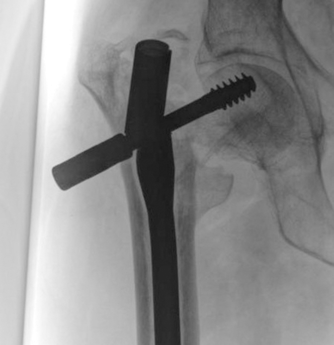
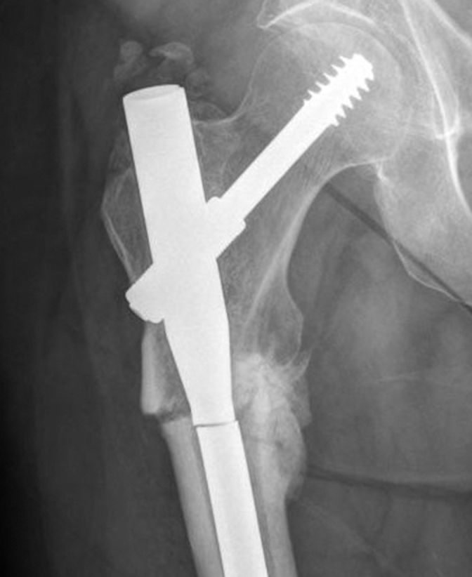
There were 19 patients with 22 broken nails between 2004 and 2013. There was one case of bilateral nail failure and two cases of recurrent ipsilateral breakage. Mean age was 70.4 years (range 55–88 years). The site of nail failure was either at the lag screw aperture in the barrel (55%; Figure 1) or distal barrel taper (45%; Figure 2). In all but one case, failure was an insidious process due to fatigue failure. In the remaining case, failure was preceded by a fall. The mean time to fracture was 10 months (range: 2.5–23 months). Twenty nails which fractured were Intramedullary Hip Screw (Smith and Nephew, Cordova, TN, USA) and two were Affixus® Hip Fracture Nails (DePuy Orthopaedics Inc, Warsaw, IN, USA). Mean nail diameter was 13.6 mm (range 12–14mm). Nine fractures in eight patients were pathological (tumour: lymphoma, 3; renal 2 (bilateral); breast 2; benign 1). All procedures for pathological fractures were for displaced fractures and were not prophylactic nailings. No atypical fractures secondary to bisphosphonates were seen.
Figure 1.

Anteroposterior radiograph showing fracture of nail through barrel lag screw aperture
Figure 2.

Nail breakage just distal to barrel taper
In the vast majority of cases, nails implanted for subtrochanteric fractures underwent failure. This is also reflected in the higher proportion of long nails in the broken nail cohort (86% versus 7.5%) Non-union and malreduction was a feature of all failures with the exception of one. In this case, the fracture was undisplaced but failed to unite. Revision procedures included nine repeat intramedullary nails, five proximal femoral replacements, four fixed-angle device, two long dynamic hip screw, one hemiarthroplasty and one total hip replacement. Four of the repeat nailings required further revision. All of the patients treated with fixed-angle devices achieved union. All three of the pathological fractures treated with revision nailing failed to heal and required further surgery.
There were 209 patients in our control group, who underwent fixation with a cephalomedullary nail between November 2009 and November 2011, in whom there was no fixation device fracture (Table 1). From this group, there were five patients for whom the ASA was not known and two for whom there was no recorded age. They were thus excluded from certain aspects of the analysis (mean age, ASA ratio and logistic regression). When compared with the broken-nail group, those who suffered nail fracture were significantly younger (70 years vs. 80 years, P<0.0005). Mean age difference was 9.2 years. Eighty-six per cent of nail breakage cases involved subtrochanteric fractures.
Table 1.
Univariate analysis of features of broken and intact nail cohorts
| Intact nail | Broken nail | P value | |
|---|---|---|---|
| Patients (n) | 209 | 22 | |
| Long nail (%) | 37.5 | 86 | |
| Male (%) | 30 | 27 | 1.0 (χ2 test) |
| Mean age (years) | 79.6 | 70.4 | < 0.0005 (t test) |
| ASA score I or II (%) | 38 (79/209) | 59 (13/22) | 0.067 (χ2) |
| Confirmed pathological fracture (%) | 5 (11/209) | 41 (9/22) | < 0.0001 (χ2) |
| Subtrochanteric fracture (%) | 17% (36/209) | 95% (21/22) | < 0.0001 (χ2test) |
ASA = American Society of Anesthesiologists
On logistical regression analysis subtrochanteric fractures were strongly and independently associated with fixation breakage (P<0.0001; Table 2). Confirmed pathological fracture (P=0.026) and lower ASA (P=0.004) were also independent risk factors. Age was not independently associated with nail fracture.
Table 2.
Logistic regression predictors of failure
| Odds ratio | 95% CI | P value | |
|---|---|---|---|
| Age (decades) | 0.7 | 0.4–1.2 | 0.20 |
| ASA score | 0.2 | 0.05–0.6 | 0.004 |
| Subtrochanteric fracture | 163 | 15–1716 | < 0.0001 |
| Confirmed pathological fracture | 17.5 | 3–114 | 0.003 |
ASA = American Society of Anesthesiologists; CI = confidence interval
To reduce confounders, a separate sub-analysis was performed comparing only intramedullary hip screw devices used to treat subtrochanteric fractures. Results were similar, with only younger age being associated with nail fracture (70.8 vs. 78.3, P=0.01; Table 3).
Table 3.
Sub-analysis of IMHS devices used in subtrochanteric fractures only
| Intact nail | Broken nail | P value | |
|---|---|---|---|
| Patients (n) | 36 | 20 | |
| Male (%) | 36 | 26 | 0.82 (χ2test) |
| Mean age (years) | 78.3 | 70.8 | 0.01 (t test) |
| ASA I or II (%) | 36 (13/23) | 45 (9/11) | 0.28 (χ2test) |
Discussion
No previous studies have identified patient factors associated with cephalomedullary nail breakage. Using the largest series to date (22 cases), we show that failure is strongly associated with lower ASA, pathological and subtrochanteric fractures. Fortunately, nail failure remains a relatively rare phenomenon. A crude estimate of nail breakage rate can be calculated from our data. In our series, there were 22 failures in 10 years. This corresponds to two failures a year. In two years, 209 nails were implanted. With approximately 100 nails implanted annually, our breakage rate can be estimated at 2%. One must be mindful, however, that this is based on the assumption that the rate of nailing has not changed over the relevant time frame, which may not be true. Nonetheless, this figure is consistent with the 0.2–5.6% rate observed in the literature.4,5,6 This figure is also comparable with the 1.3% (3/212) fracture rate observed by Abram et al in their recent UK study.6 It is, however, a catastrophic event; 23% (5/22) underwent proximal femoral replacement and 9% (2/22) complex arthroplasty procedures for salvage. It is thus important to identify those most at risk. Closer outpatient follow-up for these patients until bony union has been achieved has cost implications. The benefits of carrying out a planned revision procedure prior to nail fracture may, however, outweigh the technical difficulties in performing revision for a broken implant, together with the emergency admission required and pain experienced with nail fracture.
The concentration of subtrochanteric fractures within the failure group causes some concern and suggests that this group deserves special attention; 21 of the 22 fractured nails were implanted in subtrochanteric fractures over a 10-year period. In 2 years, there were 38 subtrochanteric nail fixations. This corresponds approximately to 200 (5 × 38) such fractures in 10 years. On a similar estimation, the failure rate for the cephalomedullary nail in subtrochanteric fractures could be as high as 10% (21/200).
The risk engendered by the subtrochanteric fracture may be explained by the observations of Abram et al.6 The group identified radiographic features of Gamma nail fixation which portend fixation failure in peri-trochanteric fractures. Their 16 cases of failure encompassed all modes of failure including implant cut out and subsidence. There were only three cases of nail breakage. However, similar to our study, non-union was a feature of the cases of failure, hence their finding are instructive. They identified three fixation points for the nail in the proximal femur that were conducive to implant survival. This configuration provides robust fixation either side of the fracture in the case of peri-trochanteric fractures. In the case of the subtrochanteric fracture, the fixation poles are all proximal to the fracture. There cannot be three-point fixation across the fracture as described for peri-trochanteric fractures. This may explain the strong link seen between the subtrochanteric fracture pattern and nail breakage.
Consistent with our in vivo observations Bauer et al reported that fracture stability also plays a role in implant fracture.9 The median fatigue limit (MLF) is defined as the minimum load that results in implant failure if loaded with 500,000 cycles. The group found that without the support of the calcar, nail breakage failure occurs at a 28% lower MLF compared to fractures with the calcar intact. Subtrochanteric fractures lack a medial support and therefore fall into this genre of fractures with regard to failure.
Those patients in whom the implant survived have the same mean age as the UK national mean age for hip fracture patients according to large studies.10 Those with implant fracture are on average 10 years younger. The links between both youth and low ASA suggest that nail failure may be a more prominent feature of moderate to high demand patients. Fatigue failure as observed in our study occurs when a material is subjected to repeated cycles of load which are below the ultimate tensile strength of the material. Youth and improved ASA may be associated with more loading cycles in the postoperative months prior to union. This is all the more so now that the NICE guidance explicitly states that all fixation devices should allow immediate postoperative full weight bearing.1 A link with age and postoperative steps has been observed in arthroplasty populations. Schmalzried et al reported that younger patients walked significantly further postoperatively than those over 65.11 The nail may thus be exposed to more cycles prior to union.
Healthier patients may also subject implants to a greater number of steps simply because of their improved survivorship. However, if longevity was the only factor, one would expect a much greater proportion of women in the failure group, given that their survival after hip fracture is greater than that for men. This is not the case. Intriguingly, on multivariate analysis, age was not a predictor for fracture when adjustment was made for ASA and other potential confounders. This suggests that physiological age and activity level may be more important than numerical age.
In our study, 41% of implant breakages occurred in patients with confirmed pathological fractures. In a 2-year period there were 11 such fractures. This extrapolates to approximately 55 pathological fractures in 10 years. We observed nine nail breakages following fixation of pathological fractures over the 10-year surveillance period. Hence, the estimated nail breakage rate for pathological fractures may be as high as 16% (9/55). Advances in oncology have meant that even patients with bony metastases are enjoying greater survivorship. It can no longer be assumed that, in this cohort, fixation devices will invariably out-survive the patients. Reoperation in this group is complex and intrusive when the patient’s health is often declining. Endoprosthetic reconstruction has been shown to be associated with fewer treatment failures, greater implant durability and a 50% reduction in all-cause failure when compared with internal fixation.12,13 A major complication rate of 12% has been reported in conversion of failed internal fixation for pathological proximal femur fracture to arthroplasty.14
While there was a high proportion of long nails in the breakage cohort, we believe this to reflect a high frequency of subtrochanteric and pathological fractures in this group. Fracture pattern determined whether the long nail was used in preference to the short. Long nails are mandatory in cases of pathological fracture.
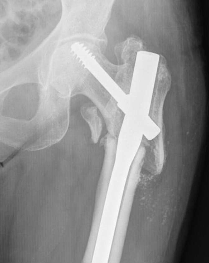
We observed that the nails were most vulnerable to fracture at the lag screw aperture and at or just distal to the distal barrel taper (Figure 3). The proximal nail is narrowest in cross-section at the site of the cephalic lag screw.15 In addition, it is subjected to the torque of the patient load against the cephalic lag screw, in the absence of the calcar. The stresses in the subtrochanteric region are the highest in the human body at 1200 lb/square inch.7 In absence of the calcar, this stress is borne by the implant. In 18 of our 22 cases, nail breakage was preceded by fracture of the distal locking screws. This ‘self-dynamisation’ and subsequent nail fracture has previously been described as a standard mode of failure.16,17 Biomechanical analysis has shown that the predominant axial load is transferred to the distal nail and therefore the distal locking screws.18,19 Twelve of the fractures in our series were fixed in varus misalignment (mean 9 degrees, range 4–15 degrees). A deficient medial cortex with high bending forces in the subtrochanteric region accentuated by varus misalignment is known to lead to a significant risk of failure and non-union.20
Figure 3.

Subtrochanteric femoral fracture fixed in varus angulation
The second largest series of 14 broken nails described by Giannoudis et al observed similar trends to ours.7 They reported that varus misalignment was a common feature of nail breakage due to non-union. In addition, they observed that subtrochanteric fractures were over-represented in the broken nail cohort. Indeed, all their cases involved this fracture pattern. They also noted breakage of the distal locking screws to be a predictor of nail fracture. Their series excluded all pathological fractures and hypertrophic non-unions. In addition, there was no control group from which to identify specific risk factors for fracture.
Our study is limited by its retrospective nature. Hence, it is possible that some episodes of nail fracture may have been overlooked. Some cases may have been lost due to patient migration, although this cohort tends not to migrate. Radiographic parameters of the intact nails were not measured. We did not record the seniority of the surgical team and data on body mass index, weight or further comorbidities were not available. However, surgery was carried out by broadly the same group of surgeons so one would expect similar adequacy of reduction and fixation. Our inclusion period for nail breakage was a 10-year window from 2004 to 2013 but, in order to recruit a sufficiently powered control group of nails, a much shorter time frame was necessary, from 2009 to 2011. There was thus overlap but no precise chronological matching. However, we felt justified in comparing metachronous groups on the grounds that over the periods compared there had been no clinically significant change in clinical practice, surgical technique or implant design.
We considered that all nails included were sufficiently similar in design for meaningful evaluation of the risk factors for breakage to be made. All nails included in the study were intramedullary hip screws (IMHS, Smith & Nephew), with the exception of two Affixus nails. Both the IMHS and Affixus nail are intramedullary devices with cephalic elements that comprise a partially threaded lag screw. The Affixus nail differs from the IMHS in that it has a dominant (inferior) and subordinate (superior) lag screw. The IMHS carries a single lag screw. None of the nails was the structurally distinct proximal femoral nail (anti-rotation), where the cephalic component is a blade, so we considered that there was sufficient congruity in design for the inclusion of both nails in the study.
Conclusion
The risk of implant fatigue failure is highest in young patients of good health. Younger patients with lower ASA score (I/II) and/or subtrochanteric fracture may benefit from review until fracture union to avoid the rare but catastrophic event of gamma nail failure. Risk of intramedullary nail failure in pathological femoral fractures is high. Other treatment options, such as proximal femoral replacement, should be considered.
References
- 1.National Institute for Health and Care Excellence. Hip Fracture: Management. (Clinical Guideline 124) London: NICE; 2011, last updated March 2014 https://www.nice.org.uk/guidance/cg124 (cited August 2016). [PubMed] [Google Scholar]
- 2.Kokoroghiannis C, Aktselis I, Deligeorgis A et al. Evolving concepts of stability and intramedullary fixation of intertrochanteric fractures: a review. Injury 2012; : 686–693. [DOI] [PubMed] [Google Scholar]
- 3.National Hip Fracture Database. Annual Report 2015. London: Royal College of Physicians; http://www.nhfd.co.uk/2015report (cited August 2016). [Google Scholar]
- 4.Alvarez DB, Aparicio JP, Fernández EL et al. Implant breakage, a rare complication with the Gamma nail. A review of 843 fractures of the proximal femur treated with a Gamma nail. Acta Orthop Belg 2004; :435–443. [PubMed] [Google Scholar]
- 5.Iwakura T, Niikura T, Lee SY et al. Breakage of a third generation gamma nail: a case report and review of the literature. Case Rep Orthop 2013; : 172352. [DOI] [PMC free article] [PubMed] [Google Scholar]
- 6.Abram SG, Pollard TC, Andrade AJ. Inadequate ‘three-point’ proximal fixation predicts failure of the Gamma nail. Bone Joint J 2013; : 825–830. [DOI] [PubMed] [Google Scholar]
- 7.Giannoudis PV, Ahmad MA, Mineo GV et al. Subtrochanteric fracture non-unions with implant failure managed with the ‘Diamond’ concept. Injury 2013; : S76–S81. [DOI] [PubMed] [Google Scholar]
- 8.Parker M, Johansen A. Hip fracture. BMJ 2006; : 27–30. [DOI] [PMC free article] [PubMed] [Google Scholar]
- 9.Eberle S, Bauer C, Gerber C et al. The stability of a hip fracture determines the fatigue of an intramedullary nail. Proc Inst Mech Eng H 2010; : 577–584. [DOI] [PubMed] [Google Scholar]
- 10.Maxwell MJ, Moran CG, Moppett IK. Development and validation of a preoperative scoring system to predict 30 day mortality in patients undergoing hip fracture surgery. Br J Anaesth 2008; : 511–517. [DOI] [PubMed] [Google Scholar]
- 11.Schmalzried TP, Szuszczewicz ES, Northfield MR et al. Quantitative assessment of walking activity after total hip or knee replacement. JBJS Am 1998; : 54–59. [PubMed] [Google Scholar]
- 12.Steensma M, Boland PJ, Morris CD et al. Endoprosthetic treatment is more durable for pathologic proximal femur fractures. Clin Orthop Relat Res 2012; : 920–926. [DOI] [PMC free article] [PubMed] [Google Scholar]
- 13.Wedin R, Bauer HC. Surgical treatment of skeletal metastatic lesions of the proximal femur: endoprosthesis or reconstruction nail? J Bone Joint Surg Br 2005; : 1,653–1,657. [DOI] [PubMed] [Google Scholar]
- 14.Jacofsky DJ, Haidukewych GJ, Zhang H et al. Complications and results of arthroplasty for salvage of failed treatment of malignant pathologic fractures of the hip. Clin Orthop Relat Res 2004; : 52–56. [DOI] [PubMed] [Google Scholar]
- 15.Zafiropoulos G, Pratt DJ. Fractured Gamma nail. Injury 1994: : 331–336. [DOI] [PubMed] [Google Scholar]
- 16.Pervez H, Parker MJ. Results of the long Gamma nail for complex proximal femoral fractures. Injury 2001; : 704–707. [DOI] [PubMed] [Google Scholar]
- 17.Bojan AJ, Beimel C, Speitling A et al. 3066 consecutive Gamma Nails: 12 years experience at a single centre. BMC Musculoskelet Disord 2010; : 133. [DOI] [PMC free article] [PubMed] [Google Scholar]
- 18.Rosenblum SF, Zuckerman JD, Kummer F et al. A biomechanical evaluation of the gamma nail. J Bone Joint Surg Br 1992; : 352–357. [DOI] [PubMed] [Google Scholar]
- 19.Mahomed N, Harrington I, Kellam J et al. Biomechanical analysis of the gamma nail and sliding hip screw. Clin Orthop 1994; : 280–288. [PubMed] [Google Scholar]
- 20.Shukla S, Johnston P, Ahmad MA et al. Outcome of traumatic subtrochanteric femoral fractures fixed using cephalomedullary nails. Injury 2007; : 1286–1293. [DOI] [PubMed] [Google Scholar]


