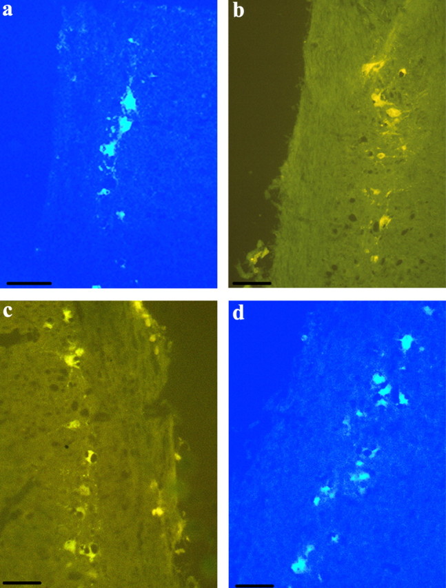
Fig. 3.

Photomicrographs of labeled motoneurons in the spinal cord after the im injection of the retrograde transport fluorescent tracer TB or HM. a, Motoneurons after TB was injected into the SH of a T-implanted male. b, HM was injected into the SC of a nonimplanted female. c, HM was injected into the PEC of a nonimplanted female. d, TB was injected into the ITB of a T-implanted female. Reference bar, 40 μm.
