Pulmonary nodules are blurred by respiratory motion during a fludeoxyglucose F 18 (FDG) positron emission tomography (PET) scan. Current scans have limited ability to assess subcentimeter nodules (1). Devices to mitigate respiratory motion by gating the PET acquisition have achieved limited application. Data-driven gating (DDG) is a novel software technique to detect respiratory motion within PET data, using the static phase for reconstruction, without any additional hardware or radiation dose (2–4).
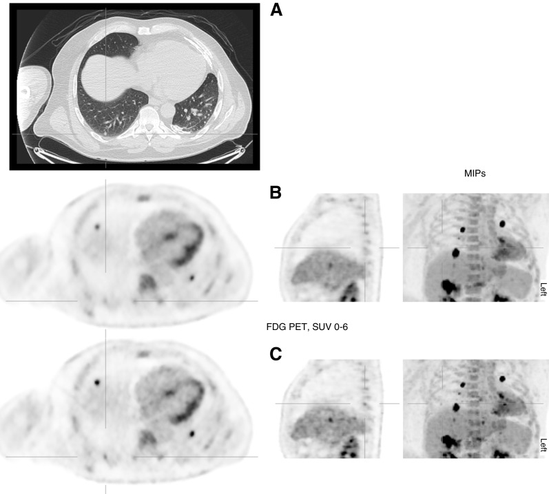
A 60-year-old man underwent FDG imaging to stage colorectal cancer (GE Discovery 690 PET/computed tomography [CT]; GE Healthcare, Milwaukee, WI). On the CT component, a 6-mm pulmonary nodule was identified posteriorly in the right lower lobe (Figure 1A). Using our routine reconstruction (5), this nodule is indistinct from background activity. The greatest Standardized Uptake Value in the volume of interest (SUVmax) is 1.9 and relates to the chest wall (Figure 1B). After retrospective DDG reconstruction, the nodule appears FDG-avid, with SUVmax 2.8 (Figure 1C) now greater than mediastinal blood pool. Uptake within other small pulmonary and hepatic metastases also became more conspicuous, being smaller with increased SUVmax.
Figure 1.
Selected fludeoxyglucose F 18 (FDG) positron emission tomography (PET)–computed tomography images showing standard and data-driven gating (DDG) reconstructions. All PET images are shown on a Standardized Uptake Value (SUV) scale of 0–6. Multiple metastases are present, seen well on the maximal intensity projection (MIP), where the difference resulting from DDG is also well displayed. (A) Axial computed tomography, lung window: 6-mm pulmonary nodule posteriorly in the right lower lobe. (B) Corresponding axial, sagittal, and frontal MIPs of the PET, standard reconstruction. The nodule is not discernible from background uptake in the nearby thoracic wall. (C) Similar PET images from the DDG reconstruction. The nodule is now identifiably FDG avid, with SUVmax 2.8. Small hepatic metastases are also more conspicuous.
Although not altering management for this patient, this illustrates that DDG may identify solitary or additional metastases that could benefit patient care. This new technology requires validation, but it promises to enhance the role of FDG PET/CT detection and assessment of small nodules. It could also improve the characterization, quantitative assessment, and risk prediction of larger pulmonary nodules (1).
Acknowledgments
Acknowledgment
The authors are grateful to Hugo Arques and Ribale Chebib at GE Healthcare for their assistance with DDG processing.
Footnotes
D.R.M. is supported by Cancer Research UK Oxford Centre C5255/A18085.
Author Contributions: N.C.D.M. identified the case; the manuscript was written, edited, and approved by all authors.
Originally Published in Press as DOI: 10.1164/rccm.201607-1371IM on October 18, 2016
Author disclosures are available with the text of this article at www.atsjournals.org.
References
- 1.Callister MEJ, Baldwin DR, Akram AR, Barnard S, Cane P, Draffan J, Franks K, Gleeson F, Graham R, Malhotra P, et al. British Thoracic Society Pulmonary Nodule Guideline Development Group; British Thoracic Society Standards of Care Committee. British Thoracic Society guidelines for the investigation and management of pulmonary nodules. Thorax. 2015;70:ii1–ii54. doi: 10.1136/thoraxjnl-2015-207168. [DOI] [PubMed] [Google Scholar]
- 2.Büther F, Ernst I, Dawood M, Kraxner P, Schäfers M, Schober O, Schäfers KP. Detection of respiratory tumour motion using intrinsic list mode-driven gating in positron emission tomography. Eur J Nucl Med Mol Imaging. 2010;37:2315–2327. doi: 10.1007/s00259-010-1533-y. [DOI] [PubMed] [Google Scholar]
- 3.Büther F, Vehren T, Schäfers K, Schäfers M. Impact of data-driven respiratory gating in clinical PET. Radiology. 2016;281:229–238. doi: 10.1148/radiol.2016152067. [DOI] [PubMed] [Google Scholar]
- 4.Kesner AL, Chung JH, Lind KE, Kwak JJ, Lynch D, Burckhardt D, Koo PJ. Validation of software gating: a practical technology for respiratory motion correction in PET. Radiology. 2016;281:239–48. doi: 10.1148/radiol.2016152105. [DOI] [PubMed] [Google Scholar]
- 5.Teoh EJ, McGowan DR, Macpherson RE, Bradley KM, Gleeson FV. Phantom and clinical evaluation of the Bayesian penalized likelihood reconstruction algorithm Q.Clear on an LYSO PET/CT System. J Nucl Med. 2015;56:1447–1452. doi: 10.2967/jnumed.115.159301. [DOI] [PMC free article] [PubMed] [Google Scholar]


