
Figure 4.
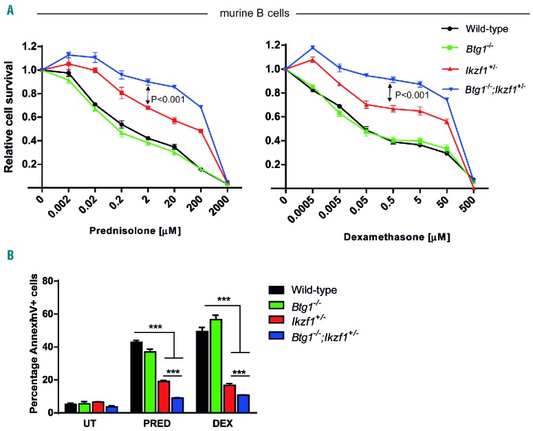
Glucocorticoid resistance of B cells isolated from Btg1 knockout mice intercrossed with haplodeficient Ikzf1 animals. (A) Splenic B cells isolated from wild-type, Ikzf1+/−, Btg1−/− and Btg1−/−;Ikzf1+/− mice were activated by LPS for 48 hours and subsequently treated in vitro for 48 hours with increasing concentrations of prednisolone (PRED, left panel) or dexamethasone (DEX, right panel) and analyzed by MTS assay (n=6). All values were normalized to untreated (UT) B cells. Error bars represent ± standard error of the mean (SEM). P-values were calculated based on the differences of the best-fit curve using two-way ANOVA. (B) AnnexinV/7-AAD staining of WT, Ikzf1+/−, Btg1−/− and Btg1−/−;Ikzf1+/− B-lymphocytes after 2 mM prednisolone or 5 mM dexamethasone treatment for 48 hours (n=4). The fraction of AnnexinV-positive cells was determined. Data represent means, and error bars represent SEM. P-values (two-sided t-test) are indicated. ***P<0.001.