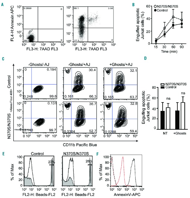
Figure 1.
Efferocytosis in Gaucher macrophages. (A) Jurkat cells were UV-irradiated, incubated at 37°C for 4 h, stained for annexin V/7AAD and analyzed by flow cytometry. Early apoptotic cells represent 66.1% of the population. (B) Control and Gaucher macrophages were co-cultured with GFP-labeled apoptotic Jurkat cells for different time intervals. Non-efferocytosed apoptotic Jurkat cells were removed by washing with PBS, and macrophages were detached and analyzed by flow cytometry. Three independent experiments were performed on samples from three different patients with Gaucher disease (GD). (C–D) GFP-labeled apoptotic Jurkat cells (AJ) were added to macrophages both with and without the addition of erythrocyte ghosts for 60 min. They were detached after extensive washing, stained for CD11b, and analyzed by flow cytometry. The graph represents data from six independent experiments. (E) 2 μm FluoSpheres sulfate beads, coated with PtdSer, were added to macrophages for 60 min and analyzed by flow cytometry. (F) 5 μm glass beads were coated with PtdSer and PtdCho, stained with annexin V and analyzed by flow cytometery. Dashed red and black lines represent unstained coated beads, while solid red and black lines indicate stained beads coated with PtdCho and PtdCho-PtdSer.

