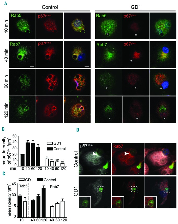
Figure 4.

p67phox recruitment to phagosomes is diminished in Gaucher macrophages. (A) Control and Gaucher macrophages were co-cultured with PtdSer-coated beads and fixed at different time points. Cells were co-stained with p67phox (red) and Rab5 (green) or Rab7 (green). DAPI (blue) staining demonstrates the position of the nucleus. Images are representative of 25 pictures taken at each time point in four different individuals with genotype N370S/N370S, two with N370S/L444P mutations and six controls. (B, C) The intensity of each protein in the phagosome was measured using IMARIS software. (D) GFP-labeled apoptotic Jurkat cells were added to control and Gaucher macrophages for 60 min, washed, fixed and stained for p67phox (white), Rab7 (red) and apoptotic Jurkat cells (green). Images represent 40 pictures taken in four independent experiments (63× magnification, scale bars: 5 μm). Insets show higher magnifications of the areas outlined in the images.
