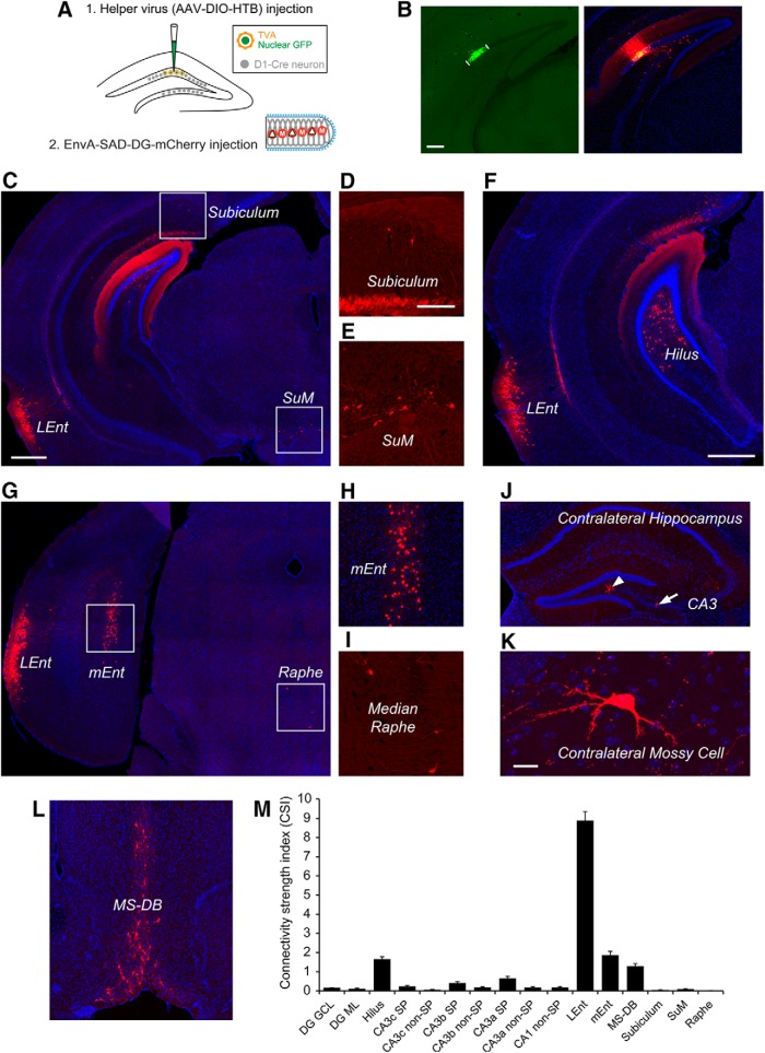
Figure 5.
Cre-dependent rabies tracing of circuit connections to dentate granule cells. A, The schematic illustrates the use of Cre dependent helper AAV and a D1-Cre transgenic mouse, in which the dentate granule cell layer expresses Cre recombinase (Lemberger et al., 2007) for Cre-dependent monosynaptic rabies tracing. B, Spatially restricted iontophoretic injection of the helper AAV (AAV-DIO-HTB) in the temporal DG of the D1-Cre mouse (left), followed by EnvA-SADΔG-mCherry rabies infection (right). The short white lines indicate restricted GFP-expressing helper AAV infection in the granule cell layer. Scale bar, 200 μm. C–L, Dentate granule cells receive very strong input from lateral and medial EC (C, F–H), extensive input from mossy cells at the temporal hilus (F), and inputs from medial septum (MS-DB) (L). Other long range inputs come from the subiculum, supramammillary nucleus (SuM), median raphe nucleus (D, E, I) as well as contralateral mossy cells (J and K). Scale bars in C (500 μm) applies to G, J, L; in D (200 μm) applies to E, H, I; in F, 500 μm; in K, 20 μm. M, The graph shows quantitative strengths of specific circuit connections to targeted dentate granule cells. The input CSI is defined as the ratio of the number of labeled presynaptic neurons in a specified structure versus the number of starter dentate granule neurons. Data are presented as mean ± SE. See Table 2 for more information.

