Figure 7.

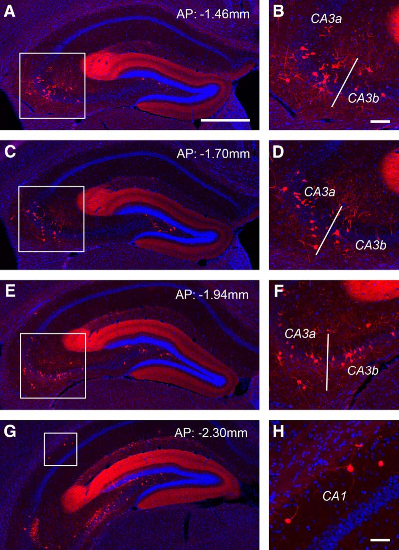
Monosynaptic rabies tracing reveals back projections from CA3 and CA1 to dentate granule cells. A–F, The targeted dentate granule cells receive extensive noncanonical inputs from CA3 in the septal directed hippocampus. AP numbers indicate the positions of the coronal sections relative to the bregma landmark. The example shows that putative CA3a/b excitatory neurons in the pyramidal cell layer and inhibitory neurons outside the pyramidal cell layer provide extensive inputs to dentate granule cells. The fimbria (indicate by the white bar) divides the distal CA3 (CA3a) and the middle CA3 subfield (CA3b). Panels on the right show enlarged views of the white box regions in the left panels. Scale bars, in A (500 μm) applies to A, C, E, G.; in B (100 μm) applies to B, D, F. G, H, Putative CA1 inhibitory neurons are also found projecting to dentate granule cells. Scale bar in H, 50 μm.
