Abstract
A variety of interventional management approaches exist for the treatment of acute pulmonary embolism (PE). However, when PE is accompanied by residual right heart thrombus, the best therapeutic options are less clear. We describe a novel combined technique of percutaneous aspiration of unstable right atrial thrombus followed by ultrasound-directed thrombolysis of massive PE.
Keywords: pulmonary embolism, thrombus aspiration, thrombolysis, EKOS, AngioVac
Hemodynamically unstable patients with acute pulmonary embolism (PE) have significant mortality, with reported rates of in-hospital death of nearly 25%.1 Systemic or catheter-directed thrombolysis is life-saving therapy in patients who present with massive PE.2,3 Even after thrombolysis, patients are at risk of developing pulmonary hypertension or persistent hypoxia due to residual thrombus. Management of unstable patients with residual, unstable thrombus proximal to the pulmonary arterial vasculature is controversial. We present a unique case of a patient with massive PE with progressive instability who underwent percutaneous aspiration of a large, acute right atrial (RA) thrombus followed by ultrasound-directed thrombolysis.
Case Presentation
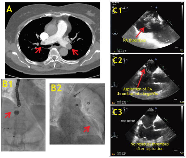
A 70-year-old female with a history of hypertension underwent biopsy of a 9.0 × 15.0 cm right retroperitoneal mass adherent to the mesentery, ileum, and colon, diagnosed by computed tomography (CT). On postoperative day 1, she developed dizziness and acute dyspnea and was in cardiogenic shock, with a systemic blood pressure of 75/60 requiring intravenous norepinephrine and a heart rate of 120 bpm. She had significant hypoxia with an oxygen saturation of 85% despite treatment with high-flow oxygen via face mask. Electrocardiogram demonstrated sinus tachycardia and contrast chest CT revealed filling defects involving the left and right pulmonary arteries extending to segmental and subsegmental branches (Figure 1). The patient was started on systemic heparin and transferred to the intensive care unit. Transthoracic echocardiography (TTE) revealed the presence of a mobile, multilobar right atrial (RA) thrombus extending from the inferior vena cava (IVC). The patient experienced progressive hemodynamic instability likely as a result of ongoing embolic events from the RA thrombus. Cardiothoracic surgery was consulted and felt the patient was too high risk for surgery. The patient was emergently brought to the cardiac catheterization laboratory for percutaneous aspiration of the thrombus.
FIGURE 1.
(A) Computed tomography of the pulmonary embolism demonstrating bilateral filling defects. (B) Fluoroscopic image showing AngioVac aspiration catheter [B1] and EKOS catheter [B2]. (C) Transesophageal echocardiographic images showing unstable, mobile RA thrombus before [C1], during [C2], and after [C3] AngioVac aspiration.
After intubation, transesophageal echocardiography (TEE) confirmed the presence of a large, multilobar RA thrombus- in-transit extending from the IVC with intermittent prolapse into the right ventricle (Figure 1). A 26 Fr sheath with a DrySeal hemostatic valve (Gore Medical) was inserted into the right common femoral vein and a 17 Fr Bio-Medicus reinfusion sheath (Medtronic) was placed into the left common femoral vein. Using a 0.035˝ Lunderquist Extra-Stiff wire (Cook Medical), the AngioVac aspiration catheter (AngioDynamics) was advanced through the 26 Fr sheath under fluoroscopic and TEE guidance to the junction of the IVC and RA. The aspiration and reinfusion sheaths were connected to cannulas to form a standard extracorporeal circuit with a centrifugal pump to achieve flows of approximately 3 L/minute. Aspiration of the thrombus into the AngioVac catheter resulted in abrupt cessation of flows due to complete obstruction of the tip with thrombus. The AngioVac catheter was then removed entirely from the sheath to reveal large, acute thrombus (Figure 2). Several passes of the AngioVac catheter into the IVC-RA junction resulted in complete removal of the RA thrombus as visualized by TEE (Figure 1).
FIGURE 2.
(A) Thrombus evacuated using AngioVac aspiration catheter. (B) Transthoracic echocardiographic images showing right-ventricular (RV) focused view immediately post procedure noting significant RV dilation [B1] and at 1-week follow-up [B2] with normalization of RV size.
Following thrombus aspiration, a pigtail catheter was advanced into both pulmonary arteries for pressure measurement and angiography. Main pulmonary artery pressure was 65/35 mm Hg; there was a proximal thrombus in the left pulmonary artery and multiple distal thrombi in the right pulmonary artery. Over a 0.035˝ Wholey wire (Covidien), the EKOS infusion catheter system (EKOS Corporation) was advanced into the left pulmonary artery to provide ultrasound-assisted thrombolysis. A 2 mg bolus of tenecteplase was administered through the catheter followed by infusion of 1 mg/hour for 12 hours. TTE revealed severe right ventricular dysfunction with a right ventricular/left ventricular size ratio of 1.2. After 12 hours, pulmonary artery pressure was 50/25 mm Hg. Based on these findings, an additional 12 hours of ultrasound-assisted thrombolysis was continued. Norepinephrine was weaned off and the patient was extubated soon thereafter. After placement of an IVC filter, the patient was discharged on postoperative day 8 with systemic anticoagulation (apixaban). At 1 week post discharge, she had no physical limitations, no dyspnea on exertion, and TTE demonstrated normal right ventricular function with a right ventricular/left ventricular size ratio of 0.7 (Figure 2).
Discussion
Systemic thrombolytic therapy has been the mainstay of treatment for massive PE; however, the risk of major bleeding and intracranial hemorrhage is significant.2,5 Catheter-directed thrombolysis allows for localized delivery of the thrombolytic agent, resulting in a lower overall dose, reduced risk of bleeding, and improved outcomes.6–8 For patients who present with simultaneous unstable venous or right-heart thrombus and massive PE, insertion of interventional equipment into the right heart and pulmonary artery may result in dislodgment of thrombotic material and further hemodynamic instability. A variety of management strategies exist to treat patients with acute life-threatening thrombi-in-transit including anticoagulation, systemic thrombolysis, and thrombectomy.9 The AngioVac system utilizes a venous drainage cannula with a balloon-actuated, funnel-shaped distal tip for removal of thrombus. This drainage system connects to a centrifugal pump and filter, which feed back to the body through a reinfusion cannula. Percutaneous thrombectomy using the AngioVac system is less invasive and more rapidly available compared with surgical intervention and has a lower bleeding risk compared with systemic thrombolysis.3,10
Conclusion
To the best of our knowledge, we present the first report of a combined technique of percutaneous evacuation of an RA thrombus using the AngioVac system followed by ultrasound-assisted thrombolysis of massive PE using an EKOS catheter. A combination of percutaneous therapies is often necessary for patients with massive PE and residual life-threatening venous thrombus, as described in this case.
Footnotes
Disclosure: The authors have completed and returned the ICMJE Form for Disclosure of Potential Conflicts of Interest. The authors report no conflicts of interest regarding the content herein.
References
- 1.Casazza F, Becattini C, Bongarzoni A, et al. Clinical features and short term outcomes of patients with acute pulmonary embolism. The Italian Pulmonary Embolism Registry (IPER) Thromb Res. 2012;130:847–852. doi: 10.1016/j.thromres.2012.08.292. [DOI] [PubMed] [Google Scholar]
- 2.Kearon C, Akl EA, Comerota AJ, et al. Antithrombotic therapy for VTE disease: antithrombotic therapy and prevention of thrombosis, 9th ed: American College of Chest Physicians Evidence-Based Clinical Practice Guidelines. Chest. 2012;141:e419S–e494S. doi: 10.1378/chest.11-2301. [DOI] [PMC free article] [PubMed] [Google Scholar]
- 3.Donaldson CW, Baker JN, Narayan RL, et al. Thrombectomy using suction filtration and veno-venous bypass: single center experience with a novel device. Catheter Cardiovasc Interv. 2015;86:E81–E87. doi: 10.1002/ccd.25583. [DOI] [PubMed] [Google Scholar]
- 4.Jaff MR, McMurtry MS, Archer SL, et al. Management of massive and submassive pulmonary embolism, iliofemoral deep vein thrombosis, and chronic thromboembolic pulmonary hypertension: a scientific statement from the American Heart Association. Circulation. 2011;123:1788–1830. doi: 10.1161/CIR.0b013e318214914f. [DOI] [PubMed] [Google Scholar]
- 5.Wan S, Quinlan DJ, Agnelli G, Eikelboom JW. Thrombolysis compared with heparin for the initial treatment of pulmonary embolism: a meta-analysis of the randomized controlled trials. Circulation. 2004;110:744–749. doi: 10.1161/01.CIR.0000137826.09715.9C. [DOI] [PubMed] [Google Scholar]
- 6.Piazza G, Hohlfelder B, Jaff MR, et al. A prospective, single-arm, multicenter trial of ultrasound-facilitated, catheter-directed, low-dose fibrinolysis for acute massive and submassive pulmonary embolism: the SEATTLE II study. JACC Cardiovasc Interv. 2015;8:1382–1392. doi: 10.1016/j.jcin.2015.04.020. [DOI] [PubMed] [Google Scholar]
- 7.Kuo WT, Banerjee A, Kim PS, et al. Pulmonary embolism response to fragmentation, embolectomy, and catheter thrombolysis (PERFECT): initial results from a prospective multicenter registry. Chest. 2015;148:667–673. doi: 10.1378/chest.15-0119. [DOI] [PubMed] [Google Scholar]
- 8.Kucher N, Boekstegers P, Müller OJ, et al. Randomized, controlled trial of ultrasound-assisted catheter-directed thrombolysis for acute intermediate-risk pulmonary embolism. Circulation. 2014;129:479–486. doi: 10.1161/CIRCULATIONAHA.113.005544. [DOI] [PubMed] [Google Scholar]
- 9.Athappan G, Sengodan P, Chacko P, Gandhi S. Comparative efficacy of different modalities for treatment of right heart thrombi in transit: a pooled analysis. Vasc Med. 2015;20:131–138. doi: 10.1177/1358863X15569009. [DOI] [PubMed] [Google Scholar]
- 10.Salsamendi J, Doshi M, Bhatia S, et al. Single center experience with the AngioVac aspiration system. Cardiovasc Intervent Radiol. 2015;38:998–1004. doi: 10.1007/s00270-015-1152-x. [DOI] [PubMed] [Google Scholar]


