

Summary
The increasingly rapid pace of research in the field of bioinspired drug delivery systems is revealing the promise of cell membrane-based nanovesicles for biomedical applications. Those cell membrane-based nanoparticles combine the natural functionalities of cell plasma membranes and the bioengineering flexibility of synthetic nanomaterials, and such versatility provides a means of designing exciting new drug formulations for personalized treatment in future nanomedicine.
Keywords: Nanobiotechnology, Nanovesicles, Biomaterial, Bio-inspired synthesis, Cell membrane
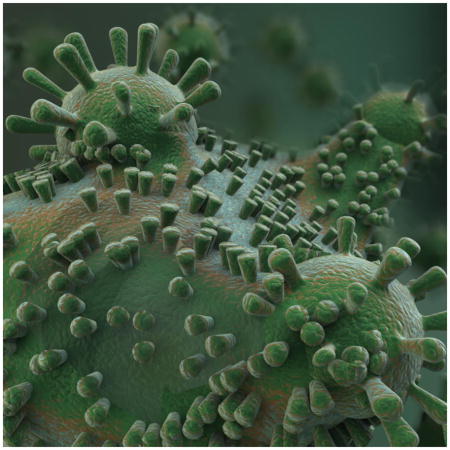
Graphical Abstract
With the advance of nanotechnology, biomaterial, biology and other fields of science, researchers pursure biocompatible nanomaterials akin to live creature that can circumvent biological barrier and specifically transport cargos to target sites. To fabricate nanovesicles with good biodegradability and novel biological functions, scientists increasingly draw lessons from nature for inspiration[1–3]. In recent years, nanovesicles based on bio-inspired synthesis have been developed, aiming at simulating unique properties of natural organism/structures/biosynthesis pathways [4]. These biomimetic delivery vehicles whose morphology, surface properties and size imitate natural organism such as red bood cells (RBC) [5], exosomes[6] or pathogens[7, 8], possess special functions to enhance delivery to target tissues or cell populations. Compared with conventional drug delivery nanosystems, cell membrane-based nanovehicles [5–7] with the specific properties of originating cells by maintaining the topologically/physiologically active form of cell membrane proteins are a promising nanobiotechnology for a variety of biomedical applications, including targeted delivery of therapeutics, tissue regeneration and theranostics.
For instance, researchers have exploited biological knowledge about the naturally occurring nanoscale extracellular nanovesicles named exosomes, that are secreted out of cell plasma membrane and trigger intercellular communication by transferring the cargoes of endogenous mRNA, miRNA, or protein from one cell to another[9], to develop as a chemotherapeutic drug delivery nanoplatform[6]. Generally, exosomes are useful for the delivery of therapeutic genes and functional proteins that are difficult to synthesize. The drug-loaded exosome-mimetic nanovesicles [6] generated from cell plasma membrane using a mini-extruder have biophysicochemical properties similar to those of exosomes, and they can be easily produced and isolated through polycarbonate filters with relatively small pore sizes. Thus, exosome-mimetic nanovesiclesare promising drug delivery systems for both endogenous and exogenous cargos.
Other than using natural cell membrane, cell plasma membrane can also be genetically/chemically engineered to display various active proteins and thereby be endowed with more specific features. Inspired by the natural budding processes of enveloped viruses that release from cell membrane and acquire lipid-envelope with viral surface glycoproteins, a similar synthesis strategy was adopted to produce enveloped virus-mimetic vesicles (VMV)[7], that firstly anchored genetically engineered viral proteins on cellular surface, followed by the formation of micron-sized vesicles displaying viral enveloped glycoproteins along with chemical surfactants embedding into plasma membrane [10]. Subsequently, the protein-displayed vesicles were further prepared to be uniform nanosized vesicles by entrapping surfactants into lipid membrane. Notably, it was found that chemical surfactants could significantly alter stability, uniformity, and the size distribution of vesicles. Futhermore, it confers vesicles important suface properties to form virus-like morphology. In general, VMV is comprised of phospholipid originated from cellular plasma membrane, surfactants that enable manipulation of the size of nanoparticles, and recombinant viral protein which are accurately guided to the plasma membrane via signal peptide sorting (Fig. 1). VMV platform is capable of effectively eliciting antibodies against live enveloped virus and in an influenza model, protects from a lethal dose of enveloped viral challenge. Thus, it can be envisioned that this approach has great potential to be applied not only for the design of antigen delivery system against a broad range of enveloped viruses, including potentially dangerous ones, but also for the rapid development of diagnostic enzyme immunoassay (EIA) kits of infectious lethal enveloped virus.
Figure 1.

Schematic showing the antigen delivery system exhibiting biomembrane-directed viral protein antigen. The model cell lines are engineered to stably express viral protein fragments or whole trimeric enveloped glycoprotein (HA protein) on the cellular plasma membrane, which is then vesiculated with the help of surfactants to further produce size-controlled virus-mimetic vesicles (VMVs, 50–150 nm) for effective vaccination: viral antigen genes are transfected into cells (A) and the cells stably expressing antigen are acquired after selection of puromycin (B). The antigenic proteins are firstly located in endoplasmic reticulum (C), plasma membrane or Golgi complex (D), and then fused with cell plasma membrane (E) via the route of signal peptide sorting. The engineered cells are incubated with sodium deoxycholate to produce major cell membrane vesicles (MCV, 500–2500 nm) (F), and then the purified MCVs are further mixed with sodium deoxycholate and triton-X100 under ultrasonication to produce nano-sized VMVs (G). Mice vaccinated with VMV-HA are able to survive after being exposed to a lethal dose of mouse-adapted H1N1 virus (H). Adapted from Ref. 7 with permission.
Rather than employing single-component natural or bio-modified cell plasma membrane to fabricate nanovesicles, many groups have attempted to acquire and purify cell plasma membrane to encapsulate synthetic nanoparticles. Those cell plasma membrane-coated nanoparticles combine the natural functionalities of cell plasma membranes and the bioengineering flexibility of synthetic nanomaterials. Such versatility provides a means of designing nanoparticle coatings and capsules with customized content and release characteristics from the surfaces of cell plasma membranes. What is perhaps most fascinating about these cell membrane-based drug delivery systems is the natural targeting ability of the producing cells that makes the exogenous engineering of targeting moieties unnecessary.
Many hybrid nanosystems have been successfully developedto undergo “natural” biological interactions with critical properties of long circulating half-life, good biocompatibility or cell targeting. Hu et al.[11] developed a RBC membrane-coated nanovehiclethat mimics many of the biological functions of RBCwith the complex immunomodulatory proteins by mixing nanocarrier with RBC vesicles and then physically squeezing them many times through polycarbonate filters with defined-scale pores. The cellular ghosts on camouflaged nanoparticles may be harvested from mesenchymal stem cells[12], cancer cell membrane[13], platelets, etc. Hu et al.[14] also demonstrated that nanocarriers coated with the plasma membrane of platelets could escape from the body's immune surveillance, and possess platelet-mimetic binding capabilities which guide them to targeted tissues and specific cell/micro organism population. Platelet membrane coating made the particles effectively attach to collagen protein, and thereby target location of injurys in blood vessels, enabling the effective releaseof an anti-restenosis agent to repair damaged blood vessels. Because platelets tend to cluster around bacteria such as methicillin-resistant Staphylococcus aureus in the blood, platelet-membrane-coated nanoparticles offer a hope of boosting the effectiveness of antibiotics to solve severe complications of infection.
Taken together, there is a huge potential of exploiting cell membrane-based nanovesicles to fabricate smart delivery systems [15]. These delivery platforms may also be able to achieve the goal of precisely targeted therapy in the way that could be easily manipulated, effective and biocompatible. However, like all newborn technologies, their clinical translation may still encounter significant challenges, including the low production yield and the difficulty of purification, as well as the demonstration of the stability with common storage methods. In addition, it is of note that the synthetic substrates and allograft membranes in nanovesicle formulation may cause immunogenicity and safety concerns. In the future, advances in personalized medicine to engineer patient’s own cells (e.g. autologous dendritic cells, T lymphocytes, mesenchymal stem cells, erythroid progenitors cells[16] and cancer cells) may involve lentivirus mediated generation of bioengineered nanovesicles towards the goal of preparing therapeutic bioinspired nanoparticles.
Highlights.
Nanovesicles based on bio-inspired synthesis aim at simulating unique properties of natural organism/structures/biosynthesis pathways
Biomimetic delivery vehicles possess special functions of red bood cells, exosomes or pathogens to enhance delivery to target tissues or cell populations
Enveloped virus-like vesicle (VMV) contains phospholipid originated from cellular plasma membrane, surfactant to manipulate the size of nanoparticles, and recombinant viral protein guided to the plasma membrane via signal peptide sorting, for antigen delivery
Acknowledgments
This work was supported by the MOST of China (2014CB744503 and 2013CB733802), the NSFC under Grant No. 81422023, 81371596, 51273165, and U1505221, the Program for New Century Excellent Talents in University (NCET-13-0502), the Fundamental Research Funds for the Central Universities, China (20720150206 and 20720150141), and the Intramural Research Program, National Institute of Biomedical Imaging and Bioengineering, National Institutes of Health.
Biographies

Pengfei Zhang obtained his M.S. in the Center for Molecular Imaging and Translational Medicine (CMITM) at Xiamen University under the supervision of Prof. Gang Liu. Currently he is a PhD student in the same group. His research interests focus on developing biomimetic nanomaterials for drug and antigen delivery and molecular imaging.
Gang Liu received his MD degree from North Sichuan Medical College (China) in 2002 and PhD degree from Sichuan University (China) in 2009. Subsequently, he focused his training on nanomedicine and molecular imaging at the National Institutes of Biomedical Imaging and Bioengineering (NIBIB), National Institutes of Health (NIH) under the supervision of Dr. Xiaoyuan Chen (2009–2011). In 2012, he joined the Center for Molecular Imaging and Translational Medicine (CMITM), School of Public Health, Xiamen University. Currently he is a Professor of Biomedical and Bioengineering and his research interests include biomaterials, theranostics, and molecular imaging. His scientific work has been published as 90 papers in prestigious journals (Adv. Mater., Nat. Commun., PNAS, etc.), 7 invited book chapters, and 11 patents.
Xiaoyuan Chen received his B.S. in 1993 and M.S. in 1996 from Nanjing University, China. He then moved to the United States and obtained his Ph.D. in chemistry from the University of Idaho in 1999. After two quick postdocs at Syracuse University and Washington University in St. Louis, he joined the University of Southern California as an Assistant Professor of Radiology. He then moved to Stanford University in 2004. He was promoted to Associate Professor in 2008, and in the summer of 2009 he joined the intramural research program of the National Institute of Biomedical Imaging and Bioengineering (NIBIB), at the National Institutes of Health, as a Senior Investigator and Chief of the Laboratory of Molecular Imaging and Nanomedicine (LOMIN). Dr. Chen has published over 500 papers, 4 books, and numerous book chapters. He is the founding editor of the journal Theranostics, and sits on the editorial board of over 10 peer-reviewed journals.
Footnotes
Publisher's Disclaimer: This is a PDF file of an unedited manuscript that has been accepted for publication. As a service to our customers we are providing this early version of the manuscript. The manuscript will undergo copyediting, typesetting, and review of the resulting proof before it is published in its final citable form. Please note that during the production process errors may be discovered which could affect the content, and all legal disclaimers that apply to the journal pertain.
References
- 1.Meyer RA, Sunshine JC, Green JJ. Trends Biotechnol. 2015;33:514–524. doi: 10.1016/j.tibtech.2015.07.001. [DOI] [PMC free article] [PubMed] [Google Scholar]
- 2.Grinthal A, Kang SH, Epstein AK, Aizenberg M, Khan M, Aizenberg J. Nano Today. 2012;7:35–52. [Google Scholar]
- 3.Sealy C. Nano Today. 2006;1:10–10. [Google Scholar]
- 4.Lu Y, Dong L, Zhang LC, Su YD, Yu SH. Nano Today. 2012;7:297–315. [Google Scholar]
- 5.Hu CMJ, Zhang L, Aryal S, Cheung C, Fang RH, Zhang LF. Proc Natl Acad Sci USA. 2011;108:10980–10985. doi: 10.1073/pnas.1106634108. [DOI] [PMC free article] [PubMed] [Google Scholar]
- 6.Jang SC, Kim OY, Yoon CM, Choi DS, Roh TY, Park J, Nilsson J, Lotvall J, Kim YK, Gho YS. ACS nano. 2013;7:7698–7710. doi: 10.1021/nn402232g. [DOI] [PubMed] [Google Scholar]
- 7.Zhang PF, Chen YX, Zeng Y, Shen CG, Li R, Guo ZD, Li SW, Zheng QB, Chu CC, Wang ZT, Zheng ZZ, Tian R, Ge SX, Zhang XZ, Xia NS, Liu G, Chen XY. P Natl Acad Sci USA. 2015;112:E6129–E6138. doi: 10.1073/pnas.1505799112. [DOI] [PMC free article] [PubMed] [Google Scholar]
- 8.Somiya M, Kuroda S. Adv Drug Deliver Rev. 2015;95:77–89. doi: 10.1016/j.addr.2015.10.003. [DOI] [PubMed] [Google Scholar]
- 9.Alvarez-Erviti L, Seow YQ, Yin HF, Betts C, Lakhal S, Wood MJA. Nat Biotechnol. 2011;29:341–345. doi: 10.1038/nbt.1807. [DOI] [PubMed] [Google Scholar]
- 10.Hildebrand A, Neubert R, Garidel P, Blume A. Langmuir. 2002;18:2836–2847. [Google Scholar]
- 11.Hu CMJ, Fang RH, Luk BT, Zhang LF. Nat Nanotechnol. 2013;8:933–938. doi: 10.1038/nnano.2013.254. [DOI] [PMC free article] [PubMed] [Google Scholar]
- 12.Furman NET, Lupu-Haber Y, Bronshtein T, Kaneti L, Letko N, Weinstein E, Baruch L, Machluf M. Nano Lett. 2013;13:3248–3255. doi: 10.1021/nl401376w. [DOI] [PubMed] [Google Scholar]
- 13.Fang RH, Hu CMJ, Luk BT, Gao WW, Copp JA, Tai YY, O'Connor DE, Zhang LF. Nano Lett. 2014;14:2181–2188. doi: 10.1021/nl500618u. [DOI] [PMC free article] [PubMed] [Google Scholar]
- 14.Hu CMJ, Fang RH, Wang KC, Luk BT, Thamphiwatana S, Dehaini D, Nguyen P, Angsantikul P, Wen CH, Kroll AV, Carpenter C, Ramesh M, Qu V, Patel SH, Zhu J, Shi W, Hofman FM, Chen TC, Gao WW, Zhang K, Chien S, Zhang LF. Nature. 2015;526:118–121. doi: 10.1038/nature15373. [DOI] [PMC free article] [PubMed] [Google Scholar]
- 15.Yoo JW, Irvine DJ, Discher DE, Mitragotri S. Nat Rev Drug Discov. 2011;10:521–535. doi: 10.1038/nrd3499. [DOI] [PubMed] [Google Scholar]
- 16.Shi JH, Kundrat L, Pishesha N, Bilate A, Theile C, Maruyama T, Dougan SK, Ploegh HL, Lodish HF. Proc Natl Acad Sci USA. 2014;111:10131–10136. doi: 10.1073/pnas.1409861111. [DOI] [PMC free article] [PubMed] [Google Scholar]