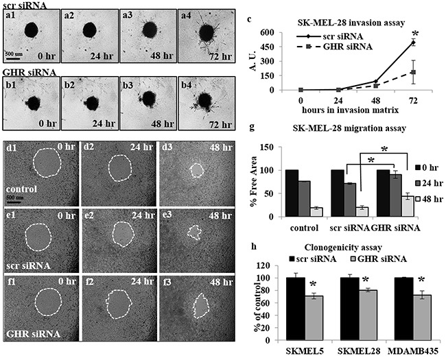
Figure 1. Growth hormone receptor knock-down (GHRKD) attenuates invasion, migration and clonogenicity in human melanoma cells.

a-c. SK-MEL-28 cells transfected with scramble (scr) (a1-a4) or GHR-siRNA (b1-b4) were seeded onto U-bottom 96-well plates at 5000 cells/well and allowed to form a spheroid. A hydrogel invasion matrix was added above the spheroid and cells were monitored for up to 72 hr. in presence of 50 ng/mL hGH. Total pixels representing structural extensions from the spheroid were calculated using ImageJ software and reflected the invasive ability of the melanoma cells (c). A significant decrease in spheroid invasion was noted following GHRKD. d-g. SK-MEL-28 cells transfected with scr- (e1-e3) or GHR-siRNA (f1-f3) as well as un-transfected controls (d1-d3) were allowed to migrate into a 0.68 mm circular spot at the center of the well, in presence of 50 ng/mL hGH for up to 48 hr. The percentage free area was calculated using ImageJ software and reflected the decrease/inhibition in migration (g). A significant decrease in migration was noted following GHRKD. Similar results for migration and invasion assays with MALME-3M, MDA-MB-435 and SK-MEL-5 cells are presented in Supplementary Figure 2. h. SK-MEL-5, SK-MEL-28 and MDA-MB-435 cells transfected with 20 nM scramble or GHR-siRNA were allowed to form colonies on soft agar for 7 days in presence of 50 ng/mL hGH. The cells were lysed at the end time point and total DNA was quantified using a fluorescent readout. A significant decrease in total number of colonies was noted following GHRKD. [*, p < 0.05, Students t-test, n = 3].
