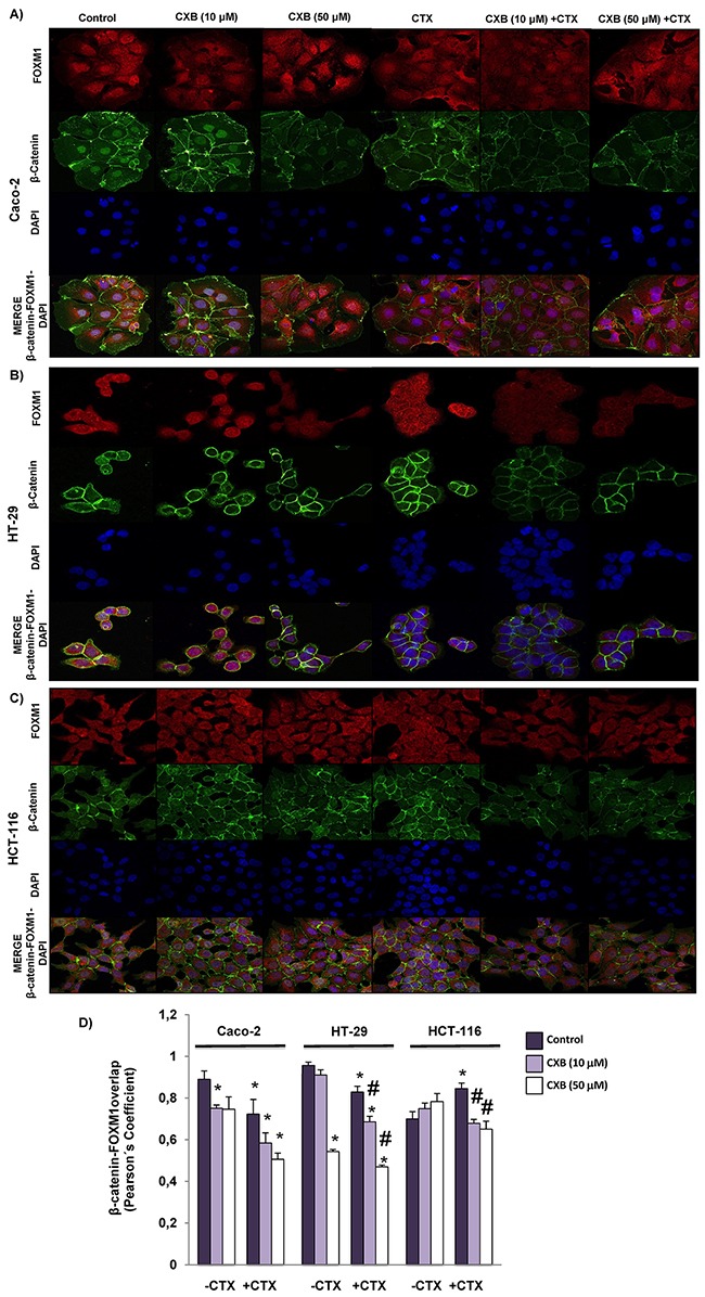
Figure 6. Combined Celecoxib/Cetuximab treatment impairs FOXM1-β-catenin interaction in colorectal cancer cells.

To determine subcellular localization of β-catenin and FOXM1, in A. Caco-2, B. HT-29 and C. HCT-116, cells were exposed for 6 h to celecoxib (CXB, 10 μM or 50 μM) alone or in combination with cetuximab (CTX, 100 μg/ml) and then, cells were stained for β-catenin (green) and FOXM1 (red) immunofluorescence, and counterstained with DAPI (blue). Merged images for β-catenin, FOXM1 and DAPI staining are also shown. Final magnification: X400 (Caco-2), X600 (HCT-116 and HT-29) D. Pearson's coefficient analysis was performed for the co-localization in cell nuclei of β-catenin and FOXM1. Data are means ± SEM of three independent experiments (*p <0.05, compared with the control; # p<0.05, compared with cetuximab-treated cells).
