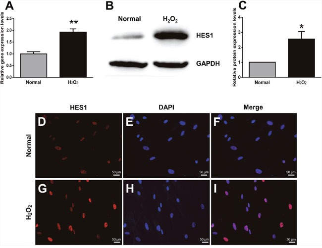
Figure 4. HES1 expression was up-regulated at both mRNA and protein levels in HTMCs under oxidative stress.

The qPCR showed an enhanced HES1 expression at mRNA levels in HTMCs subjected to the 2 h-treatment of H2O2 (A). Representative western blots showed an increased protein expression of HES1 induced by the 2 h-oxidative stress (B). The intensities of HES1 protein bands were normalized to those of the internal standard GAPDH, and the relative HES1 protein expression levels were shown in (C). HES1 immunostaining confirmed the trend of its expression changes under norm (D) and oxidative stress (G). The cell nuclei under both conditions were stained with DAPI (E and H). The HES1 staining was colocalized with DAPI staining (F and I). The data were presented as mean ± SEM (n = 3 per group for each experiment, each experiment was repeated 3 times; * p < 0.05, ** p < 0.01, as compared with normal control).
