Abstract
This is the first identified case of Mycobacterium bovis bacillus Calmette-Guérin (BCG)-derived cutaneous tuberculosis that localizes at a place different from the vaccination site in hosts without immune deficiency. A healthy baby with a developing abscess is described. A multiplex PCR identified the abscess as originating from M. bovis BCG Tokyo 172.
CASE REPORT
A 6-month-old girl with no previous significant medical problems developed an abscess in her left humerus. She had been vaccinated with Mycobacterium bovis bacillus Calmette-Guérin (BCG) Tokyo 172 at the age of 4 months. At 5 months, a red and swelling nodule appeared in her left humerus at a place different from the vaccination site. At 6 months, she visited our hospital. A red, fluctuating, and swollen abscess about 2 cm from the region of the vaccination was found. The abscess measured 10 by 15 mm. We found no swelling of the lymph nodes. The patient was afebrile and did not have respiratory symptoms. There was no abnormal chest radiographic finding. Skin testing with purified protein derivative tuberculin was positive with 10- by 7-mm erythema, without an induration or surrounding erythema. There was no family history of a similar reaction to BCG vaccination. The history of the girl's grandmother revealed pulmonary tuberculosis in her twenties. In addition, the possibility of close contact with other tuberculosis patients could not be excluded.
We punctured the abscess and submitted the pus for conventional microbial culture, mycobacterial culture, amplification of mycobacterial nucleic acid using an AMPLICOR MTB microwell plate assay kit (Roche Diagnostic Systems, Branchburg, N.J.), and microscopic examination of a smear. Conventional microbial culture failed to detect any significant pathogens. The smear examination was negative, but amplification of nucleic acid using a primer for a Mycobacterium tuberculosis complex-specific region of the 16S rRNA gene (KY172-T3) was positive for M. tuberculosis complexes. A 4-week mycobacterial culture grew an M. tuberculosis complex colony. This colony showed sensitivity to isoniazid, rifampin, ethambutol, and streptomycin and resistance to pyrazinamide, suggesting that the colony may have derived from BCG Tokyo 172. However, we could not clearly distinguish the origin among M. tuberculosis complex strains (M. tuberculosis, M. bovis BCG, or other strains of M. bovis).
For the purpose of quick and accurate identification of the sample, we ran multiplex PCR on the sample by a simple modification of the method of Bedwell et al. (1). In 1999, a DNA microarray clarified that 16 regions in BCG strains, named RD1 to RD16, were different from those in M. tuberculosis H37Rv and that some regions differed among BCG strains (2). RD1 is not present in all M. bovis BCG strains, but it is present in other strains of the M. tuberculosis complex. For M. bovis BCG strains, Talbot et al. designed primers to amplify the complement sequence of RD1 (9). Another PCR procedure based on the intergenic region named SenX3-RegX3 was reported to be able to distinguish M. bovis BCG from other M. tuberculosis complex strains. This method can also divide BCG strains into three groups according to the PCR product length (5). Recently, Bedwell and colleagues developed the novel multiplex PCR, which could distinguish BCG substrains (1). Since PCR products for SenX3-RegX3 of M. bovis, except BCG strains, and those for RD16 of BCG Tokyo 172 were very close in size, we modified the original multiplex PCR method of Bedwell et al. by simply excluding primers for RD16. Thus, our multiplex PCR contains primers for RD1, the complement sequence of RD1, RD2, RD8, RD14, and SenX3-RegX3, which were designed as previously described (1).
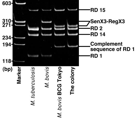
To identify the origin of the abscess, DNA was extracted from the colony. The multiplex PCR amplification was performed as previously described (1). M. tuberculosis Aoyama B (obtained from the National Institute of Infectious Diseases, Tokyo, Japan) and M. bovis Ushi 10 (obtained from the Japanese Association of Veterinary Biologics) showed a 150-bp RD1 product, whereas both M. bovis BCG Tokyo 172 and the colony showed 200-bp bands as the complement sequence of RD1, indicating the deletion of RD1 (Fig. 1). The RD2 and SenX3-RegX3 PCR products from M. tuberculosis were similar in length and appeared as one broad band (Fig. 1). The SenX3-RegX3 product from M. bovis was different from the others, whereas SenX3-RegX3 products from M. bovis BCG Tokyo 172 and the colony showed similar lengths. Behr et al. have shown that some M. bovis BCG strains are missing RD2, RD8, and RD14 (2), but not M. bovis BCG Tokyo 172, and we obtained similar results from M. bovis BCG Tokyo 172 and the colony (Fig. 1). Moreover, the colony showed susceptibility to thiophene-2-carboxylic acid hydrazide (TCH). M. tuberculosis is resistant to TCH, whereas M. bovis is susceptible to TCH. Taken together, we concluded that the colony originated from M. bovis BCG and, among M. bovis BCG strains, from BCG Tokyo 172.
FIG. 1.
Agarose gel electrophoresis of the multiplex PCR products. PCR products of M. bovis BCG Tokyo 172 and the colony showed similar bands.
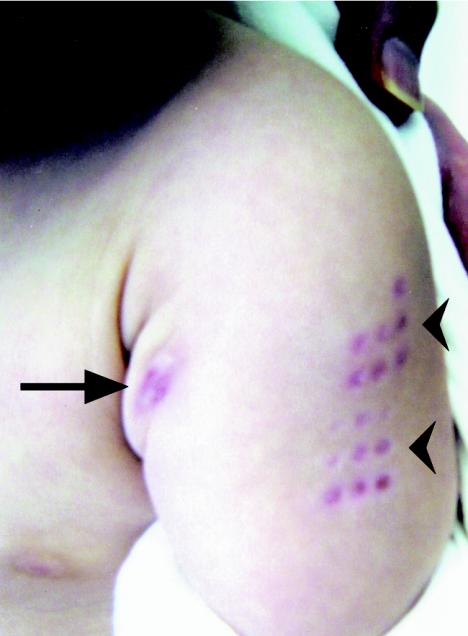
The abscess was sterilized and treated with gentamicin sulfate ointment daily, but the abscess swelled and collapsed several times. We began oral treatment with 10 mg of isoniazid (dry syrup)/kg of body weight/day. This treatment induced improvement of the lesion within the first 2 weeks, but the family interrupted the treatment for the subsequent 10 days. This interruption resulted in renewed swelling of the abscess (Fig. 2). We restarted the treatment. Two months after resumption, the skin lesion became clear, and medication continued for another month. No relapse has been observed after 1 year of follow-up.
FIG. 2.
BCG-derived cutaneous tuberculosis. Interruption of isoniazid treatment resulted in renewed swelling of the abscess. Arrow indicates the abscess. Arrowheads indicate a BCG vaccination scar.
BCG is a live attenuated strain of M. bovis that was first used for immunization against tuberculosis in 1921, and it has been considered safe. Localized abscesses, regional lymphadenopathy, and disseminated disease in immunocompromised hosts are uncommon but well-recognized complications (8). This patient showed no symptoms of immune deficiency. An abscess at the region of BCG vaccination has been reported for healthy hosts (6, 7). To our knowledge, a cutaneous abscess, which proved to be derived from BCG, localized at a place different from the vaccination site in a healthy host has not been reported.
M. tuberculosis complex includes M. tuberculosis, M. bovis, M. africanum, M. microti, M. canetti, M. caprae, and M. pennipedii (3). M. bovis BCG is an attenuated strain of M. bovis that is used for vaccination. TCH susceptibility can distinguish M. bovis from M. tuberculosis, but discrimination between M. bovis BCG and other strains of M. bovis has been difficult (2). Here a multiplex PCR clearly showed the difference between M. bovis BCG and other strains of M. bovis. Furthermore, this method can subdivide BCG strains. In addition to vaccination against tuberculosis, BCG is an effective agent for therapy of superficial transitional cell carcinoma of the urinary bladder. Intravesical instillation of BCG has been widely used, and several BCG strains have been used for this therapy. Various BCG-related complications have been reported after intravesical instillation (4). BCG is composed of phenotypically different daughter strains because of continuous passage, and differential degrees of attenuation of virulence among BCG daughter strains have been reported (2). To identify the incidence of complications and virulence among BCG strains, applying the multiplex PCR to BCG-derived complications might be effective.
In summary, we reported a healthy baby with cutaneous tuberculosis at a place different from the BCG vaccination site. Multiplex PCR identified the origin as M. bovis BCG Tokyo 172. These days, several BCG strains are used, and the extension of BCG utilization has increased the incidence of BCG-related complications. To identify the exact origin of these complications rapidly, quick and accurate methods are required. We found that the multiplex PCR was easy and effective for identification of clinical samples.
Acknowledgments
We thank James P. Butler for review of the manuscript.
This study was supported by a research grant from the Kanae Foundation for Life and Sociomedical Science to S.E.
REFERENCES
- 1.Bedwell, J., S. K. Kairo, M. A. Behr, and J. A. Bygraves. 2001. Identification of substrains of BCG vaccine using multiplex PCR. Vaccine 19:2146-2151. [DOI] [PubMed] [Google Scholar]
- 2.Behr, M. A., M. A. Wilson, W. P. Gill, H. Salamon, G. K. Schoolnik, S. Rane, and P. M. Small. 1999. Comparative genomics of BCG vaccines by whole-genome DNA microarray. Science 284:1520-1523. [DOI] [PubMed] [Google Scholar]
- 3.Cousins, D. V., R. Bastida, A. Cataldi, V. Quse, S. Redrobe, S. Dow, P. Dulgnan, A. Murray, C. Dupont, N. Ahmed, D. M. Collins, W. R. Butler, D. Dawson, D. Rodriguez, J. Loureiro, M. I. Romano, A. Alito, M. Zumarraga, and A. Bernardelli. 2003. Tuberculosis in seals caused by a novel member of the Mycobacterium tuberculosis complex: Mycobacterium pinnipedii sp. nov. Int. J. Syst. Evol. Microbiol. 53:1305-1314. [DOI] [PubMed] [Google Scholar]
- 4.Lamm, D. L., V. D. Stogdill, B. J. Stogdill, and R. G. Crispen. 1986. Complications of bacillus Calmette-Guérin immunotherapy in 1,278 patients with bladder cancer. J. Urol. 135:272-274. [DOI] [PubMed] [Google Scholar]
- 5.Magdalena, J., P. Supply, and C. Locht. 1998. Specific differentiation between Mycobacterium bovis BCG and virulent strains of the Mycobacterium tuberculosis complex. J. Clin. Microbiol. 36:2471-2476. [DOI] [PMC free article] [PubMed] [Google Scholar]
- 6.Murphy, P. D., D. L. Mayers, N. F. Brock, and K. F. Wagner. 1989. Cure of bacille Calmette-Guérin vaccination abscess with erythromycin. Rev. Infect. Dis. 11:335-337. [DOI] [PubMed] [Google Scholar]
- 7.Singh, G., and M. Singh. 1984. Erythromycin for BCG cold abscess. Lancet ii:979. [DOI] [PubMed] [Google Scholar]
- 8.Talbot, E. A., M. D. Perkins, S. F. Silva, and R. Frothingham. 1997. Disseminated bacille Calmette-Guérin disease after vaccination: case report and review. Clin. Infect. Dis. 24:1139-1146. [DOI] [PubMed] [Google Scholar]
- 9.Talbot, E. A., D. L. Williams, and R. Frothingham. 1997. PCR identification of Mycobacterium bovis BCG. J. Clin. Microbiol. 35:566-569. [DOI] [PMC free article] [PubMed] [Google Scholar]


