Abstract
The pathogen of frogs Amphibiocystidium ranae was recently described as a new genus. Due to its spherical shape, containing hundred of endospores, it was thought to be closely related to the pathogens of fish, mammals, and birds known as Dermocystidium spp., Rhinosporidium seeberi, and Sphaerothecum destruens in the Mesomycetozoea, but further studies were not conducted to confirm this relationship. To investigate its phylogenetic affinities, total genomic DNA was extracted from samples collected from infected frogs containing multiple cysts (sporangia) and endospores. The universal primers NS1 and NS8, used to amplify the 18S small-subunit rRNA by PCR, yielded ≈1,770-bp amplicons. Sequencing and basic local alignment search tool analyses indicated that the 18S small-subunit rRNA of A. ranae from both Rana esculenta and Rana lessonae was closely related to all of the above organisms. Our phylogenetic analysis placed this pathogen of frogs as the sister group to the genus Dermocystidium and closely related to Rhinosporidium. These data strongly supported the placement of the genus Amphibiocystidium within the mesomycetozoeans, which is in agreement with the phenotypic features that A. ranae shares with the other members of this class. Interestingly, during this study Dermocystidium percae did not group within the Dermocystidium spp. from fish; rather, it was found to be the sister group to Sphaerothecum destruens. This finding suggests that D. percae could well be a member of the genus Sphaerothecum or perhaps represents a new genus.
In Italy, Rana esculenta complex water frogs constitute mixed populations of a nonhybrid taxon and hemiclonally reproducing hybrids that are directly analogous to the well-studied central European Rana lessonae/Rana esculenta systems (3, 7, 16, 17). Since 1999, a significantly high incidence of Amphibiocystidium ranae was observed in the parental species, whose frequency has decreased (50%) relative to the hybrid Rana esculenta (12). The skin lesions were observed as small regular hemispherical elevations between 3 and 5 mm in diameter that in some cases became ulcerated. The elevations were observed as single or multiple skin lesions on the infected frogs. Histopathologically, those studies reported several ovoid, U-shape, and/or spherical cysts (sporangia in some mesomycetozoeans) of 100 to 600 μm in diameter, containing 2- to 6-μm-diameter endospores (2). In the vicinity of these cysts, an inflammatory infiltrated composed by lymphocytes, macrophages, and other leukocytes was always present (2, 9, 12).
The phenotypic features of Dermocystidium ranae were recently determined from samples collected in a population of Rana esculenta in central Italy (12). Based on the ultrastructural characteristics of this spherical pathogen, it was found that the so-called Dermocystidium specie in frogs have some features not found in its homologous pathogens of fish, both of which were for a long time classified in the genus Dermocystidium. Thus, the epithet Amphibiocystidium ranae was introduced (12). This paper deals with the phylogenetic analysis carried out on the 18S small-subunit rRNA gene of two samples of A. ranae collected from R. esculenta and R. lessonae. During this analysis, A. ranae was found to be the sister group to Dermocystidium but not far away from the genus Rhinosporidium. These data support the taxonomic link previously suggested for this new mesomycetozoean (12).
MATERIALS AND METHODS
Sample collection.
Metamorphosed water frogs of similar sizes were collected from Italy and Switzerland during the year 2003. After capture, the animals were clinically examined with a stereomicroscope and skin biopsies, from elevated hemispherical lesions of the skin, were performed for histopathological examination by light and electron microscopy and for total genomic DNA isolation. Following the protocol of Pascolini et al. (12), only samples containing large cysts were selected. All procedures were carried out under general anesthesia with a 0.05% solution of 3-aminobenzoic acid ethyl ester (MS-222, Sigma-Aldrich, St. Louis, Mo.), following federal guidelines and institutional policies. The frogs were released into their habitats after the skin biopsies and phalanx removal for species determination (3).
Comparative analyses by transmission electron microscopy and light microscopy.
The collected tissue samples were fixed in 2.5% glutaraldehyde, postfixed in 1% osmium tetroxide, dehydrated in graded ethanols, and embedded in Epon-Araldite. Semithin sections were stained with toluidine blue and observed by light microscopy. Ultrathin sections were counterstained with uranyl acetate and lead citrate and examined with a Philips 400 transmission electron microscopy at 60 kV. Samples for transmission electron microscopy from Dermocystidium salmonis were not available. Some samples were also fixed in 10% formaldehyde, embedded in paraffin, sectioned, stained with hematoxylin and eosin and examined under light microscopy. Tissue samples infected with R. seeberi (human) and Dermocystidium salmonis (fish) were obtained from previous studies (10).
DNA extraction, PCR protocol, and sequencing of A. ranae 18S small-subunit rRNA.
Since A. ranae is intractable to culture, its genomic DNA was directly extracted from the hemispherical skin lesions containing cysts with endospores, from infected Rana esculenta (A. ranae 1; from Italy) and Rana lessonae from Switzerland (A. ranae 2). The proper identification of A. ranae from the collected biopsies was done according to the morphological characteristics recently proposed for this pathogen by Pascolini et al. (12). For genomic DNA isolation, the tissues embedded in paraffin were processed as follows: 10-μm sections were deparaffinized twice in xylene and centrifuged at high speed, and the pellet was washed with 95% and 70% ethanol. Tissues were then dried, and the genomic DNA was extracted following the protocol of the Wizard genomic DNA purification kit (Promega, Madison, Wis.). The extracted DNA was used to amplify the 18S small-subunit rRNA by PCR with the NS1 and NS8 universal primers (6). The PCR protocol consisted of an initial activation of the Taq Gold polymerase (Applied Biosystems, Foster City, Calif.) at 95°C for 10 min, 40 cycles of 1 min at 94°C, 2 min at 50°C, and 3 min at 70°C, with a last extension cycle of 72°C for 7 min. The amplicons were run on 0.8% agarose gels stained with ethidium bromide and visualized on a Bio-Rad Gel Doc 1000 with Multi-Analyst version 1.0.2 (Bio-Rad, Hercules, Calif.).
The amplicons obtained by PCR were cloned into a pCR 2.1-TOPO plasmid (Invitrogen, Carlsbad, Calif.), purified with a S.N.A.P. miniprep kit protocol (Invitrogen), and the resulting clones were sequenced in both directions with BigDye Terminator chemistry in an ABI Prism 310 genetic analyzer apparatus (Perkin Elmer, Norwalk, Conn.). The sequences of at least 10 clones were edited and aligned with Sequence Analysis and Sequence Navigator software (Applied Biosystems/Perkin Elmer).
Phylogenetic analysis.
At least 10 clones from each of the two samples (A. ranae 1 and A. ranae 2; see above) containing the amplicons of A. ranae 18S small-subunit rRNA sequences, were aligned by visual examination with each other and then with the other 19 sequences from several mesomycetozoeans. A chorallochytrean and two choanoflagellates, previously analyzed by Mendoza et al. (10) and Arkush et al. (1) with the Sequence Navigator (Perkin Elmer/Applied Biosystems), were also included. Phylogenetic and molecular evolutionary analyses were conducted with the computer software programs PAUP (Phylogenetic Analysis Using Parsimony, version 3.1; D. L. Swofford, Illinois Natural History Survey, Champaign, Ill.) and MEGA version 2.1 (Sudhir Kumar, Koichiro Tamura, Ingrid B. Jakobsen, and Masatoshi Nei, MEGA2 Molecular Evolutionary Genetics Analysis software, Arizona State University, Tempe, Ariz.). Neighbor-joining and parsimony analyses (heuristic) of the 18S small-subunit rRNA sequences with maximum-likelihood multiple hit correction and 1,000 bootstrap-resampled data set were used to assess branch support.
Nucleotide sequence accession numbers.
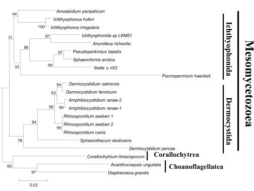
The edited sequences of A. ranae 18S small-subunit rRNA were deposited in GenBank under accession numbers AY550245 (A. ranae 1; see Fig. 5) and AY692319 (A. ranae 2; Fig. 5).
FIG. 5.
Phylogenetic relationship between the 18S small-subunit rRNA of 18 mesomycetozoeans, one coralochytrean, and two choanoflagellates previously studied by Mendoza et al. (25). Multiple hit correction and 1,000 bootstrap-resampled data sets were used to assess branch support. The genus Rhinosporidium (R. canis from a dog and R. seeberi from humans) and Dermocystidium spp. from fish are both in two different well-supported groups. In this tree, Amphibiocystidium ranae is the sister group to D. fennicum and D. salmonis. In turn, both sister groups are linked to R. seeberi. The numbers above the branches are percentages of bootstrap-resampled data sets as obtained by neighbor joining. Corallochytrium limacisporum, Acanthocoepsis unguilata, and Diaphanoeca grandis were used as the outgroup. The scale bar represents evolutionary distance in substitutions per nucleotide.
RESULTS
Clinical and histopathological findings.
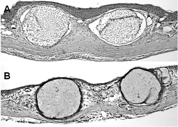
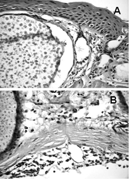
Elevated hemispherical skin lesions usually in the ventral and toe and, less frequently, in dorsal skin were observed in the collected Rana spp. One sample of R. esculenta with only ventral lesions and a sample of R. lessonae with swellings in the inguinal, toe, and dorsal areas were processed for both histopathological and molecular analyses. Histopathological examination revealed several cysts of ≈400 μm in diameter filled with ≈2.0- to ≈7.0 μm-diameter endospores subjacent to the epithelium, which in R. lessonae appeared less hyperplasic (Fig. 1A and B). Details of the cysts with endospores typical of A. ranae in both R. esculenta and R. lessonae are shown in Fig. 2A and B. Inflammatory cells, including neutrophils, eosinophils, and macrophages, were also evident in the subcutaneous tissues surrounding the cysts in both R. esculenta and R. lessonae (Fig. 2A and B).
FIG. 1.
Hematoxylin- and eosin-stained micrographs of the skin infected by Amphibiocystidium ranae in Rana esculenta (A) and Rana lessonae (B). The intradermal cysts were located subjacent to the epidermis, which is more hyperplasic in R. esculenta than in R. lessonae. The A. ranae endospores appear more developed in the skin sample of R. esculenta than in R. lessonae. A, ×15; B, ×10.
FIG. 2.
Detail of cysts and endospores of Amphibiocystidium ranae in infected Rana esculenta (A) and Rana lessonae (B). Inflammatory cells, such as eosinophils and neutrophils, are also observed near the cysts. Hematoxylin and eosin, ×40. Note that the endospores within the cysts of A. ranae in R. esculenta (A) appear more developed than in R. lessonae (B).
Comparative transmission electron microscopy and light microscopy analyses.
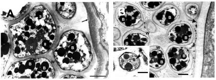
Comparative analyses of R. seeberi and A. ranae with their transmission electron microscopy images showed that these two mesomycetozoeans have several striking ultramicroscopic features in common (Fig. 3). The sporangium's cell wall (cyst) at maturity in both organisms is formed of a thin outer and a thick inner electron-dense layer, and both harbor several hundred endospores (Fig. 3). The endospores possess a thick outer wall with a capsule-like material and a number of electron-dense bodies. Nuclei were also found in some endospores of both microbes (Fig. 3, lower section of panel B). Tissue containing Dermocystidium salmonis was not available for transmission electron microscopy analysis.
FIG. 3.
(A) Transmission electron micrograph of Amphibiocystidium ranae mature sporangium (cyst) containing endospores from an infected frog. The arrow points to the sporangium cell wall; B depicts multiple electron dense bodies; N, nucleus; W, capsule of the endospores. Bar, 2 μm. (B) Rhinosporidium seeberi from a Sri Lankan man with rhinosporidiosis. The arrow points to the mature sporangium cell wall. B, electron-dense bodies; W, endospore wall. Bar, 2 μm. Note the striking ultrastructural similarities between these two pathogenic mesomycetozoeans. The left lower insert in panel B shows a transmission electron micrograph of R. seeberi endospores with a prominent nucleus and a nucleolus (arrowhead). Bar, 2.5 μm.
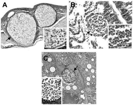
Fixed paraffin-embedded tissues from A. ranae, D. salmonis, and R. seeberi were hematoxylin and eosin stained and evaluated microscopically. This study showed that A. ranae possesses several microscopic features in common with both Dermocystidium sp. and R. seeberi (Fig. 4). Microscopically, all three specimens have spherical shapes at different stages of development. Mature sporangia, measuring between ≈200 and ≈400 μm in diameter, were the biggest and harbored hundred of endospores. The sporangia's cell wall at mature stages was observed in this stain as a thin layer (Fig. 4, arrows). The endospores of A. ranae and D. salmonis in hematoxylin and eosin stains shared more features in common between them than with the endospores of R. seeberi. For instance, both A. ranae and D. salmonis have endospores with a solitary vesicle, whereas R. seeberi's endospores possess multiple small vesicles (Fig. 4, lower panels). Table 1 illustrates the comparative phenotypic and other important features that A. ranae shared with its closest relatives.
FIG. 4.
Hematoxylin- and eosin-stained micrographs depicting the spherical phenotypic characteristic (arrows) of Amphibiocystidium ranae from an infected frog (A, ×20), Dermocystidium salmonis from an infected salmon (B, ×20), and Rhinosporidium seeberi from an infected human (C, ×10). The three panels illustrate the typical spherical phenotype with endospores in these pathogens. The morphological features of the endospores from each organism are depicted in the lower section of panels A, B, and C. Note the similarities between the endospores of A. ranae and D. salmonis, a feature that contrasts with those in R. seeberi (right lower corner, panels A and B, and left lower corner, panel C), ×40.
TABLE 1.
Main clinical, parasitic, life cycle, and morphological similarities and differences between Amphibiocystidium ranae, Dermocystidium salmonis, and Rhinosporidium seeberia
| Parameter | A. ranae | D. salmonis | R. seeberi |
|---|---|---|---|
| Clinical features | Elevated hemispherical skin lesions | Round lesion of the gills | Cutaneous and/or mucocutaneous polyps |
| Parasitic stage similarities | Oval to spherical cysts with endospores (sporangia) | Oval or spherical cysts (sporangia) with endospores | Spherical sporangia with endospores and different size immature forms |
| Parasitic stage differences | Usually cysts located under the skin | Usually cysts in the gills of the infected hosts | Usually sporangia (cysts) within infected polyps |
| Host | Frog pathogen (other amphibians?) | Fish pathogen | Mammalian and bird pathogen |
| Development of uniflagellated zoospores | Absent | Present in all known Dermocystidium spp. | Absent |
| Cyst cell wall (sporangia) | Spherical thin outer and thick inner layers (TEM and LM) | Spherical thin outer layer (only LM) | Spherical thin outer and thick inner layers (TEM and LM) |
| Endospores | Encapsulate endospores with solitary vesicles | Endospores with solitary vesicles (only LM) | Encapsulate endospores with multiple vesicles |
LM, light microscopy; TEM, transmission electron microscopy.
Phylogenetic analysis of the A. ranae 18S small-subunit rRNA.
The NS1 and NS8 primers amplified ≈1,770 bp, nearly the full length of A. ranae's 18S small-subunit rRNA gene from infected R. esculenta (A. ranae 1) and R. lessonae (A ranae 2). Some clones containing the 18S small-subunit rRNA of R. esculenta and R. lessonae were not fully sequenced. Beside the 18S small-subunit rRNA amplified from A. ranae and Rana spp. no other amplicons were detected during this study. Basic Local Alignment Search Tool (BLAST) analysis of the ≈1,770 bp revealed that the 18S small-subunit rRNA from A. ranae 1 and 2 were both closely related to all members of the order Dermocystida and to some extent to the members of the order Ichthyophonida in the class Mesomycetozoea (Ichthyosporea). Of particular interest was its close association, in this analysis, with the genera Dermocystidium and Rhinosporidium. Neighbor-joining and parsimony analyses showed very similar rooted and unrooted phylogenetic trees. In these trees, the two strains of A. ranae, used during this analysis, were consistently the sister group to Dermocystidium spp. (Fig. 5). Although in some trees A. ranae also appeared as the sister group to R. seeberi and R. canis (data not shown), the position of A. ranae as the sister taxon to Dermocystidium was not well supported in parsimony and distance analyses. Of the three Dermocystidium species used in this study (D. fennicum, D. salmonis, and D. percae), only two were sister taxa to A. ranae. Dermocystidium percae was more closely related to Sphaerothecum destruens than to well known Dermocystidium species in all phylogenetic trees.
DISCUSSION
Morphologically A. ranae possesses several features in common with members of the genera Rhinosporidium and Dermocystidium and to some extent S. destruens. These phenotypic similarities include striking ultramicroscopic and microscopic features in their spherical sporangia, cell wall, and the development of numerous endospores within the mature sporangia (Table 1, Fig. 1, 2, and 3). Interestingly, Dermocystidium spp. are characterized by the development of uniflagellated zoospores in vitro and in nature (11). In contrast, despite numerous attempts to induce zoospore production, so far the development of flagellated cells has not yet been possible in A. ranae or R. seeberi (10, 12). As previously discussed for the genus Rhinosporidium (10), it is possible that these two microbes lost their ability to develop flagellated cells, which suggests a closer taxonomic link between them than to the other members of this order characterized by the development of uniflagellated zoospores (Table 1) (1, 11).
Another morphological similarity between the members of the order Dermocystida is the production of spherical phenotypes containing hundreds of endospores within mature sporangia. Although A. ranae cysts from R. esculenta elicited a prominent skin hyperplasia, a feature absent in R. lessonae, cysts of A. ranae in both R. esculenta and R. lessonae showed identical spherical shapes. One possible explanation is that the cysts of R. esculenta used in this study may be older than those in R. lessonae (Fig. 1). Thus, the skin lesions observed in R. esculenta could be more chronic than the one observed in R. lessonae. This observation is supported by the fact that the endospores within the cysts of R. lessonae appeared less developed than those in R. esculenta (Fig. 1B; Fig. 2A and B). On histological preparations A. ranae seems to have more features in common with the endospores in the genus Dermocystidium that those found in R. seeberi. For instance, A. ranae and Dermocystidium spp. endospores have single vesicles (12; this study), whereas R. seeberi harbors multiple vesicles within the endospores (Fig. 3). Intriguingly, Broz and Privora (2) noted that wet preparations of A. ranae endospores from R. temporaria have multiple vesicles remarkably similar to those found in R. seeberi. These phenotypic similarities are in agreement with their phylogenetic preferences. This fact and our phylogenetic analyses indicate that A. ranae is taxonomically and phylogenetically close to the genera Dermocystidium and Rhinosporidium but not far away from S. destruens.
In the past 5 years, new understanding of the phylogenetics of the order Dermocystida have emerged (1, 10, 12). These studies have been of paramount importance to relocate new organisms that had long been considered part of the fungi or protistal microbes within the class Mesomycetozoea (1, 4, 10, 14). The proposals of Arkush et al. (1) and Pascolini et al. (12) served to clarify much of the controversy surrounding these and similar aquatic pathogenic microbes long referred to as Dermocystidium spp. Arkush et al. (1) suggested that the genus Sphaerothecum, formerly known as the “rosette agent,” is different from the typical Dermocystidium spp. but closer to the so-called Dermocystidium-like intracellular microbes and that most probably they belonged or are closely related to S. destruens.
Pascolini et al. (12) gave details on the genus Dermocystidium in amphibians. Based on morphological features and host specificities, they created the genus Amphibiocystidium and presented data indicating that Dermocystidium spp. in frogs such as: D. pusula, D. ranae, Dermosporidium spp., and Dermomycoides spp. should all be considered synonyms of A. ranae. Based on host predilections, they also separated A. ranae from Dermocystidium spp. of fish. Our phylogenetic data strongly support their proposal and indicate that A. ranae is the sister taxon to both Dermocystidum and Rhinosporidium. Moreover, these three pathogens have enough host, phenotypic, and phylogenetic differences to stand as their own genus (4, 8, 10, 14). Our phylogenetic and phenotypic data, collected from samples of R. esculenta and R. lessonae, suggest that A. ranae may be the etiologic agent of the skin-forming cysts with endospores in Rana spp. in these and other geographic locations. However, the true host range of A. ranae within Rana spp. and other amphibians is unknown. Conversely, due to the fact that A. ranae is intractable to culture, experimental infection to reproduce the disease in Rana spp. has not yet been possible. Thus, the assessment of host susceptibility of Rana spp. to A. ranae infection would be a challenge.
One interesting finding during our phylogenetic analysis was the placement of D. percae as the sister taxon to S. destruens and far away from the genus Dermocystidium. Based on our phylogenetic trees, we speculate that D. percae could well be a species of the genus Sphaerothecum or perhaps represents a new genus by itself. The fact that D. percae is an intracellular organism in some stages of its cell cycle, a feature in common with S. destruens, suggests that these two organisms are not only phylogenetically linked but also share physiological and phenotypic features (1, 13). Phylogenetic analysis by Pekkarinen et al. (13) showed S. destruens as the outgroup linked to D. percae and to members of the order Ichthyophonida. In our trees, S. destruens was the sister group to Dermocystidium, Amphibiocystidium, and Rhinosporidium spp., but D. percae was always the sister group to S. destruens, even in unrooted trees. Our phylogenetic study suggested that D. percae has a strong link to S. destruens and that is not a member of the genus Dermocystidium. Based on morphological, phylogenetic, and host specificities between these two taxa, the creation of a new genus might be justifiable in this case.
Pekkarinen et al. (13) reported that D. percae has giant elongate-cylindrical cysts (>580 μm in diameter). Giant elongate cysts, similar to that in D. percae, have been previously reported in carp in the Czech Republic (5), in cultured eels in Scotland (18), and in unarmored threespine stickleback fish in Germany (15). Due to their typical elongate cysts with endospores, it was believed that their infections were caused by species of the genus Dermocystidium (9). However, ultrastructural features in previous studies also showed a strong link to the morphological features described for D. percae by Pekkarinen et al. (13) and different from the spherical-oval sporangia typical of the genus Dermocystidium. It would be very interesting to investigate the true phylogenetic affinities of organisms with giant elongate-cylindrical cysts during parasitic stages traditionally attributed to the genus Dermocystidium. Meanwhile, the epithet Dermotheca is suggested for organisms such as D. percae with giant elongate-cylindrical shapes and with phylogenetic affinities to S. destruens.
Acknowledgments
We thank Kathy Hoag for suggestions and critical review of the manuscript.
REFERENCES
- 1.Arkush, K. D., L. Mendoza, M. A. Adkinson, and R. P. Hedrick. 2003. Observations on the life stages of Sphaerothecum destruens n. g., n. sp., a mesomycetozoean fish pathogen formerly referred to as the Rosette Agent. J. Eukaryot. Microbiol. 50:430-438. [DOI] [PubMed] [Google Scholar]
- 2.Bro█, O., and M. Privora. 1951. Two skin parasites of Rana temporaria: Dermocystidium ranae Guyènot Naville and Dermosporidium granulosum n. sp. Parasitology 42:65-69. [DOI] [PubMed] [Google Scholar]
- 3.Bucci, S., M. Ragghianti, F. Guerrini, V. Cerrini, A. Morosi, M. Mossone, R. Pascolini, and G. Mancino. 2000. Negative environmental factors and biodiversity: the case of the hybridogenetic green frog system from Lake Trasimeno. Ital. J. Zool. 67:365-370. [Google Scholar]
- 4.Cavalier-Smith, T. 1998. Neomonada and the origin of animals and fungi, p. 375-407. In G. H. Coombs, K. Vickerman, M. A. Sleigh, and A. Warren (ed.), Evolutionary relationships among protozoa. Chapman and Hall, London, England.
- 5.Cervinka, S., J. Vitovec, J. Lom, J. Hoška, and F. Kub. 1974. Dermocystidiosis—a gill disease of the carp due to Dermocystidium cyprinid. J. Fish Biol. 6:689-699. [Google Scholar]
- 6.Gargas, A., and P. T. DePriest. 1996. A nomenclature for fungal PCR primers with examples from intron-containing small-subunit rRNA. Mycologia 88:745-748. [Google Scholar]
- 7.Graf, J. D., and P. Pelaz. 1989. Evolutionary genetics of the Rana esculenta complex, p. 289-302. In R. M. Dawley and J. P. Bogart (ed.), Evolution and ecology of unisexual vertebrates, bulletin 466. New York State Museum, Albany, N.Y.
- 8.Herr, R. A., L. Ajello, J. W. Taylor, S. N. Arseculeratne, and L. Mendoza. 1999. Phylogenetic analysis of Rhinosporidium seeberi's 18S small-subunit ribosomal DNA groups this pathogen among members of the protoctistant Mesomycetozoa clade. J. Clin. Microbiol. 37:2750-2754. [DOI] [PMC free article] [PubMed] [Google Scholar]
- 9.Hoffman, G. L. 1998. Parasites of North American freshwater fish, 2nd ed., p. 89-91. Comstock Publishing Associates, London, England.
- 10.Mendoza, L., J. W. Taylor, and L. Ajello. 2002. The class Mesomycetozoea: a heterogeneous group of microorganisms at the animal-fungal boundary. Annu. Rev. Microbiol. 56:315-344. [DOI] [PubMed] [Google Scholar]
- 11.Olson, R. E., C. F. Dungan, and R. A. Holt. 1991. Water-borne transmission of Dermocystidium salmonis in the laboratory. Dis. Aquatic Organisms 12:41-48. [Google Scholar]
- 12.Pascolini, R., P. Daszak, A. Cunningham, A. Fagotti, S. Tei, D. Vagnetti, S. Bucci, and I. Di Rosa. 2003. Parasitism by Dermocystidium ranae in a population of Rana esculenta complex in Central Italy and description of of Amphibiocystidium n. gen. Dis. Aquat. Org. 56:65-74. [DOI] [PubMed] [Google Scholar]
- 13.Pekkarinen, M., J. Lom, C. A. Murphy, M. A. Ragan, and I. Dyková. 2003. Phylogenetic position and ultrastructure of two Dermocystidium species (Ichthyosporea) from the common perch (Perca fluviatilis). Acta Protozool. 42:287-307. [Google Scholar]
- 14.Ragan, M. A., C. L. Goggin, R. J. Cawthorn, L. Cerenius, A. V. C. Jamienson, S. M. Plourdes, T. G. Tand, K. Soderhall, and R. R. Gutell. 1996. A novel clade of protistes near the animal-fungal divergence. Proc. Natl. Acad. Sci. USA 93:11907-11912. [DOI] [PMC free article] [PubMed] [Google Scholar]
- 15.Scheer, D. 1957. Die fishparasitarën arten der gattung Dermocystidium (Haplosporidia?). Z. Fisch. Hilfswiss. 6:127-134. [Google Scholar]
- 16.Uzzell, T., and H. Hotz. 1979. Electrophoretic and morphological evidence for the two forms of green frogs (Rana esculenta complex) in peninsular Italy (Amphibia, Salientia). Mitt. Zool. Mus. Berlin 55:13-27. [Google Scholar]
- 17.Uzzell, T. 1983. An immunological survey of Italian water frogs Salentia:Ranidae. Herpetologica 39:225-234. [Google Scholar]
- 18.Wootten, R., and A. H. McVicar. 1982. Dermocystidium from cultured eels, Anguila anguilla L., in Scotland. J. Fish Dis. 5:215-222. [Google Scholar]


