

Abstract
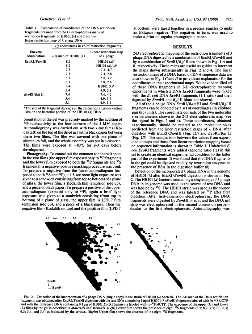
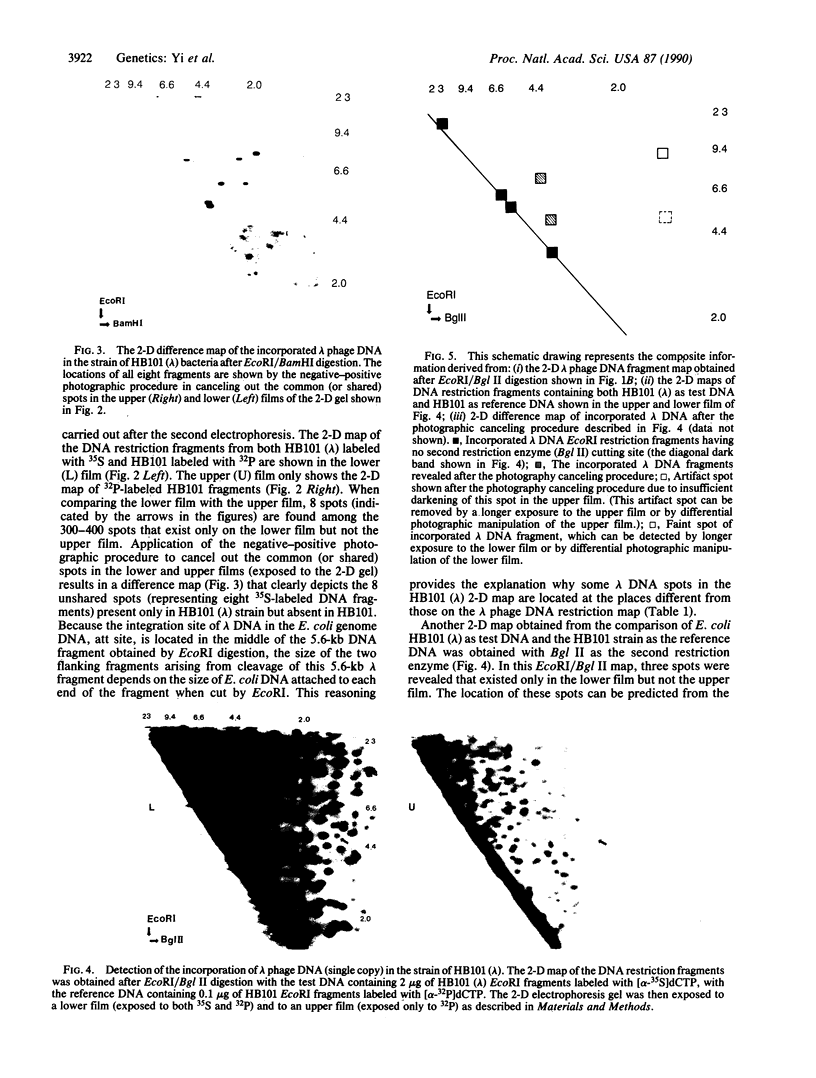
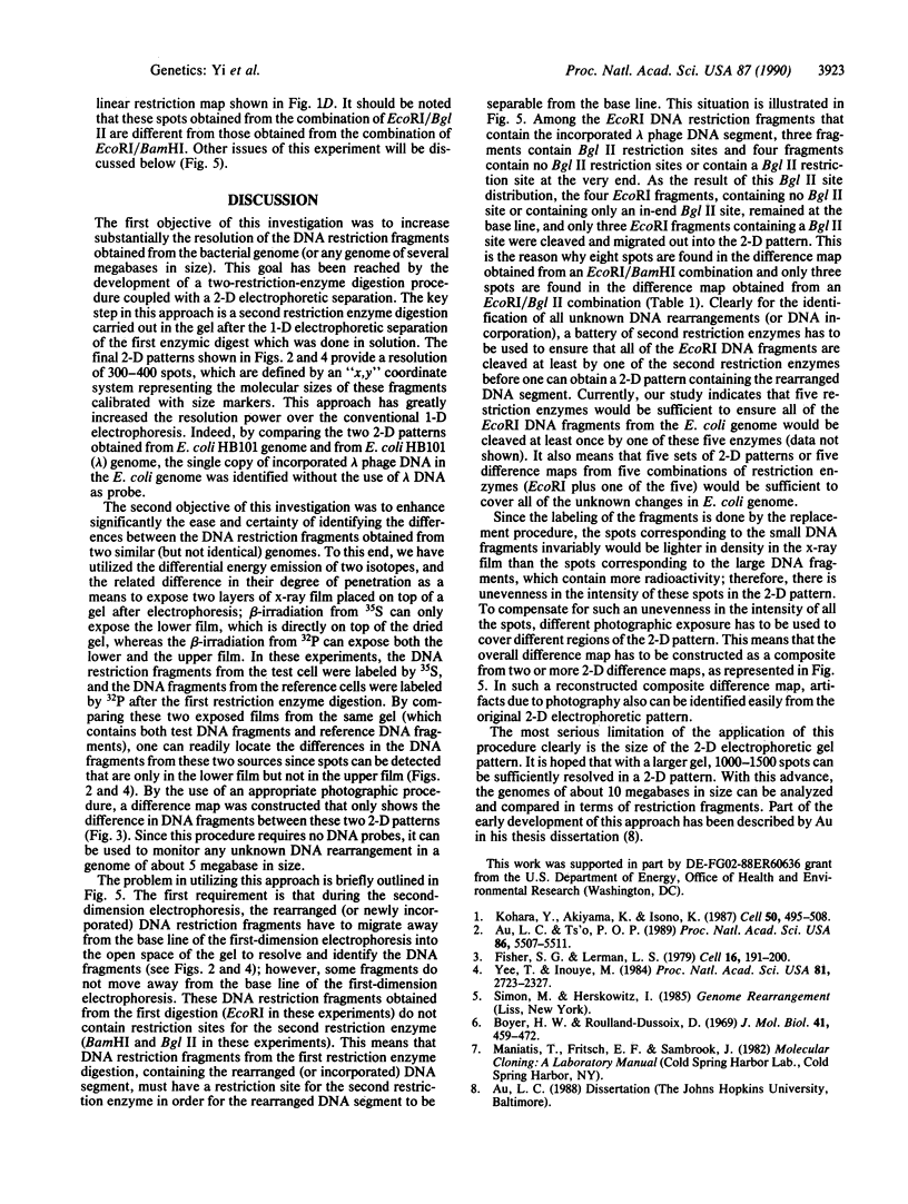
A probe-free method was developed to detect DNA rearrangement in bacteria based on the electrophoretic separation of twice-digested restriction fragments of genomic DNA into a two-dimensional (2-D) pattern. The first restriction enzyme digestion was done in solution, followed by electrophoresis of the restriction fragments in one dimension. A second restriction enzyme digestion was carried out in situ in the gel, followed by electrophoresis in a second dimension perpendicular to the first electrophoresis. The 2-D pattern provides for the resolution of 300-400 spots, which are defined and indexed by an "x,y" coordinate system with size markers. This approach has greatly increased the resolution power over conventional one-dimensional (1-D) electrophoresis. To study DNA rearrangement, a 2-D pattern from a test strain was compared with the 2-D pattern from a reference strain. After the first digestion, genomic DNA fragments from the test strain were labeled with 35S, while those from the reference strain were labeled with 35P. This was done to utilize the difference in the energy emission of 35S and 32P isotopes for autoradiography when two x-ray films were exposed simultaneously on top of the gel after the 2-D electrophoresis. The irradiation from the decay of 35S exposed only the lower film, whereas the irradiation from the decay of 32P exposed both the lower and upper films. Different DNA fragments existed in the test DNA compared with the reference DNA can be identified unambiguously by the differential two 2-D patterns produced on two films upon exposure to the 35S and 32P fragments in the same gel. An appropriate photographic procedure further simplified the process, allowing only the difference in DNA fragments between these two patterns to be shown in the map. We have utilized the difference map obtained from Escherichia coli strains HB101 and HB101 (lambda) genomic DNA to show the incorporation of one copy of phage lambda DNA without the use of a lambda DNA probe. This is the same test system that was used previously.
Full text
PDF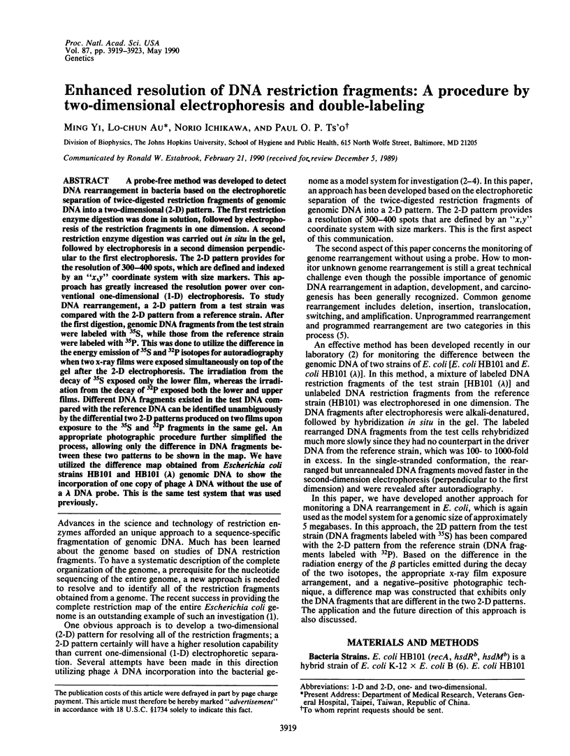

Images in this article
Selected References
These references are in PubMed. This may not be the complete list of references from this article.
- Au L. C., Ts'o P. O. A general method for detecting rearrangements in a bacterial genome. Proc Natl Acad Sci U S A. 1989 Jul;86(14):5507–5511. doi: 10.1073/pnas.86.14.5507. [DOI] [PMC free article] [PubMed] [Google Scholar]
- Boyer H. W., Roulland-Dussoix D. A complementation analysis of the restriction and modification of DNA in Escherichia coli. J Mol Biol. 1969 May 14;41(3):459–472. doi: 10.1016/0022-2836(69)90288-5. [DOI] [PubMed] [Google Scholar]
- Fischer S. G., Lerman L. S. Length-independent separation of DNA restriction fragments in two-dimensional gel electrophoresis. Cell. 1979 Jan;16(1):191–200. doi: 10.1016/0092-8674(79)90200-9. [DOI] [PubMed] [Google Scholar]
- Kohara Y., Akiyama K., Isono K. The physical map of the whole E. coli chromosome: application of a new strategy for rapid analysis and sorting of a large genomic library. Cell. 1987 Jul 31;50(3):495–508. doi: 10.1016/0092-8674(87)90503-4. [DOI] [PubMed] [Google Scholar]
- Yee T., Inouye M. Two-dimensional S1 nuclease heteroduplex mapping: detection of rearrangements in bacterial genomes. Proc Natl Acad Sci U S A. 1984 May;81(9):2723–2727. doi: 10.1073/pnas.81.9.2723. [DOI] [PMC free article] [PubMed] [Google Scholar]