Abstract
Human metapneumovirus (hMPV) is associated with acute respiratory tract disease especially in young children. Using a quantitative real-time TaqMan PCR, we analyzed the replication kinetics of hMPV in different cell lines. Our results indicate that hMPV replicates slightly more efficiently in LLC-MK2 than in Vero cells and poorly in HEp-2 cells.
The human metapneumovirus (hMPV) is a newly discovered member of the family Paramyxoviridae responsible for acute respiratory tract infections in young children, elderly patients, and immunocompromised hosts (4, 12). This negative-sense RNA virus, which can be classified into two genotypes (A and B) (1, 3, 9, 10), has been preliminarily assigned to the genus Metapneumovirus within the subfamily Pneumovirinae (11). The clinical syndromes associated with this viral infection encompass mild respiratory problems and acute wheezing as well as bronchiolitis and pneumonia (4, 5, 8, 13). Serological studies from The Netherlands and elsewhere have indicated that virtually all children are infected by the age of 5 and that hMPV has been circulating in humans for at least 50 years (7, 12, 14).
hMPV was only recently identified due to its slow growth in a few permissive cell lines, notably tertiary monkey kidney cells. The viral cytopathic effects (CPE) may vary from syncytia resembling those caused by human respiratory syncytial virus to rounding of refringent cells with subsequent detachment from the monolayer. Since some reports have suggested that hMPV might also replicate efficiently in HEp-2 (6) and Vero cells (1), we sought to analyze the replication kinetics of hMPV in different cell lines using a quantitative real-time PCR assay.
Specimens and cell lines.
Frozen (−80°C) nasopharyngeal aspirates collected from young children in 2002 (5) were inoculated in 24-well plates on the continuous human laryngeal carcinoma (HEp-2), African green monkey kidney (Vero), and rhesus monkey kidney (LLC-MK2) cell lines. The infections were made with 100 μl of previously reported hMPV PCR-positive specimens (5) (in triplicate) in 1 ml of Opti-MEM I medium (Invitrogen GIBCO, Burlington, Ontario, Canada) containing 2 μg of trypsin (Sigma, Oakville, Ontario, Canada) per ml and antibiotics. Plates were incubated at 37°C with 5% CO2 and examined for CPE every other day for 22 days. At the time of each of those examinations, an aliquot of 200 μl of cell culture supernatant was collected and frozen at −80°C, and then the culture was replenished with an equal volume of fresh medium containing 5 μg of trypsin per ml.
RNA extraction and cDNA synthesis.
Viral RNA was extracted from 200 μl of infected cell culture supernatants by using a QIAamp viral RNA minikit (Qiagen, Mississauga, Ontario, Canada) according to the manufacturer's protocol and then eluted into a final volume of 40 μl. cDNA was synthesized by using 10 μl of RNA eluate, random hexamer primers (Amersham Pharmacia Biotech, Baie d'Urfé, Québec, Canada), and an Omniscript reverse transcriptase kit (Qiagen) according to the manufacturer's instructions.
Real-time PCR.
The real-time quantitative PCR for hMPV was developed in a LightCycler instrument (Roche Diagnostics, Laval, Québec, Canada). The primers and TaqMan probe for the L (polymerase) gene of hMPV were based on the sequence of the prototype strain 00-1 from The Netherlands (GenBank accession number af371337). The sequences were as follows: forward primer, 5′-GTTGCCATAGAGAATCCTGTTA-3′; reverse primer, 5′-CCAATTACTAAACCAATTGCTTACCCA-3′; and TaqMan probe, 5′-(6-FAM)CGAGCATGTTAGACTCAAAAATGCA(TAMRA-PH)-3′. Each 20-μl reaction mixture included 2 μl of cDNA and 18 μl of a reaction mixture containing 4 mM MgCl2, 0.4 μM hMPV primer (each), 0.6 μM TaqMan probe, 2 μl of the LightCycler hybridization probe mixture (Roche Diagnostics), and 0.6 μl of dimethyl sulfoxide. Cycling conditions included a 30-s denaturation step at 95°C and then 60 PCR cycles consisting of 5 s at 95°C, 5 s at 56°C, and 25 s at 72°C. Fluorescence measurements were performed at each cycle (at the end of amplification) in the F1 channel, and a threshold cycle value for each sample was calculated by determining the point at which the fluorescence exceeded a threshold limit. For quantification, a standard curve was constructed using serial dilutions (from 102 to 109 copies) of a transcribed hMPV plasmid. The quantitative real-time PCR assay for hMPV had a lower limit of detection of 100 copies, with intra- and interassay coefficients of variability of 18.7 and 19.6%, respectively, when 104 copies of the hMPV plasmid were used.
Table 1 summarizes the replication kinetics of five hMPV strains based on CPE development and quantitative real-time PCR. Of the five nasopharyngeal aspirate specimens tested, all showed CPE on LLC-MK2 cells, three showed CPE on Vero cells, and none showed CPE on HEp-2 cells. The CPE observed on LLC-MK2 cells consisted of round refringent cells with subsequent detachment from the monolayer and clusters of small syncytia. In contrast, the CPE on Vero cells was indistinguishable from that caused by other viruses (e.g., enteroviruses) and consisted of cell rounding and death. Initial CPE appeared sooner on LLC-MK2 than Vero cells for almost all strains, as well as maximal CPE (Table 1).
TABLE 1.
Replication kinetics of five hMPV strains in three cell lines
| Clinical specimen | No. of replicates with CPEa (days to onset of CPE)/total
|
Maximum hMPV viral loadb (days to peak viral load)
|
||||
|---|---|---|---|---|---|---|
| LLC-MK2 | Vero | HEp-2 | LLC-MK2 | Vero | HEp-2 | |
| 85126 | 3/3 (8-12) | 3/3 (16) | 0/3 | 7.5 × 10⁸ (18) | 6.6 × 10⁴ (20) | 5.9 × 10⁵ (6) |
| 85525 | 3/3 (8-10) | 3/3 (8-10) | 0/3 | 3.2 × 10⁸ (14) | 5.2 × 10⁸ (16) | 1.1 × 10⁶ (14) |
| 85728 | 1/3 (14) | 1/3 (16) | 0/3 | 6.0 × 10⁸ (20) | 3.4 × 10⁹ (20) | 0 |
| 91728 | 1/3 (8) | 0/3 | 0/3 | 8.0 × 10⁸ (18) | 8.6 × 10⁷ (20) | 0 |
| 91749 | 2/3 (10-12) | 0/3 | 0/3 | 5.5 × 10⁸ (16) | 0 | 0 |
CPE obtained from three replicates of nasopharyngeal samples.
Mean copy number per 200 μl of supernatant from positive replicates.
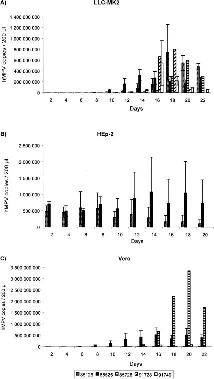
Using real-time PCR, hMPV RNA was detected in five of five, four of five, and two of five samples from supernatants of LLC-MK2, Vero, and HEp-2 cells, respectively. Peak hMPV RNA load was detected at a slightly earlier time in LLC-MK2 cells for four strains, whereas maximum detection occurred simultaneously in LLC-MK2 and Vero cells for the other strain (Table 1). Peak viral loads were higher in supernatants of LLC-MK2 cells for three strains whereas, Vero cells had higher loads for two strains. For one strain (specimen 91728), hMPV RNA was detected even though no CPE was observed on Vero cells, as it occurred on HEp-2 cells for the two positive strains. Maximum CPE corresponded mostly to maximum viral load for LLC-MK2 and Vero cells. HEp-2 cells supported only low and variable RNA replication for two strains without an evident peak in viral load, as found in LLC-MK2 and Vero cells (Fig. 1).
FIG. 1.
Replication kinetics of five hMPV strains over a period of 22 days as determined by real-time PCR on three cell lines. Data are mean viral loads (number of copies per 200 μl of supernatant), and error bars indicate standard deviations for strains with at least two positive replicates. Note that the y-axis scales are different for the three cell lines.
LLC-MK2 cells allow slightly more consistent viral replication than Vero cells, as shown by the fact that more strains exhibited both earlier and maximum CPE. In addition, replication kinetics were better in three of five (60%) strains when real-time PCR was used, although viral load was sometimes higher in Vero cells. As reported previously (2, 12), the addition of trypsin allowed faster development of CPE in both LLC-MK2 and Vero cells (data not shown). HMPV detection in HEp-2 cells based on visualization of CPE appears inadequate. Moreover, HEp-2 cells do not permit efficient and sustained viral replication in our study, contrasting with previous data (6). This could be due to early cell death when these cells are kept confluent for ≥1 week. There are some limitations to our study. First, only hMPV group A strains were evaluated, and thus, the replication kinetics of group B strains remain unknown. Second, there was some variability in viral load assessment and CPE development between different replicates of the same specimen (Table 1). This may be related to extended freezing of some samples.
In conclusion, our results indicate that hMPV replicates slightly more efficiently in LLC-MK2 than Vero cells but much more poorly in HEp-2 cells, based on CPE development and viral load measurement. These findings should be confirmed by testing a larger number of clinical specimens and additional cell lines. Nevertheless, neither cell line tested in our study allows a timely detection of this important respiratory pathogen in a clinical setting. Future research should be aimed at comparing the performance of different laboratory methods (real-time PCR, antigen detection, and cell culture) for the accurate and rapid diagnosis of hMPV in various clinical specimens.
REFERENCES
- 1.Biacchesi, S., M. H. Skiadopoulos, G. Boivin, C. T. Hanson, B. R. Murphy, P. L. Collins, and U. J. Buchholz. 2003. Genetic diversity between human metapneumovirus subgroups. Virology 315:1-9. [DOI] [PubMed] [Google Scholar]
- 2.Biacchesi, S., M. H. Skiadopoulos, K. C. Tran, B. R. Murphy, P. L. Collins, and U. J. Buchholz. 2004. Recovery of human metapneumovirus from cDNA: optimization of growth in vitro and expression of additional genes. Virology 321:247-259. [DOI] [PubMed] [Google Scholar]
- 3.Boivin, G., I. Mackay, T. P. Sloots, S. Madhi, F. Freymuth, D. Wolf, Y. Shemer-Avni, H. Ludewick, G. C. Gray, and E. LeBlanc. 2004. Global genetic diversity of human metapneumovirus fusion gene. Emerg. Infect. Dis. 10:1154-1157. [DOI] [PMC free article] [PubMed] [Google Scholar]
- 4.Boivin, G., Y. Abed, G. Pelletier, L. Ruel, D. Moisan, S. Cote, T. C. Peret, D. D. Erdman, and L. J. Anderson. 2002. Virological features and clinical manifestations associated with human metapneumovirus: a new paramyxovirus responsible for acute respiratory-tract infections in all age groups. J. Infect. Dis. 186:1330-1334. [DOI] [PubMed] [Google Scholar]
- 5.Boivin, G., G. De Serres, S. Cote, R. Gilca, Y. Abed, L. Rochette, M. G. Bergeron, and P. Dery. 2003. Human metapneumovirus infections in hospitalized children. Emerg. Infect. Dis. 9:634-640. [DOI] [PMC free article] [PubMed] [Google Scholar]
- 6.Chan, P. K., J. S. Tam, C. W. Lam, E. Chan, A. Wu, C. K. Li, T. A. Buckley, K. C. Ng, G. M. Joynt, F. W. Cheng, K. F. To, N. Lee, D. S. Hui, J. L. Cheung, I. Chu, E. Liu, S. S. Chung, and J. J. Sung. 2003. Human metapneumovirus detection in patients with severe acute respiratory syndrome. Emerg. Infect. Dis. 9:1058-1063. [DOI] [PMC free article] [PubMed] [Google Scholar]
- 7.Ebihara, T., R. Endo, H. Kikuta, N. Ishiguro, M. Yoshioka, X. Ma, and K. Kobayashi. 2003. Seroprevalence of human metapneumovirus in Japan. J. Med. Virol. 70:281-283. [DOI] [PubMed] [Google Scholar]
- 8.Jartti, T., B. van den Hoogen, R. P. Garofalo, A. D. Osterhaus, and O. Ruuskanen. 2002. Metapneumovirus and acute wheezing in children. Lancet 360:1393-1394. [DOI] [PMC free article] [PubMed] [Google Scholar]
- 9.Peret, T. C., Y. Abed, L. J. Anderson, D. D. Erdman, and G. Boivin. 2004. Sequence polymorphism of the predicted human metapneumovirus G glycoprotein. J. Gen. Virol. 85:679-686. [DOI] [PubMed] [Google Scholar]
- 10.Skiadopoulos, M. H., S. Biacchesi, U. J. Buchholz, J. M. Riggs, S. R. Surman, E. Amaro-Carambot, J. M. McAuliffe, W. R. Elkins, M. St Claire, P. L. Collins, and B. R. Murphy. 2004. The two major human metapneumovirus genetic lineages are highly related antigenically, and the fusion (F) protein is a major contributor to this antigenic relatedness. J. Virol. 78:6927-6937. [DOI] [PMC free article] [PubMed] [Google Scholar]
- 11.van den Hoogen, B. G., T. M. Bestebroer, A. D. Osterhaus, and R. A. Fouchier. 2002. Analysis of the genomic sequence of a human metapneumovirus. Virology 295:119-132. [DOI] [PubMed] [Google Scholar]
- 12.van den Hoogen, B. G., J. C. de Jong, J. Groen, T. Kuiken, R. de Groot, R. A. Fouchier, and A. D. Osterhaus. 2001. A newly discovered human pneumovirus isolated from young children with respiratory tract disease. Nat. Med. 7:719-724. [DOI] [PMC free article] [PubMed] [Google Scholar]
- 13.van den Hoogen, B. G., G. J. van Doornum, J. C. Fockens, J. J. Cornelissen, W. E. Beyer, R. de Groot, A. D. Osterhaus, and R. A. Fouchier. 2003. Prevalence and clinical symptoms of human metapneumovirus infection in hospitalized patients. J. Infect. Dis. 188:1571-1577. [DOI] [PubMed] [Google Scholar]
- 14.Wolf, D. G., Z. Zakay-Rones, A. Fadeela, D. Greenberg, and R. Dagan. 2003. High seroprevalence of human metapneumovirus among young children in Israel. J. Infect. Dis. 188:1865-1867. [DOI] [PubMed] [Google Scholar]


