Abstract
Psoriasin (S100A7) is an 11-kDa small calcium binding protein initially isolated from psoriatic skin lesions. It belongs to the S100 family of proteins which play an important role in a range of cell functions including proliferation, differentiation, migration and apoptosis. Aberrant Psoriasin expression has been implicated in a range of cancers and is often associated with poor prognosis. This study examined the role of Psoriasin on pancreatic cancer cell functions and the implication in progression of the disease. Expression of Psoriasin was determined in a cohort of pancreatic tissues comprised of 126 pancreatic tumours and 114 adjacent non-tumour pancreatic tissues. Knockdown and overexpression of Psoriasin in pancreatic cancer cells was performed using specifically constructed plasmids, which either had anti-Psoriasin ribozyme transgene or the full length human Psoriasin coding sequence. Psoriasin knockdown and overexpression was verified using conventional RT-PCR and qPCR. The effect of manipulating Psoriasin expression on pancreatic cancer cell functions was assessed using several in vitro cell function assays. Local invasive pancreatic cancers extended beyond the pancreas expressed higher levels of Psoriasin transcripts compared with the cancers confined to the pancreas. Primary tumours with distant metastases exhibited a reduced expression of Psoriasin. Psoriasin overexpression cell lines exhibited significantly increased growth and migration compared to control cells. In addition, Psoriasin overexpression resulted in increased pancreatic cancer cell invasion which was associated with upregulation of matrix metalloproteinase-2 (MMP-2) and MMP-9. Overexpression of Psoriasin also promoted aggregation and survival of pancreatic cancer cells when they lost anchorage. Taken together, higher expression of Psoriasin was associated with local invasion in pancreatic cancers. Psoriasin expression is associated with pancreatic cancer cell growth, migration, cell-matrix adhesion, and invasion via regulation of MMPs. As such, the proposed implications of Psoriasin in invasion, disease progression and as a potential therapeutic target warrant further investigation.
Keywords: Psoriasin, S100A7, pancreatic cancer, adhesion, invasion, metastasis
Introduction
Psoriasin, also known as S100A7, is a member of the S100 protein family of calcium binding proteins and was first identified as being overexpressed in psoriatic skin lesions (1). It is a 11.4-kDa secretory protein encoded on chromosome 1q21, clustered alongside 16 other members of the S100 family within a region known as the epidermal differentiation complex (EDC) (2,3). The S100 family encodes both cytoplasmic and secreted proteins that share EF-hand helix-loop-helix domains, important for their role as calcium binding proteins (4). Psoriasin S100 proteins are widely expressed in numerous cell types localised to the cytoplasm and/or nucleus, or in some cases are secreted. Due to their role in calcium binding and signalling, they are involved in numerous cell functions including proliferation, differentiation, migration and apoptosis.
In addition to its overexpression in psoriatic skin lesions, aberrant Psoriasin expression has been implicated in a range of human diseases including cancer. Interestingly, Psoriasin is a chemotactic factor for keratinocytes and leukocytes (5–7), and is also upregulated and excreted from cells of the epidermis during inflammation. Additionally, Psoriasin has been implicated as an antimicrobial peptide, selectively killing E. coli on the surface of the skin (8). Therefore, Psoriasin may be utilised, via the host immune response, as a selective chemotactic factor both in psoriasis and cancer (9).
The expression of Psoriasin during carcinogenesis has been studied in detail, and its overexpression has been linked to a number of cancers including breast (10), prostate (11), skin (12), head and neck (13), bladder (14) and lung cancer (15). It is also often associated with poor prognosis. For example in breast cancer, Psoriasin expression correlates with features of poor prognosis, including oestrogen receptor (ER) and progesterone receptor (PR) negativity, HER2 positivity, and the presence of lymphocytic infiltration (16–18). The precise role of Psoriasin in cancer remains unclear. One hypothesis links Psoriasin expression with stimulation of angiogenesis regulated by VEGF (19). In the development of breast cancer, Psoriasin regulates epidermal growth factor (EGF)-induced phosphorylation of the EGF receptor (EGFR), actin remodelling and NF-κβ mediated matrix metalloproteinase-9 (MMP-9) secretion, promoting tumour development and metastasis (20). These results suggest Psoriasin plays an important role in carcinoma development.
Pancreatic cancer is the fourth leading cause of cancer deaths in Western countries and carries a very poor prognosis due to delayed diagnosis and lack of effective treatments. The majority of patients present with metastasis at diagnosis; 25% with local metastasis and 55% with regional metastasis (21). The most common sites of metastasis are the liver, followed by the peritoneum, lung and pleura, bones and adrenal glands. Outcome for patients is poor with a 5-year survival rate of <5% (22). In comparison to the studies of other malignancies, pancreatic cancer requires more intensive study for understanding genetic and molecular machinery which is utilised by cancerous cells during disease progression and metastasis. This study aimed to examine the role of Psoriasin in pancreatic cancer with a focus for its involvement in local invasion and distant metastasis.
Materials and nethods
Materials and cell lines
Pancreatic cell lines (MIA-PaCa-2 and PANC-1) were purchased from the American Type Culture Collection (ATCC, Rockville, MD, USA). Both cell lines are derived from primary tumours of pancreatic cancer. MIA-PaCa-2 was isolated from a pancreatic carcinoma while PANC-1 was derived from an epithelioid carcinoma of pancreas. The pancreatic cancer cells were maintained in Dulbecco's modified Eagle's medium (DMEM)-F12 medium, supplemented with 10% fetal calf serum (FCS) and antibiotics. LP-9 mesothelial cells were purchased from the Coriell Institute for Medical Research (Camden, NJ, USA) and maintained in M199 medium supplemented with 15% FCS and antibiotics. Matrigel (BD Matrigel Basement Membrane Matrix) was obtained from BD Biosciences (Oxford, UK).
Construction of ribozyme transgenes targeting human Psoriasin and Psoriasin overexpression vectors
Anti-human Psoriasin hammerhead ribozymes were designed based on the secondary structure of the mRNA generated using Zuker's RNA Mfold program (23). The ribozymes were synthesized and cloned into pEF6/V5-His-TOPO plasmid vector (Invitrogen, Paisley, UK). The primers used for ribozyme synthesis are shown in Table I. Similarly, full-length human Psoriasin coding sequence amplified from normal human prostate tissues was cloned into the same vector. Constructed plasmids were extracted using the Genelute Plasmid Mini-Prep kit (Sigma-Aldrich, Poole, UK). Ribozyme transgenes, overexpression constructs, and control plasmids were transfected into MIA-PaCa-2 and PANC-1 cells by electroporation (Gene Pulser Xcell; Bio-Rad, Hertfordshire, UK). Stable transfectants were obtained and verified following two weeks selection using blasticidin (5 μg/ml, Melford Laboratories Ltd., Suffolk, UK). The cells were then cultured in DMEM with blasticidin at a lower concentration (0.5 μg/ml) to maintain plasmid expression.
Table I.
Primer sequences.
| Gene | Forward | Reverse |
|---|---|---|
| Psoriasin | 5′-GAGGTCCATAATAGGCATGA | 5′-AGCAAGGACAGAAACTCAGA |
| RAGE | 5′-GATCCAGGATGAGGGGATTT | 5′-GCTACTGCTCCACCTTCTGG |
| Psoriasin (expression) | 5′-ATGAGCAACACTCAAGCTG | 5′-ACTGGCTGCCCCCGGAACA |
| Psoriasin (QPCR) | 5′-TGTGACAAAAAGGGCACAAA | 5′-ACTGAACCTGACCGTACACCCAGCAAGGACAGAAACTC |
| CK19 (QPCR) | 5′-CAGGTCCGAGGTTACTGAC | 5′-ACTGAACCTGACCGTACACCGTTTCTGCCAGTGTGTCTTC |
| Cyclin D1 | 5′-CGGTGTCCTACTTCAAATGT | 5′-ACCTCCTCCTCCTCCTCT |
| p27Kip1 | 5′-GGAATAAGGAAGCGACCTG | 5′-CCGTCTGAAACATTTTCTTC |
| Caspase 3 | 5′-GGCGTGTCATAAAATACCAG | 5′-ACAAAGCGACTGGATGAA |
| MMP9 | 5′-AACTACGACCGGGACAAG | 5′-ATTCACGTCGTCCTTATGC |
| GAPDH | 5′-GGCTGCTTTTAACTCTGGTA | 5′-AGCAAGGACAGAAACTCAGA |
| Psoriasin (ribozyme) | 5′-CTGCAGTCACAGGCACTAAGGAAGTTGGGCTGATGAGTCCGTGAGGA | 5′-ACTAGTGGCTGGTGTTTGACATTTCGTCCTCACGGACT |
RNA isolation, reverse transcription-PCR (RT-PCR) and quantitative PCR (qPCR)
Total RNA was isolated using TRI reagent (Sigma-Aldrich). First strand cDNA synthesis was undertaken using the Precision nanoScript Reverse Transcription kit (Primer Design, Southampton, UK). PCR was performed using GoTaq Green MasterMix (Promega, Dorset, UK). Cycling conditions were as follows: 94°C for 5 min followed by 36 cycles of 94°C for 30 sec, 55°C for 30 sec, and 72°C for 40 sec. This was followed by a final extension of 72°C for 10 min. Products were visualised using 2% agarose gels stained with SYBR Safe (Invitrogen).
QPCR was performed using the Icycler IQ5 system (Bio-Rad, Hammel Hempsted, UK). Pancreatic cancer cell cDNA samples were examined for Psoriasin transcript expression, alongside a set of standards and negative controls. The Amplifluor system (Intergen Inc., Oxford, UK) and qPCR mastermix (Bio-Rad) were used. Psoriasin primer pairs were designed using Beacon design software (Premier Biosoft, Palo Alto, CA, USA), whereby the reverse primer contained an additional Z sequence (5′-ACTGAACCTGACCGTACA) complementary to the universal Z probe (Intergen Inc.). Reaction conditions were as follows: 94°C for 12 min, followed by 90 cycles of 94°C for 15 sec, 55°C for 40 sec and 72°C for 20 sec.
Quantification of Psoriasin transcripts in human pancreatic cancer
Human pancreatic tissue was collected from patients undergoing surgical resection of pancreatic tumours at the Beijing Cancer Hospital. The specimens retrieved comprised 126 pancreatic tumours and 114 along with matched adjacent normal pancreatic tissues over a period from 2002 to 2011 with follow-up information up to 2013 (median, 12 months, IQR, 6–20.5 months). The tissues were stored immediately after surgery at −80°C until use. Clinicopathological factors, including age, sex, histological type, TNM stage, lymph node metastasis, distant metastases and embolism, were recorded and stored in the patient database. The protocol and procedure were reviewed and approved by the Beijing Cancer Hospital Research Ethics Committee and written consent was obtained from all patients involved. Total RNA extraction was then performed using TRI reagent (Sigma-Aldrich). Psoriasin transcripts were determined using QPCR together with an internal control keratin 19 (CK19). Normalised Psoriasin transcript levels are shown in Table II. Primer sequences are provided in Table I.
Table II.
Psoriasin transcript levels in pancreatic cancer.
| N | Mean ± SEM (copies/25 ngRNA) | p-value | |
|---|---|---|---|
| Clinical samples | |||
| Tumour | 126 | 162.9±75.8 | 0.32 |
| Normal | 114 | 1,7793±17,660 | |
| Histology | |||
| Adeno | 108 | 134.7±79 | |
| Ductal | 4 | 981±981 | 0.45 vs adeno |
| Others | 14 | 147±147 | 0.94 vs adeno |
| T staging | |||
| 1 | 2 | 1.77±1.77 | |
| 2 | 19 | 0.847±0.799 | |
| 3 | 73 | 184.5±97.6 | 0.064 vs T2 |
| 4 | 10 | 644±640 | |
| T1-2 | 21 | ||
| T3-4 | 83 | 240±114 | 0.04 |
| Node status | |||
| Node-negative | 46 | 272±166 | |
| Node-positive | 4 | 1.77±1.77 | 0.11 |
| Metastasis | |||
| Yes | 9 | 0.00014±0.00010 | |
| No | 117 | 175.5±81.5 | 0.033 |
| Clinical outcome | |||
| Died | 89 | 90.1±64.3 | |
| Alive | 26 | 402±283 | 0.29 |
| Embolism | |||
| Yes | 35 | 212±162 | |
| No | 73 | 179±105 | 0.87 |
In vitro cell growth assay
A standard growth assay was undertaken as previously described (15,24). Cells were plated into a 96-well plate (3,000 cells/well) and growth was assessed after 1, 3 and 5 days. Crystal violet was used to stain the cells, and absorbance was determined at a wavelength of 540 nm using a spectrophotometer (Elx800; Bio-Tek, Bedfordshire, UK).
Electric cell-substrate impedance sensing (ECIS) based cellular migration assays
An ECIS-Ztheta instrument (Applied Biophysics Ltd.; Troy, NJ, USA) was used with a 96-well 96W1E microarrays and wounding module. Pancreatic cancer cells were seeded at 40,000 cells per well and cell adhesion was tracked immediately over a range of frequencies from 1,000 to 64,000 Hz. Once the cells reached confluence, a current of 2,600 μA was passed across each well for 20 sec to produce a reproducible wound, after which cell migration was automatically tracked.
Cell-matrix adhesion assay
A total of 20,000 cells were added to each well of a 96-well plate previously prepared by coating with Matrigel (5 μg/well). The cells were incubated at 37°C in 5% CO2 for 45 min and the medium was then discarded. Non-adherent cells were washed off using balanced salt solution (BSS) buffer. Adherent cells were then fixed in 4% formalin, stained with crystal violet and absorbance determined at 540 nm.
Cell-cell adhesion assay
LP-9 mesothelial cells were trypsinised and transferred to a 96-well plate at 30,000 cells per well and grown to confluence over a 24-h period. Pancreatic cancer cells were stained with 10 μg/ml DiI membrane stain (1,1′-dioctadecyl-3,3,3′,3′-tetramethylindo-carbocyanine perchlorate, Life Technologies) before being added at 20,000 cells per well to the mesothelial cell mono-layer. Pancreatic cancer cells were left to adhere for 40 min before non-adherent cells were washed off using BSS buffer. Adherent cells were then fixed (4% formalin) and images were taken at 549 excitation/565 emission and phase contrast using an EVOS FL auto (Life Technologies), before the total number of adherent pancreatic cancer cells was counted.
In vitro invasion assay
Transwell inserts with 8 μm pore size (Greiner Bio-One, Gloucester, UK) were coated with 50 μg Matrigel and air-dried before being rehydrated. A total of 20,000 cells were added to each insert. Following 72-h incubation, cells that had migrated through the matrix and pores were fixed (4% formalin), stained in crystal violet and absorbance determined at 540 nm.
Gelatin zymography assay
Pancreatic cancer cells (3×10⁶) were seeded into a 75-cm2 flask. Following overnight culture, cells were washed twice with serum-free DMEM and cultured in serum-free DMEM for 6 h. The conditioned medium was then collected by centrifugation to remove any cells. Samples were prepared in non-reducing sample buffer (containing 0.625 mM Tris-HCl, 10% glycerol, 2% SDS, and 2% bromophenol blue). Each sample (30 μl) was loaded into each lane and separated by 10% sodium dodecyl sulfate polyacrylamide gel electrophoresis (SDS-PAGE) on gels containing 1 mg/ml gelatine (Sigma-Aldrich). The gel was then renatured at room temperature in washing buffer (containing 2.5% Triton X-100 and 0.02% NaN3), before being incubated in a buffer containing 50 mM Tris-HCl (pH 7.6), 5 mM CaCl2, and 0.02% NaN3 for 72 h before staining with Coomassie blue. The clear bands of gelatin digested by MMPs was documented and analysed using densitometry.
Cell aggregation assay
Cell aggregation was measured over a time course of 3 h, at 1 h intervals. MiaPa-Ca-1 cells with varying Psoriasin expression were harvested using trypsin. A small amount was aliquoted for initial measurements. The remaining solution was left at 37°C, on a tilt table, to prevent adherence. The collected samples were kept on ice to prevent additional aggregation. Cells were aliquoted out at 1, 2 and 3 h. Cell aggregation was measured using a Neubauer haemocytometer (Celeromics, Cambridge, UK) under a light microscope, with total individual cells counted followed by the number of cell groups.
Anoikis assay
Cells (250,000–500,000) per experiment were transferred to a fresh UC and spun down in a centrifuge at 1700 rpm for 5 min and washed with ice-cold PBS and re-centrifuged. The cells were suspended in 250 μl of Annexin V binding (Invitrogen) buffer per 250,000 cells. This solution (100 μl) was mixed with 5 μl of pre-mixed staining solution (0.2 μl propidium iodide, 0.8 μl Annexin V-FITC, 4 μl Annexin V binding buffer) (Invitrogen) and left in the dark, at room temperature, for 30 min. It was then diluted with 400 μl of filtered PBS and kept on ice. The remaining cell solution was incubated at 37°C at 5% CO2 and the process repeated at 1 and 2 h. The samples were then run through a flow cytometer (Cyflow, Sysmex UK Ltd.).
Western blot analysis
Equal amount of total proteins of each cell line was separated on an SDS-PAGE gel. The proteins were then electrically transferred on to a nitrocellulose membrane and probed with anti-caspase 3 and GAPDH antibodies (Santa Cruz Biotechnology, CA, USA), respectively, and also corresponding peroxidase conjugated secondary antibodies (Sigma-Aldrich). Images of the protein bands were developed using a chemiluminescence detection kit (Luminata, Millipore) and a UVITech imager (UVITech, Inc.).
Statistical analysis
Statistical analysis was performed using SPSS18 (SPSS Inc., Chicago, IL, USA). The t-test and Mann-Whitney U test were used for normally distributed and non-normally distributed data respectively. Kaplan-Meier analysis was used to analyse the correlation with patient survival. Differences were considered statistically significant at p<0.05.
Results
Expression of Psoriasin in pancreatic cancer, and evaluation of Psoriasin levels in cell lines
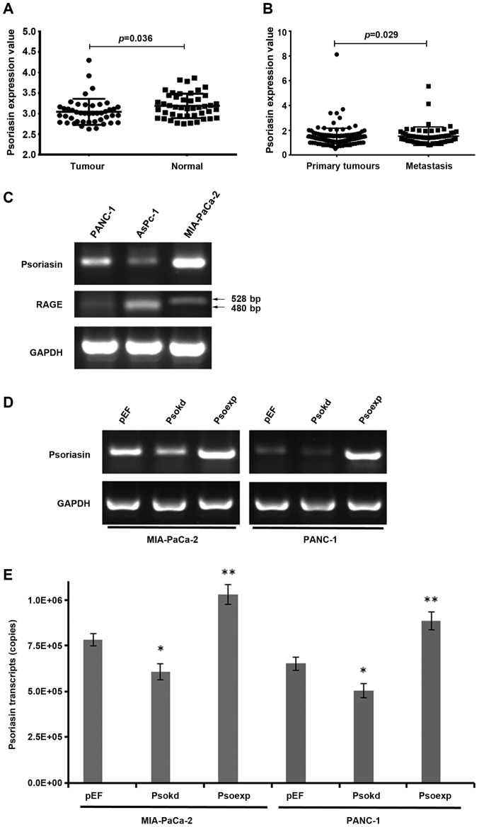
In the search of gene expression array data base at PubMed, a reduction was seen in the expression of Psoriasin in pancreatic cancers in comparison with the paired adjacent normal pancreatic tissues (GDS4336) (Fig. 1A). We further determined the transcript levels of Psoriasin in a cohort of 126 pancreatic tumours and 114 adjacent normal pancreatic tissues. Psoriasin expression appeared to be reduced in the tumours but did not reach a significant level in comparison with the adjacent normal tissues (Table II). An even lower expression of Psoriasin was seen in tumours with distant metastases. Interestingly, higher Psoriasin transcript levels were seen in locally invasive tumours which extended beyond the pancreas (T3 and T4) in comparison with tumours confined to the pancreas (T1 and T2) (Table II). It suggests that Psoriasin plays a significant role in regulation of invasion of pancreatic cancer cells. To clarify the involvement of Psoriasin in metastasis, we analysed the expression of Psoriasin in another gene expression array of pancreatic tumours which includes 145 primary tumours and 61 metastatic tumours (GSE71729) (25). An increased expression of Psoriasin was seen in the secondary tumours in comparison with its expression in primary tumours (Fig. 1B).
Figure 1.
Expression of Psoriasin in pancreatic cancer and pancreatic cancer cell lines with altered Psoriasin expression. (A) Reduced expression of Psoriasin was seen in a cohort of 45 pancreatic ductal adenocarcinomas in comparison with its expression in the paired adjacent normal pancreatic tissues (GDS4336) (26). (B) An elevated expression of Psoriasin was seen in metastases of pancreatic cancer compared with its expression in primary tumours (GSE71729) (25). (C) Psoriasin and RAGE transcripts were detected in PANC-1, AsPC-1 and MIA-PaCa-2 cell lines using conventional PCR. Knockdown and overexpression of Psoriasin were performed using anti-Psoriasin ribozyme transgenes and a recombinant plasmid vector carrying full-length human Psoriasin coding sequence, respectively. The altered expression of Psoriasin in the transfected pancreatic cancer cell lines (MIA-PaCa-2 and PANC-1) was verified using conventional PCR (D) and real-time PCR (E). *p<0.05, **p<0.01.
The mRNA expression of Psoriasin in three different pancreatic cancer cell lines was evaluated using RT-PCR (Fig. 1C). RAGE, the receptor of Psoriasin was detected in the three pancreatic cancer cell lines. PCR products of 528 bp were amplicons of both variant 2 and 4, while 480-bp products represented amplicons from variant 1, 3, 5, 6, 7, 8 and 9. However, exact variants expressed in these cell lines are yet to be further determined (Fig. 1C). The expression of Psoriasin in MIA-PaCa-2 is higher than that for PANC-1, and lowest in AsPc-1. Psoriasin knockdown and overexpression was verified in the MIA-PaCa-2 and PANC-1 cell lines transfected with ribozyme transgenes using RT-PCR (Fig. 1D) and qPCR (Fig. 1E). The Psoriasin-modified MIA-PaCa-2 and PANC-1 cell lines were used for subsequent experiments.
Effect of Psoriasin knockdown and overexpression on pancreatic cancer cell growth and migration
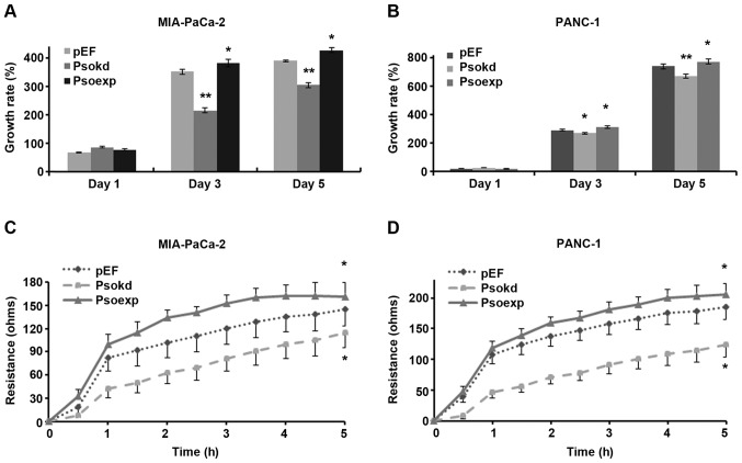
A significant decrease in cellular growth was observed in Psoriasin knockdown MIA-PaCa-2 (Fig. 2A, p<0.001) and PANC-1 (Fig. 2B, p<0.05) cell lines in comparison with controls. Conversely, overexpression of Psoriasin resulted in increased growth of MIA-PaCa-2 and PANC-1 cell lines (p<0.05).
Figure 2.
Effect of Psoriasin on proliferation and migration of pancreatic cancer cell lines. The influence on cell proliferation was determined over a period up to five days. Impact on in vitro proliferation was determined for MIA-PaCa-2 (A) and PANC-1 (B) cell lines a colorimetric method. The migration of pancreatic cancer cell lines were measured using ECIS for MIA-PaCa-2 (C) and for PANC-1 (D). Six repeats were included for each cell line and three independent experiments were performed. Growth rate was calculated against the corresponding day 0 control. *p<0.05, **p<0.01.
Psoriasin knockdown resulted in decreased migration of MIA-PaCa-2 (Fig. 2C, p<0.001) and PANC-1 (Fig. 2D, p<0.05) cell lines compared to controls. Conversely, Psoriasin overexpression resulted in increased migration of MIA-PaCa-2 (p<0.001) and PANC-1 (p<0.05) cell lines.
Influence of Psoriasin expression on pancreatic cancer cell adhesion
An in vitro cell-matrix adhesion assay was adopted to investigate the effect of Psoriasin knockdown and overexpression on the adhesive ability of pancreatic cancer cells to extracellular matrix. MIA-PaCa-2 (Fig. 3A, p<0.05) and PANC-1 (Fig. 3B, p<0.05) Psoriasin overexpressing cells exhibited decreased cell-matrix adhesion whilst Psoriasin knockdown resulted in increased adhesion to matrix (p<0.05).
Figure 3.
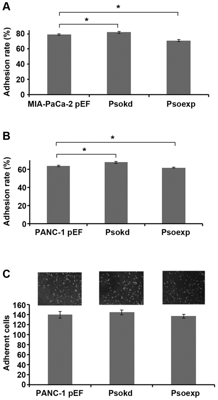
Influence on adhesion of pancreatic cancer cell lines. (A) Adhesion of MIA-PaCa-2 cells was altered by the knockdown and overexpression of Psoriasin. (B) Impact on cell adhesion of PANC-1 cell by the differential expression of Psoriasin. (C) Adhesion of PANC-1 cells to the peritoneal mesothelial cells. Adhesion rate was calculated based on the number of cells seeded. Six repeats were included for each cell line and three independent experiments were performed. *p<0.05.
According to the involvement in adhesion of pancreatic cancer cells, we were curious about a possible implication in dissemination through peritoneal cavity. Therefore, we determined the effect of Psoriasin expression on pancreatic cancer cell adhesion to mesothelial which constitute peritoneum. Neither Psoriasin overexpression nor knockdown had any significant effect on the adhesion of PANC-1 cells to peritoneal mesothelia compared to control (Fig. 3C).
Effect of Psoriasin expression on pancreatic cancer cell invasion
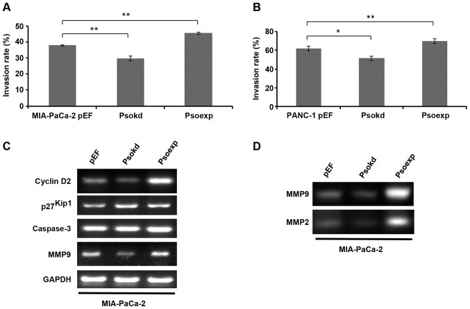
The influence of Psoriasin expression on MIA-PaCa-2 and PANC-1 cell invasion was examined. Interestingly, Psoriasin overexpression resulted in increased invasion of MIA-PaCa-2 (Fig. 4A, p<0.001) and PANC-1 (Fig. 4B, p<0.05) cell lines whilst Psoriasin knockdown resulted in decreased invasion of MIA-PaCa-2 (p<0.001) and PANC-1 (p<0.05) cells.
Figure 4.
Psoriasin and invasion of pancreatic cancer cell lines. The invasion of MIA-PaCa-2 (A) and PANC-1 (B) cell lines were determined using a Transwell invasion assay in triplicates. (C) Expression of MMP-2, p27Kip1, cyclin D2 and caspase-3 was detected in MIA-PaCa-2 cells with differential expression of Psoriasin using conventional PCR. (D) Active MMP-2 and MMP-9 were determined using gelatine zymography. Invasion rates were calculated against corresponding control in which the same number of cells were seeded at beginning of the experiments. Three independent experiments were performed. *p<0.05, **p<0.01.
At the mRNA level, MMP-9 expression was significantly increased in Psoriasin overexpression MIA-PaCa-2 cells compared to controls (Fig. 4C). Conversely, MMP-9 expression was decreased in Psoriasin knockdown MIA-PaCa-2 cells. As the results from zymography show, MMP-2 and MMP-9 activity is increased in Psoriasin overexpression MIA-PaCa-2 cells compared to control (Fig. 4D), whilst MMP-2 and MMP-9 activity is decreased in Psoriasin knockdown MIA-PaCa-2 cells. As shown in Fig. 4C, decreased Psoriasin expression resulted in the decreased expression of cyclin D2 at the mRNA level in MIA-PaCa-2 cells in which an increased level of p27Kip1 was also noted. Moreover, overexpression of Psoriasin resulted in increased cyclin D2 expression.
Influence of Psoriasin on aggregation and survival of pancreatic cancer cells
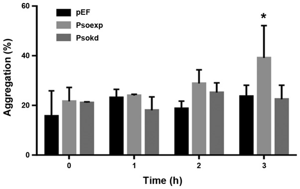
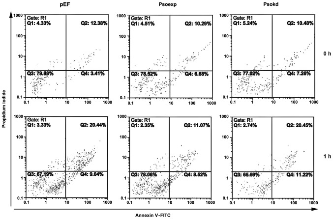
In order to evaluate how MIA-PaCa-2 cells interact with one another in suspension, a cell aggregation assay was performed. Cells with increased Psoriasin expression demonstrated an increased propensity to form cell-cell contacts whilst in suspension, reaching significance at 3 h (Fig. 5, p<0.05). To determine whether this observation had an impact on the anoikis resistance in this cell line, an apoptosis assay was performed. We observed cell survival after suspension was increased in Psoriasin overexpressing cell lines (Fig. 6). Western blot analyses of adherent cells showed an enhanced activation of caspase-3 in MIA-PaCa-2 cells as cleaved proteins in comparison with the control (Fig. 7). A reduction in the cleavage of caspase-3 was not observed in the Psoriasin overexpression cells compared with the control which also exhibited a lower level of the activation.
Figure 5.
Psoriasin and aggregation of pancreatic cancer cells. Aggregation of MIA-PaCa-2 cells was determined over a duration ≤3 h. Three independent experiments were performed. *p<0.05.
Figure 6.
Psoriasin and anoikis of pancreatic cancer cells. Apoptosis of suspension MIA-PaCa-2 cells with altered expression of Psoriasin was determined using a flow cytometric method.
Figure 7.
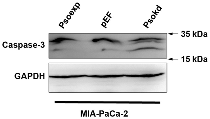
Caspase 3 in MIA-PaCa-2 cells were detected using western blot analysis.
Discussion
Psoriasin was first identified as being overexpressed in the hyperproliferative skin disease psoriasis. Numerous studies have since demonstrated Psoriasin overexpression to be associated with tumour progression. Upregulation of Psoriasin in cancer was initially identified in pre-invasive carcinomas; whether this upregulation occurs in pancreatic intra-epithelial neoplasia progression that proceeds to pancreatic adenocarcinoma remains to be elucidated. Increased Psoriasin expression has also been identified in cancers at an advanced stage. In breast cancer, Psoriasin overexpression is associated with metastasis and poor prognosis. In the present study, Psoriasin expression was determined in a cohort of pancreatic cancer patients. Psoriasin expression tended to be reduced in the pancreatic cancers compared to adjacent normal pancreatic tissues, though did not reach statistical significance. This observation is similar to findings from a GEO data set (GDS4336) (Fig. 1A) (26). A reduced expression was noted in primary tumours (n=9) with distant metastases. This finding is yet to be verified by examining of a large cohort pancreatic primary tumours with a reasonable number of primary tumours which have distant metastasis. In an analysis of a gene expression array data, an increased expression levels of Psoriasin was seen in metastases of pancreatic cancer in comparison with primary tumours (Fig. 1B). Interestingly, a greater expression of Psoriasin was evident in locally advanced pancreatic tumours. It suggests that Psoriasin may play differential roles during the development and progression of the disease, in particular, the metastatic process.
We determined the expression of Psoriasin in the cell lines AsPc-1, MIA-PaCa-2 and PANC-1, and expression appeared highest in MIA-PaCa-2 which is the most tumourigenic of the three cell lines (27). These results demonstrate that Psoriasin overexpression is associated with increased cancer cell growth in vitro, whilst knockdown inhibits growth. Interestingly, during breast cancer progression, Psoriasin has been shown to interact with Jab1 (c-jun activation-domain binding protein), which is involved in proteosomal degradation and signal transduction. Interaction between Psoriasin and Jab1 results in increased activator protein-1 (AP-1) activity, increased expression of AP-1 and HIF-1-dependent genes, and reduced expression of the cell cycle inhibitor p27Kip1, resulting in enhanced tumourigenesis and metastasis in vivo (28). It may be that in pancreatic cancer, the same mechanisms are taking place promoting tumour growth and metastasis. Jab1 is a negative regulator of p27Kip1, promoting its degradation (29). p27Kip1 normally binds to and prevents the activation of cyclin complexes controlling cell cycle progression at G1 (30). Thus, it may be that increased interaction between Psoriasin and Jab1 results in decreased p27Kip1. In line with this, an increased expression of p27Kip1 was also observed in the Psoriasin knockdown cells (Fig. 4C). We also found that Psoriasin overexpression results in increased expression of cyclin D2, which may explain the mode of action for Psoriasin-mediated promotion of cyclin activation and cell cycle progression. Additionally, differential expression of RAGE, the receptor of Psoriasin was seen in three pancreatic cancer cell lines. RAGE has 8 isoforms of protein products encoded by 9 different splicing variants in humans (listed at PubMed Gene). However, we cannot identify which variants were expressed by these cell lines. The exact role played by RAGE in mediating functions of Psoriasin in pancreatic cancer is yet to be investigated.
In this study, Psoriasin expression was inversely linked to cell-matrix adhesion. This is similar to our results observed in both prostate and lung cancer (11,15). It suggests that expression of Psoriasin is a response by cancer cells to changes of cell-matrix adhesion and reciprocally affects cell adhesion to matrix. We also found Psoriasin expression has no effect on the adhesion of pancreatic cancer cells to peritoneal mesothelial in vitro, and conclude that Psoriasin may not be required for the adhesion during peritoneal metastasis of pancreatic cancer.
Psoriasin is regarded as an inflammation-associated protein and has chemoattractant properties, promoting migration of granulocytes, lymphocytes, macrophages and monocytes. Our study showed Psoriasin overexpression results in increased migration of pancreatic cancer cells. The multi-ligand receptor for advanced glycation end-products (RAGE) has been identified as a Psoriasin receptor, and implicated in leukocyte migration and the inflammatory process. It may be that, similar to leukocyte migration, Psoriasin attaches to RAGE in order to promote cancer cell migration (5).
Further to its influences on cell growth, adhesion and migration, we also examined the effect of Psoriasin on pancreatic cancer cell invasion. It is well known that activation of MMP-2 and MMP-9 promotes the invasive and metastatic potential of pancreatic cancer cells (31,32). In our recent study of Psoriasin in prostate cancer, Psoriasin has been shown to have an effect on prostate cancer cell invasion via MMPs (11). This is subsequently confirmed herein, whereby Psoriasin overexpression in pancreatic cancer cells resulted in increased MMP-2 and MMP-9 expression and activity. Taken together, these data suggest Psoriasin regulates invasion of pancreatic cancer cells via regulation of MMPs. In a recent study, Morgan et al observed interaction between Psoriasin and the cytoplasmic domain of the integrin β6 subunit is required for αvβ6-dependent breast cancer cell invasion. Similarly, they showed upregulation of MMPs in Psoriasin overexpressing breast cancer cells (33). In line with its higher expression levels in local invasive tumours, Psoriasin promotes invasion of pancreatic cancer cells which leads to a greater local invasive expansion of the disease.
A recent report of disruption of cell anchorage inducing apoptosis led us to investigate whether Psoriasin has a role of inducing this form of anoikis resistance in pancreatic cancer cells (34). Anoikis is referred to as a programmed cell death or apoptosis which happens to adherent cells lacking anchorage (34). It can occur during the dissemination of pancreatic cancer cells through circulation and peritoneal cavity when they lose anchorage. At 3 h we saw a significant rise of cell-cell aggregates, indicating a role for Psoriasin in inducing these aggregates. Previous experiments showed a role for Psoriasin in preventing anoikis (34), a result we confirmed in our cell lines, hinting that cell-cell aggregation may contribute to preventing anoikis. Despite this, based on our results, the number of cell-cell aggregates show a significant increase only at 3 h, whereas cells display anoikis resistance at 1 h, indicating other mechanisms must be at play to compensate for the disparity. Western blot analyses of adherent cells showed an increased activation of caspase 3 in the Psoriasin knockdown MIA-PaCa-2 cells. The involvement of intrinsic and extrinsic caspase pathways and activation of caspase-3 in pancreatic cancer cells undergoing anoikis remains unclear and is yet to be investigated. Further investigation is also required to reveal molecules involved in the enhanced cell-cell adhesion and aggregation.
In conclusion, reduced expression of Psoriasin was seen in pancreatic cancers with distant metastases, whilst higher transcript levels were seen in locally advanced tumours. Psoriasin expression in pancreatic cancer cells is associated with cell growth, cell-matrix adhesion and migration. Psoriasin regulates invasion of pancreatic cancer cells via expression of MMPs. The implication of Psoriasin expression in invasion, disease progression and potential as a therapeutic target warrants further investigation.
Acknowledgments
The authors thank the support from Cancer Research Wales, Life Sciences Research Network Wales (Welsh Government's Ser Cymru Program) and Albert Huang Foundation. Ying Liu, Yanan Gu and Gang Chen are recipients of China Medical Scholarship from the Cardiff University.
References
- 1.Madsen P, Rasmussen HH, Leffers H, Honoré B, Dejgaard K, Olsen E, Kiil J, Walbum E, Andersen AH, Basse B, et al. Molecular cloning, occurrence, and expression of a novel partially secreted protein “psoriasin” that is highly up-regulated in psoriatic skin. J Invest Dermatol. 1991;97:701–712. doi: 10.1111/1523-1747.ep12484041. [DOI] [PubMed] [Google Scholar]
- 2.Hoffmann HJ, Olsen E, Etzerodt M, Madsen P, Thøgersen HC, Kruse T, Celis JE. Psoriasin binds calcium and is upregulated by calcium to levels that resemble those observed in normal skin. J Invest Dermatol. 1994;103:370–375. doi: 10.1111/1523-1747.ep12395202. [DOI] [PubMed] [Google Scholar]
- 3.Børglum AD, Flint T, Madsen P, Celis JE, Kruse TA. Refined mapping of the psoriasin gene S100A7 to chromosome 1cen-q21. Hum Genet. 1995;96:592–596. doi: 10.1007/BF00197417. [DOI] [PubMed] [Google Scholar]
- 4.Schäfer BW, Heizmann CW. The S100 family of EF-hand calcium-binding proteins: Functions and pathology. Trends Biochem Sci. 1996;21:134–140. doi: 10.1016/S0968-0004(96)80167-8. [DOI] [PubMed] [Google Scholar]
- 5.Wolf R, Howard OM, Dong H-F, Voscopoulos C, Boeshans K, Winston J, Divi R, Gunsior M, Goldsmith P, Ahvazi B, et al. Chemotactic activity of S100A7 (Psoriasin) is mediated by the receptor for advanced glycation end products and potentiates inflammation with highly homologous but functionally distinct S100A15. J Immunol. 2008;181:1499–1506. doi: 10.4049/jimmunol.181.2.1499. [DOI] [PMC free article] [PubMed] [Google Scholar]
- 6.Boniface K, Bernard FX, Garcia M, Gurney AL, Lecron JC, Morel F. IL-22 inhibits epidermal differentiation and induces proinflammatory gene expression and migration of human keratinocytes. J Immunol. 2005;174:3695–3702. doi: 10.4049/jimmunol.174.6.3695. [DOI] [PubMed] [Google Scholar]
- 7.Boniface K, Diveu C, Morel F, Pedretti N, Froger J, Ravon E, Garcia M, Venereau E, Preisser L, Guignouard E, et al. Oncostatin M secreted by skin infiltrating T lymphocytes is a potent keratinocyte activator involved in skin inflammation. J Immunol. 2007;178:4615–4622. doi: 10.4049/jimmunol.178.7.4615. [DOI] [PubMed] [Google Scholar]
- 8.Gläser R, Harder J, Lange H, Bartels J, Christophers E, Schröder JM. Antimicrobial psoriasin (S100A7) protects human skin from Escherichia coli infection. Nat Immunol. 2005;6:57–64. doi: 10.1038/ni1142. [DOI] [PubMed] [Google Scholar]
- 9.Jinquan T, Vorum H, Larsen CG, Madsen P, Rasmussen HH, Gesser B, Etzerodt M, Honoré B, Celis JE, Thestrup-Pedersen K. Psoriasin: A novel chemotactic protein. J Invest Dermatol. 1996;107:5–10. doi: 10.1111/1523-1747.ep12294284. [DOI] [PubMed] [Google Scholar]
- 10.Jiang WG, Watkins G, Douglas-Jones A, Mansel RE. Psoriasin is aberrantly expressed in human breast cancer and is related to clinical outcomes. Int J Oncol. 2004;25:81–85. [PubMed] [Google Scholar]
- 11.Ye L, Sun PH, Martin TA, Sanders AJ, Mason MD, Jiang WG. Psoriasin (S100A7) is a positive regulator of survival and invasion of prostate cancer cells. Urol Oncol. 2013;31:1576–1583. doi: 10.1016/j.urolonc.2012.05.006. [DOI] [PubMed] [Google Scholar]
- 12.Moubayed N, Weichenthal M, Harder J, Wandel E, Sticherling M, Gläser R. Psoriasin (S100A7) is significantly up-regulated in human epithelial skin tumours. J Cancer Res Clin Oncol. 2007;133:253–261. doi: 10.1007/s00432-006-0164-y. [DOI] [PubMed] [Google Scholar]
- 13.Tripathi SC, Matta A, Kaur J, Grigull J, Chauhan SS, Thakar A, Shukla NK, Duggal R, DattaGupta S, Ralhan R, et al. Nuclear S100A7 is associated with poor prognosis in head and neck cancer. PLoS One. 2010;5:e11939. doi: 10.1371/journal.pone.0011939. [DOI] [PMC free article] [PubMed] [Google Scholar]
- 14.Celis JE, Rasmussen HH, Vorum H, Madsen P, Honoré B, Wolf H, Orntoft TF. Bladder squamous cell carcinomas express psoriasin and externalize it to the urine. J Urol. 1996;155:2105–2112. doi: 10.1016/S0022-5347(01)66118-4. [DOI] [PubMed] [Google Scholar]
- 15.Hu M, Ye L, Ruge F, Zhi X, Zhang L, Jiang WG. The clinical significance of Psoriasin for non-small cell lung cancer patients and its biological impact on lung cancer cell functions. BMC Cancer. 2012;12:588. doi: 10.1186/1471-2407-12-588. [DOI] [PMC free article] [PubMed] [Google Scholar]
- 16.Enerbäck C, Porter DA, Seth P, Sgroi D, Gaudet J, Weremowicz S, Morton CC, Schnitt S, Pitts RL, Stampl J, et al. Psoriasin expression in mammary epithelial cells in vitro and in vivo. Cancer Res. 2002;62:43–47. [PubMed] [Google Scholar]
- 17.Emberley ED, Alowami S, Snell L, Murphy LC, Watson PH. S100A7 (psoriasin) expression is associated with aggressive features and alteration of Jab1 in ductal carcinoma in situ of the breast. Breast Cancer Res. 2004;6:R308–R315. doi: 10.1186/bcr791. [DOI] [PMC free article] [PubMed] [Google Scholar]
- 18.Watson PH, Leygue ER, Murphy LC. Psoriasin (S100A7) Int J Biochem Cell Biol. 1998;30:567–571. doi: 10.1016/S1357-2725(97)00066-6. [DOI] [PubMed] [Google Scholar]
- 19.Shubbar E, Vegfors J, Carlström M, Petersson S, Enerbäck C. Psoriasin (S100A7) increases the expression of ROS and VEGF and acts through RAGE to promote endothelial cell proliferation. Breast Cancer Res Treat. 2012;134:71–80. doi: 10.1007/s10549-011-1920-5. [DOI] [PubMed] [Google Scholar]
- 20.Sneh A, Deol YS, Ganju A, Shilo K, Rosol TJ, Nasser MW, Ganju RK. Differential role of psoriasin (S100A7) in estrogen receptor α positive and negative breast cancer cells occur through actin remodeling. Breast Cancer Res Treat. 2013;138:727–739. doi: 10.1007/s10549-013-2491-4. [DOI] [PMC free article] [PubMed] [Google Scholar]
- 21.Jemal A, Siegel R, Ward E, Hao Y, Xu J, Thun MJ. Cancer statistics, 2009. CA Cancer J Clin. 2009;59:225–249. doi: 10.3322/caac.20006. [DOI] [PubMed] [Google Scholar]
- 22.Gadiya M, Mori N, Cao MD, Mironchik Y, Kakkad S, Gribbestad IS, Glunde K, Krishnamachary B, Bhujwalla ZM. Phospholipase D1 and choline kinase-α are interactive targets in breast cancer. Cancer Biol Ther. 2014;15:593–601. doi: 10.4161/cbt.28165. [DOI] [PMC free article] [PubMed] [Google Scholar]
- 23.Zuker M. Mfold web server for nucleic acid folding and hybridization prediction. Nucleic Acids Res. 2003;31:3406–3415. doi: 10.1093/nar/gkg595. [DOI] [PMC free article] [PubMed] [Google Scholar]
- 24.Bonnekoh B, Wevers A, Jugert F, Merk H, Mahrle G. Colorimetric growth assay for epidermal cell cultures by their crystal violet binding capacity. Arch Dermatol Res. 1989;281:487–490. doi: 10.1007/BF00510085. [DOI] [PubMed] [Google Scholar]
- 25.Moffitt RA, Marayati R, Flate EL, Volmar KE, Loeza SG, Hoadley KA, Rashid NU, Williams LA, Eaton SC, Chung AH, et al. Virtual microdissection identifies distinct tumor- and stroma-specific subtypes of pancreatic ductal adenocarcinoma. Nat Genet. 2015;47:1168–1178. doi: 10.1038/ng.3398. [DOI] [PMC free article] [PubMed] [Google Scholar]
- 26.Zhang G, He P, Tan H, Budhu A, Gaedcke J, Ghadimi BM, Ried T, Yfantis HG, Lee DH, Maitra A, et al. Integration of metabolomics and transcriptomics revealed a fatty acid network exerting growth inhibitory effects in human pancreatic cancer. Clin Cancer Res. 2013;19:4983–4993. doi: 10.1158/1078-0432.CCR-13-0209. [DOI] [PMC free article] [PubMed] [Google Scholar]
- 27.Deer EL, González-Hernández J, Coursen JD, Shea JE, Ngatia J, Scaife CL, Firpo MA, Mulvihill SJ. Phenotype and genotype of pancreatic cancer cell lines. Pancreas. 2010;39:425–435. doi: 10.1097/MPA.0b013e3181c15963. [DOI] [PMC free article] [PubMed] [Google Scholar]
- 28.Emberley ED, Niu Y, Leygue E, Tomes L, Gietz RD, Murphy LC, Watson PH. Psoriasin interacts with Jab1 and influences breast cancer progression. Cancer Res. 2003;63:1954–1961. [PubMed] [Google Scholar]
- 29.Tomoda K, Kubota Y, Kato J. Degradation of the cyclin-dependent-kinase inhibitor p27Kip1 is instigated by Jab1. Nature. 1999;398:160–165. doi: 10.1038/18230. [DOI] [PubMed] [Google Scholar]
- 30.Sgambato A, Cittadini A, Faraglia B, Weinstein IB. Multiple functions of p27(Kip1) and its alterations in tumor cells: A review. J Cell Physiol. 2000;183:18–27. doi: 10.1002/(SICI)1097-4652(200004)183:1<18::AID-JCP3>3.0.CO;2-S. [DOI] [PubMed] [Google Scholar]
- 31.Ellenrieder V, Alber B, Lacher U, Hendler SF, Menke A, Boeck W, Wagner M, Wilda M, Friess H, Büchler M, et al. Role of MT-MMPs and MMP-2 in pancreatic cancer progression. Int J Cancer. 2000;85:14–20. doi: 10.1002/(SICI)1097-0215(20000101)85:1<14::AID-IJC3>3.0.CO;2-O. [DOI] [PubMed] [Google Scholar]
- 32.Zhang K, Chen D, Jiao X, Zhang S, Liu X, Cao J, Wu L, Wang D. Slug enhances invasion ability of pancreatic cancer cells through upregulation of matrix metalloproteinase-9 and actin cytoskeleton remodeling. Lab Invest. 2011;91:426–438. doi: 10.1038/labinvest.2010.201. [DOI] [PMC free article] [PubMed] [Google Scholar] [Retracted]
- 33.Morgan MR, Jazayeri M, Ramsay AG, Thomas GJ, Boulanger MJ, Hart IR, Marshall JF. Psoriasin (S100A7) associates with integrin β6 subunit and is required for αvβ6-dependent carcinoma cell invasion. Oncogene. 2011;30:1422–1435. doi: 10.1038/onc.2010.535. [DOI] [PubMed] [Google Scholar]
- 34.Dey KK, Sarkar S, Pal I, Das S, Dey G, Bharti R, Banik P, Roy J, Maity S, Kulavi I, et al. Mechanistic attributes of S100A7 (psoriasin) in resistance of anoikis resulting tumor progression in squamous cell carcinoma of the oral cavity. Cancer Cell Int. 2015;15:74. doi: 10.1186/s12935-015-0226-9. [DOI] [PMC free article] [PubMed] [Google Scholar] [Retracted]


