Abstract
The conserved protein complex known as Mediator conveys transcriptional signals by acting as an intermediary between transcription factors and RNA polymerase II. As a result, Mediator subunits play multiple roles in regulating developmental as well as abiotic and biotic stress pathways. In this report we identify the head domain subunits MEDIATOR18 and MEDIATOR20 as important susceptibility factors for Fusarium oxysporum infection in Arabidopsis thaliana. Mutants of MED18 and MED20 display down-regulation of genes associated with jasmonate signaling and biosynthesis while up-regulation of salicylic acid associated pathogenesis related genes and reactive oxygen producing and scavenging genes. We propose that MED18 and MED20 form a sub-domain within Mediator that controls the balance of salicylic acid and jasmonate associated defense pathways.
Introduction
Being sessile in nature, plants require finely tuned response pathways to adapt to the environment around them. Facing challenges from both abiotic and biotic stresses, plants require sensors to perceive external signals and a signaling pathway that leads to transcriptional activation through DNA-binding transcription factors (TFs). Control over which pathway in the broader plant signaling network is activated is crucial to produce the correct response and to prevent misallocation in energy production [1, 2]. One example of a finely-tuned network can be seen in plant defense signaling pathways, where successful recognition of a pathogen modulates the output of defense genes that are transcribed. For instance, it is generally accepted that the model plant Arabidopsis thaliana will activate genes from the salicylic acid (SA) associated pathway in response to infection by biotrophic pathogens whereas genes associated with the jasmonate (JA) and ethylene (ET) associated pathways are activated strongly in response to necrotrophic pathogens [3]. While defense pathways are likely to be more complex when considered as a network [4, 5], antagonistic regulation of the SA and JA/ET defense pathways allow prioritization of the defense response for maximum effectiveness against the pathogen that is invading [6].
To overcome plant defenses, plant pathogens may modify hormone signaling to create an environment suitable for colonization [7]. For instance, the bacterial pathogen Pseudomonas syringae pv. tomato (Pst) is known to affect the abscisic acid, auxin, JA, and SA signaling pathways [8–10]. Pst uses the JA-isoleucine (JA-Ile) hormone mimic coronatine to activate the JA associated transcription factor MYC2, which then activates the NAC TF genes, ANAC019, ANAC055 and ANAC072 to suppress SA biosynthesis and metabolism [11]. Similarly, isolates of the hemi-biotrophic fungal pathogen, Fusarium oxysporum have been shown to produce JA-Ile and other JA conjugates to help infect A. thaliana [12, 13]. Mutants in the JA-Ile receptor CORONATINE INSENSITIVE1 (COI1) show strong resistance to F. oxysporum isolates that produce JA-Ile in culture, while the myc2 mutant and an activation allele of the JAZ7 transcriptional repressor have enhanced resistance and susceptibility, respectively [13–17]. The resistance phenotype of coi1 was shown to be independent of SA-associated defense genes using the NahG transgene [15], however exogenous application of SA to the leaves was found to increase resistance [18]. F. oxysporum has been shown to induce JA-associated gene expression as well as tryptophan secondary metabolism during infection [19, 20]. Activation of the tryptophan metabolic pathway leads to increased auxin production. Interestingly, auxin signaling and transport genes, but not auxin biosynthetic genes have been shown to be involved in susceptibility to F. oxysporum [20]. Lastly, the ethylene receptor mutant etr1-1 and the abscisic acid (ABA) biosynthetic mutant aba2-1 show increased resistance to F. oxysporum suggesting ethylene and ABA are also required for susceptibility [14, 21].
With a complex transcriptional network to co-ordinate, plants have evolved a large number of TFs to fine tune gene expression. For instance, the A. thaliana genome contains over 1500 transcription factors [22, 23]. To process information from a large number of TFs, eukaryotes possess a protein complex called Mediator that relays the signal from TFs to RNA Polymerase II. Not surprisingly, mutations in the Mediator complex have been found to affect a wide range of plant developmental processes as well as abiotic and biotic stress responses [24, 25].
In a screen of 12 individual A. thaliana Mediator subunits, we previously identified the med25 and med8 mutants to be moderately resistant to F. oxysporum [26]. The med25 mutant was found to be partially insensitive to JA and showed a reduction in JA-associated gene expression [26, 27]. As insensitivity to JA has been linked to F. oxysporum resistance [15], the med25 mutant was also hypothesized to be resistant due to an attenuation of JA signaling. In contrast, the med8 mutant had no change in JA-associated defense gene expression and a med25 med8 double mutant led to additive resistance over the single mutations [26]. This suggested a potentially separate mechanism for susceptibility to F. oxysporum that is conferred by MED8. The MED8 subunit is predicted to be located in the Head domain of the Mediator complex and is thought to act as a linker with the “movable jaw” subunits MED18 and MED20 based on structural conservation between eukaryotic Mediator complexes [28, 29].
In this study, we examined mutants of the A. thaliana MED18 and MED20 genes and found them to be highly resistant to F. oxysporum. JA signaling genes were significantly reduced under F. oxysporum infection in med18 and med20, while expression of the SA-associated genes, PATHOGENESIS RELATED1 (PR1) and PATHOGENESIS RELATED5 (PR5) as well as several genes associated with reactive oxygen production were up-regulated. Our data suggest that the MED18-MED20 sub-module of the Mediator complex confers susceptibility to F. oxysporum and modulates crosstalk between JA- and SA-associated defense pathways.
Materials and methods
Plant growth conditions
A. thaliana Col-0, med18 and med20 seeds were sown into autoclaved University of California mix soil and kept at 4°C in the dark for 48 hours. After stratification, plants were grown at 24°C, with an 8 hour photoperiod (160 μE m-2s-1) and 60% humidity, with a night time temperature of 21°C and humidity of 70%. After 2 weeks, seedlings were gently removed from the soil and transferred to 30-well trays, and grown until the six to eight leaf stage until inoculation with F. oxysporum or treatment with hormones. SA treatment was performed according to [30]. Grafting of WT and med18 plants was performed as described [15] using young seedlings grown on ½ MS agar in long day conditions (16 hour photoperiod (160 μE m-2s-1) and 60% humidity, with a night time temperature of 21°C and humidity of 70%). Once the grafts had formed, plants were gently transferred to soil for three weeks before inoculation with F. oxysporum. Flowering time assays were also conducted under the same long day conditions, and the number of rosette leaves recorded at the initiation of flowering. The med18 seeds (salk_027178C) and med18-1 (sail_889_C08) were obtained from the Arabidopsis Biological Resource Center while the med18-1;MED18-HA seeds were a kind gift from Tesfaye Mengiste. The med20 seeds contain a C-to-T mutation in At2g28230 (MED20a) resulting in a premature stop, and were a kind gift from Xuemei Chen [31].
F. oxysporum inoculation
Plants were inoculated with F. oxysporum isolate 5176 as described previously [26]. Briefly, at 1 h after the start of the photoperiod, plants were gently uprooted and dipped for fifteen seconds in a spore suspension with a concentration of 1 x 10⁶ spores/mL in water and then replanted. Mock plants were dipped in water and replanted.
F. oxysporum colonization scoring
The β-GLUCURONIDASE (GUS) expressing F. oxysporum 5176 strain was a kind gift from U. Schumann [32]. GUS staining was performed according to [33]. Analysis of GUS staining was performed on ten plants of both WT and med18, which were gently uprooted twelve days after infection and washed in distilled water before being vacuum infiltrated in staining solution and incubated at 37°C. GUS stained roots were imaged under a compound microscope (Nikon) and scored for the presence of GUS as a percentage of the total root length using ImageJ.
Yeast-2-hybrid screening
To test the interaction between plant Mediator subunits, the full length coding sequence of MED8, MED18, MED20 and MED21 were amplified with primers listed in S1 Table from Arabidopsis Col-0 cDNA. The PCR products were cloned into the vector pCR8GW-TOPO according to the manufacturer’s instructions (Invitrogen). All plasmid constructs were confirmed by restriction analysis and sequencing. The GAL4-DNA binding domain bait constructs (DBD-MEDs) were generated by an LR recombination reaction with pCR8GW–MEDs and pDEST32. The GAL4 Activation domain prey constructs (AD-MEDs) were generated by LR recombination reaction with pDEST22 and the pCR8GW–MED plasmids. The LR recombination reaction was performed using the Gateway LR Clonase II Enzyme kit (Invitrogen) according to the manufacturer’s instructions and checked using restriction digests. The pDEST32-GFP (DBD-GFP) control vector was a kind gift from Volkan Cevik. Yeast-2-hybrid interactions were performed by transforming bait and prey plasmids into the yeast strain S. cerevisiae (MaV203). The transformed cells were plated onto synthetic complete medium minus leucine and tryptophan SC-Leu-Trp (-LW) plates before streaking onto fresh plates. Freshly streaked cultures were then resuspended in 50ul sterile water and serially diluted ten times and plated on SC-Leu-Trp (-LW) and SC-Leu-Trp-His (-LWH) plates (Sunrise Science) containing 12.5mM 3-aminotriazole (3-AT) (Sigma). The Yeast-2-Hybrid interaction experiment was repeated with separate transformations and showed the same results.
RNA sequencing
Infected root tissues of Col-0, med18 and med20 were harvested 24 h after inoculation with F. oxysporum (three independent biological replicates of 20 plants each). Total RNA was isolated using an RNeasy Plant Mini kit (Qiaqen) and RNA quality checked using a Nanodrop ND-1000 (Nanodrop) and an Agilent 2100 Bioanalyser (Agilent Biotechnologies). Library preparation and RNA sequencing on the infected root RNA samples were performed by the Australian Genome Research Facility (AGRF). Messenger RNA was selected using Poly-A tail selection prior to preparation of 100bp paired-end libraries. Sequencing was performed on an Illumina HiSeq 2000 system generating approximately 23 million raw RNAseq reads per sample. Fastq files are available at the NCBI Sequence Read Archive (SRA) under study number SRP092151. Differential expression analysis was performed using the Tuxedo analysis suite [34]. Briefly, Bowtie2 along with Tophat were used to align generated reads to the TAIR10 A. thaliana reference genome. After expressed transfrags were assembled, Cufflinks was used to quantify gene abundance and transcriptome assemblies were then merged using Cuffmerge. Statistical analysis was performed within the Cufflinks analysis with false discovery rate and correction for multiple comparisons applied using standard run parameters. Genes considered differentially expressed showed a statistically significant difference in expression values (P<0.05) and a Log2 fold change >1. Venn diagrams were produced using Venny 2.1 (http://bioinfogp.cnb.csic.es/tools/venny/index.html) and a heatmap produced using matrix2png [35].
Real-time quantitative reverse transcriptase PCR (Real time qRT-PCR) analyses
Real time qRT-PCR expression analysis was performed on hormone treated leaves as well as mock or F. oxysporum infected roots. Leaf and root tissues were collected at 24 hours after treatment for gene expression studies. Salicylic acid (SA) was obtained from Sigma-Aldrich. SA treatment was performed by lightly and evenly spraying leaves with 100μM SA. Total RNA was isolated using an RNeasy Plant Mini kit (Qiaqen) and RNA quality checked using a Nanodrop ND-1000 (Nanodrop). cDNA synthesis was performed using SuperScriptIII (Thermo Fisher) and real time quantitative reverse transcriptase PCR was performed using an ABI ViiA7 Sequence Detection System (Applied Biosystems). Each reaction contained 5 μL of SYBR Green (Applied Biosystems) and 1 μL of 3 nM of each gene-specific primer pair and 4 μL of cDNA template to a final volume of 10 μL. The PCR primer efficiency (E) of each primer pair in each individual reaction was calculated from the changes in fluorescence values (ΔRn) of each amplification plot, using LinReg PCR software [36]. Amplification plots were analyzed using a threshold of 0.20 to give a cycle threshold (Ct) value for each gene and cDNA combination. Gene expression levels relative to the Arabidopsis housekeeping genes β-ACTIN 2 (AT3G18780), β-ACTIN 3 (AT3G53750) and β-ACTIN 7 (AT1G49240) were calculated for each cDNA sample using the following equation: The gene transcript levels relative to actin = (E gene^(-Ct gene)) / (E Actin ^(-Ct Actin)). The primer pairs used in Real time RT-qPCR (S1 Table).
Results
MED18 and MED20 confer susceptibility to F. oxysporum
The Arabidopsis MED8 and MED25 genes were previously found to confer susceptibility to F. oxysporum [26]. MED25 has been shown to interact with MED16 in Arabidopsis which is located in the Tail domain of Mediator [37], while MED8 is located in the Head domain. We investigated whether the Arabidopsis head domain subunits, MED18 and MED20, which have been shown to be located adjacent to MED8 in the yeast Mediator complex [28, 29], also conferred F. oxysporum susceptibility. We inoculated med18 (salk_027178C) and med20 [31] mutants with F. oxysporum and found them to be highly resistant with less than 3% of leaves showing disease symptoms compared to approximately 40% of leaves in the wild-type A. thaliana Col-0 (Fig 1A and 1B). As previously reported [31, 38], we found that med18 and med20 plants had a strong delay in flowering time and showed a similar phenotype to the med8 mutant with approximately forty leaves being produced before flowering in long day (LD) conditions (Fig 1C; [26]). Although there are no T-DNA insertion mutants available for med20, we were able to screen a second med18 insertion mutant (previously referred to as med18-1 [39]) and a complemented med18-1 line expressing a hemagglutinin (HA)-tagged MED18 construct [39]. The med18-1 line showed complete resistance to F. oxysporum while the complemented line showed a partial restoration of susceptibility, suggesting the HA-Tag may potentially affect MED18’s role in mediating F. oxysporum susceptibility (Fig 1D).
Fig 1. MED18 and MED20 are F. oxysporum susceptibility factors.
(A) Typical disease symptoms of WT (Col-0), med18 and med20 plants 14 days after infection with F. oxysporum. (B) Average percentage of leaves showing chlorosis at 14 days after infection. Inoculations consisted of three biological replicates with each containing twenty plants. (C) Number of rosette leaves at flowering when grown under long-day conditions. Rosette leaves were counted at the first sighting of the floral bud at approximately 0.5cm in length. (D) Disease symptoms of med18-1 and med18-1;MED18-HA plants 18 days after infection with F. oxysporum. *** represents significance (p<0.001) using Student’s T-test of each mutant compared to the WT. Error bars represent standard error.
MED18 interacts with MED20 in Y2H experiments
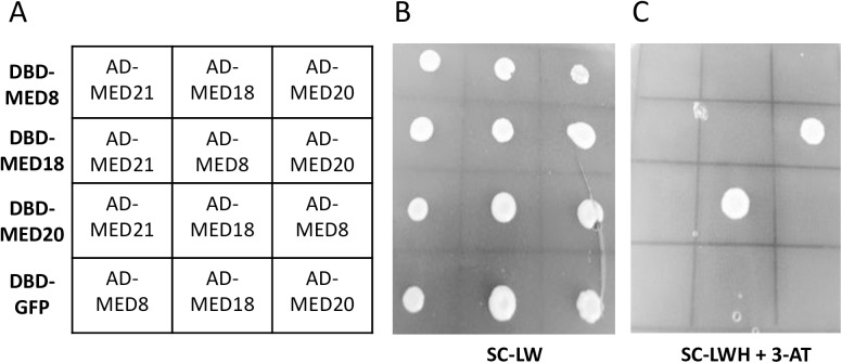
As, med8, med18 and med20 mutants have similar pathogen and developmental phenotypes, we examined whether MED8, MED18 and MED20 interact in Yeast-2-Hybrid assays. As negative controls we included GFP and MED21 which are not expected to interact with MED18 or MED20 based on the yeast Mediator architecture. We found a positive interaction between MED18 and MED20 in reciprocal GAL4-Activation Domain and GAL4-DNA-Binding Domain fusions, however we did not detect positive interactions with MED8, MED21 or GFP (Fig 2). While more research is required on the structure of the Arabidopsis Mediator complex, these results suggest that MED18/MED20 may form a subdomain of the Mediator head complex that controls both flowering time and susceptibility to F. oxysporum in Arabidopsis. Additional experiments are required to demonstrate a direct interaction between MED18 and MED20 in vivo.
Fig 2. MED18 and MED20 interact in Yeast-2-Hybrid experiments.
GAL4 DNA-Binding Domain (DBD-) plasmids and GAL4 Activation Domain (AD-) plasmids were co-transformed into S. cerevisiae and plated onto synthetic complete medium lacking Leucine and Tryptophan (SC-LW) or synthetic complete medium lacking Leucine, Tryptophan and Histidine but containing 12.5mM 3-aminotriazole (SC-LWH +3-AT). The plate layout is shown in (A) where each row contains the same DBD-MED or DBD-GFP plasmid with the co-transformed AD-MED plasmid shown in the 3x4 grid. (B) The transformants plated on SC-LW. (C) The transformants plated on SC-LWH +3-AT showing reciprocal positive interactions between MED18 and MED20 plasmids. The experiment was repeated with independent transformations and showed the same result.
med18 roots are required for F. oxysporum resistance but not the late flowering phenotype
As loss of either of MED18 or MED20 leads to similar defense and flowering phenotypes in A. thaliana, we chose to focus on MED18 to further investigate how MED18 and MED20 affect resistance to F. oxysporum. Prior to the identification of the med18 and med20 F. oxysporum resistance phenotypes, the only mutant identified to have comparable reductions in disease symptoms was a mutant in the JA-Ile receptor COI1, [13, 15, 16]. Through grafting experiments, we previously showed that the coi1 rootstock was responsible for resistance as grafts made with the coi1 rootstock showed a strong reduction in leaf symptoms regardless of the shoot genotype [15]. Thus, we performed grafting experiments with the med18 mutant to determine whether resistance is also determined by the roots.
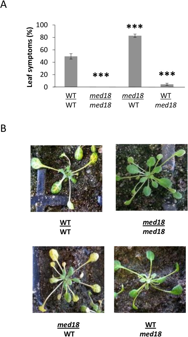
Prior to grafting, the seedlings were grown in long day conditions and therefore have an accelerated flowering time. The delay in flowering time in med18 was retained in the grafting process with grafts containing the WT Col-0 scion transitioning to flowering whereas the grafts with med18 scions remained vegetative. As a delay in flowering time had previously been associated with increased F. oxysporum resistance [26, 40] we examined whether the transition to flowering in the grafted plants affected resistance to F. oxysporum. Interestingly, despite undergoing flowering during the time of inoculation, the med18 rootstock: Col-0 scion graft was resistant to F. oxysporum infection, whereas the Col-0 rootstock: med18 scion was susceptible (Fig 3 and S1 Fig). These results show that, similar to coi1, the resistance of med18 to F. oxysporum is dependent on the root genotype. In addition, delayed flowering time and F. oxysporum resistance are spatially independent traits in the med18 mutant.
Fig 3. The resistance of med18 is mediated by the roots.
Reciprocal grafts made between the WT (Col-0) and med18 seedlings revealed that roots containing the med18 root genotype were highly resistant to infection, whereas med18/WT or WT/WT self-grafts were highly susceptible. (A) shows the average percentage of leaf chlorosis from eighteen plants per graft combination. Error bars represent standard error. (B) shows typical disease symptoms at 12 days after infection. *** represents significance (p<0.001) using Student’s T-test of each mutant compared to the WT/WT self-graft.
F. oxysporum colonization is restricted in med18 roots
As med18 roots are responsible for resistance to F. oxysporum, we determined whether there is a reduction in root colonization in med18 roots due to enhanced defenses or whether med18 roots are similarly colonized but the pathogen is not able to cause disease in the shoots. We examined root colonization of med18 with the use of a transgenic F. oxysporum constitutively expressing the β-GLUCURONIDASE (GUS) transgene [32]. Four week old WT and med18 plants were root dipped in GUS-expressing F. oxysporum and the colonization of the root tissue was examined twelve days post infection. WT (Col-0) plants start to develop leaf symptoms around seven days post infection and at twelve days post infection the majority of WT plants were showing leaf symptoms and approaching collapse. In contrast the med18 mutant plants fail to develop leaf symptoms and eventually proceed to flowering. A clear difference in root colonization was observed with WT plants showing extensive fungal colonization of the root system, whereas med18 plants showed very limited colonization, with less than 5% of the roots positively stained for GUS (Fig 4A–4F). It was observed that WT plants showing chlorosis symptoms in the leaves often possessed strong colonization of a sub-section of the root, suggesting that the entire root system does not need to be colonized. This would explain why WT plants on average had only 15% of the root system colonized. However the colonization in these regions was extensive with the GUS expression in the WT roots appearing to spread along the root vascular tissue and connect through to adjacent lateral roots and up towards the hypocotyl (Fig 4B and 4D) similar to previous reports [41] whereas colonization in the med18 line appeared to be punctate and rarely spread to adjacent lateral roots (Fig 4C and 4E). Therefore reduced leaf symptom development in the med18 mutant correlates with restricted fungal colonization relative to WT roots.
Fig 4. F. oxysporum shows restricted colonization in med18 roots.
Ten plants of each genotype were isolated twelve days post infection with a GUS-expressing strain of F. oxysporum and stained for GUS expression. Colonization of the med18 (A, C, E) and WT (B, D) root system. Infection in the med18 root system was localized to a small region in the roots and evidence of unsuccessful infection attempts were observed (red arrows). Photos shown in (C) and (D) are the lower portion of the root system seen in (A) and (B) respectively. Photos are representative of the results for each genotype. (A-D) are imaged using a Zeiss stereomicroscope at 4x zoom and (E) imaged using a Nikon compound microscope. Scale bars represent 5mm (A-D) and 100μm (E). Colonization was scored as a percentage of the total root mass and the average shown in (F). Error bars are standard error of ten individual plants. * represents significance using Student’s T-test (p<0.05).
RNA sequencing analysis of med18 and med20 reveal a common gene regulon
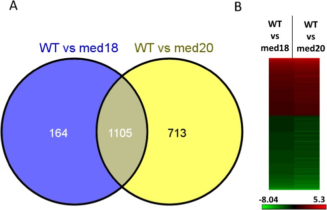
To identify genes that might be controlling resistance to F. oxysporum we conducted an RNA sequencing (RNAseq) experiment in WT, med18 and med20 roots infected with F. oxysporum. We chose to analyse early defense responses to F. oxysporum infection at one day post inoculation based on studies showing activation of defense gene expression in the roots at this time point [19]. Overall, 1269 and 1818 genes were differentially regulated (>2 fold and significant; FDR p<0.05) in med18 and med20, respectively, compared to the WT after F. oxysporum infection (S2 Table). Comparing the WT vs med18 and WT vs med20 differentially expressed gene (DEG) lists to each other indicated a significant percentage of co-regulated genes with approximately 87% of med18 DEGs and 61% of med20 DEGs showing similar patterns of expression (Fig 5A and 5B). Thus the RNAseq analysis suggests that MED18 and MED20 regulate the expression of similar gene sets in Arabidopsis.
Fig 5. MED18 and MED20 co-regulate a similar subset of genes.
(A) Venn diagram of the genes differentially expressed between WT and med18 or WT and med20. (B) Heat map of the 1105 co-regulated genes. Heat map displays the Log2 fold change of med18 or med20 compared to the WT, with red representing higher and green representing lower expression in the Mediator mutants, respectively. Scale bar shows the colour change according to the Log2 fold change.
To investigate the genes that may be leading to enhanced resistance in the med18 and med20 mutants, we performed Gene Ontology (GO) enrichment analysis on the co-expressed genes [42]. The genes induced in the WT versus both med18 and med20 included several defence and stress related GO terms that were significantly enriched including: response to chitin, response to fungus, defense response, response to hormones (JA and abscisic acid) and plant cell wall modification (S3 Table). Examining the gene lists themselves, we found PAMP triggered immunity (PTI) associated genes such as FLAGELLIN SENSITIVE 2 (FLS2), PLANT U-BOX 23 (PUB23) and MITOGEN-ACTIVATED PROTEIN KINASE 3 (MPK3) as being induced higher in the WT (Table 1). Also in the list were other genes associated with defence such as the cell wall synthases (CESA4 and CESA8), the plant defensins PDF1.4 and PDF2.2, as well as PROPEP1, which act as a precursor for the PEP1 peptide which activates defense genes and reactive oxygen species (ROS) [43]. In addition, several JA signaling genes were expressed higher in the WT relative to the mutants. These included the JA-associated transcription factor MYC2, the JASMONATE ZIM DOMAIN (JAZ) repressor proteins; JAZ1, JAZ5, JAZ7, JAZ8 and JAZ10, JA biosynthesis genes ALLENE OXIDE SYNTHASE (AOS), ALLENE OXIDE CYCLASE (AOC3), LIPOXYGENASE4 (LOX4), SULFOTRANSFERASE 2A (ST2A) which acts specifically on 11- and 12-hydroxyjasmonic acid [44], the galactolipase DONGLE, a JA-Ile-hydroxylase (CYP94B3) and VEGETATIVE STORAGE PROTEINS; VSP1 and VSP2 (Table 1). We confirmed the accuracy of the RNAseq analysis by examining the expression of MYC2, PDF1.4, PDF2.2 as well as the differentially expressed JAZ genes using RT-qPCR (S2 Fig).
Table 1. The expression of plant defense and JA-associated genes is increased in the WT relative to med18 and med20 in response to F. oxysporum infection.
Values represent the fold change in expression of the WT vs either med18 or med20 in the RNAseq experiment.
| Category | Gene | Locus Identifier | WT vs med18 | WT vs med20 |
|---|---|---|---|---|
| Pattern triggered immunity | FLAGELLIN-SENSITIVE 2 (FLS2) | AT5G46330 | 3.22 | 3.65 |
| PLANT U-BOX 23 (PUB23) | AT2G35930 | 2.02 | 2.04 | |
| MITOGEN-ACTIVATED PROTEIN KINASE 3 (MPK3) | AT3G45640 | 2.79 | 3.08 | |
| PROPEP 1 | AT5G64900 | 3.24 | 4.03 | |
| PROPEP 4 | AT5G09980 | 3.31 | 8.84 | |
| Cell wall associated defense | CELLULOSE SYNTHASE A4 (CESA4) | AT5G44030 | 2.43 | 2.60 |
| CELLULOSE SYNTHASE 8 (CESA8) | AT4G18780 | 2.21 | 2.42 | |
| Plant Defensins | PLANT DEFENSIN 1.4 (PDF1.4) | AT1G19610 | 9.66 | 2.92 |
| PLANT DEFENSIN 2.2 (PDF2.2) | AT2G02100 | 4.00 | 4.84 | |
| Jasmonate associated genes | MYC 2 | AT1G32640 | 2.34 | 2.39 |
| JASMONATE-ZIM-DOMAIN PROTEIN 1 (JAZ1) | AT1G19180 | 2.16 | 2.22 | |
| JASMONATE-ZIM-DOMAIN PROTEIN 5 (JAZ5) | AT1G17380 | 2.96 | 3.51 | |
| JASMONATE-ZIM-DOMAIN PROTEIN 7 (JAZ7) | AT2G34600 | 3.20 | 3.82 | |
| JASMONATE-ZIM-DOMAIN PROTEIN 8 (JAZ8) | AT1G30135 | 2.39 | 3.17 | |
| JASMONATE-ZIM-DOMAIN PROTEIN 10 (JAZ10) | AT5G13220 | 4.44 | 5.60 | |
| DONGLE (DGL) | AT1G05800 | 4.42 | 4.68 | |
| ALLENE OXIDE SYNTHASE (AOS) | AT5G42650 | 2.25 | 2.38 | |
| ALLENE OXIDE CYCLASE 3 (AOC3) | AT3G25780 | 2.43 | 2.96 | |
| LIPOXYGENASE 4 (LOX4) | AT1G72520 | 2.82 | 3.39 | |
| SULFOTRANSFERASE 2A (ST2A) | AT5G07010 | 2.99 | 2.53 | |
| CYTOCHROME P450, FAMILY 94, SUBFAMILY B3 (CYP94B3) | AT3G48520 | 5.29 | 6.88 | |
| VEGETATIVE STORAGE PROTEIN 1 (VSP1) | AT5G24780 | 5.01 | 7.22 | |
| VEGETATIVE STORAGE PROTEIN 2 (VSP2) | AT5G24770 | 3.61 | 3.25 |
The reduced expression of JA signaling genes such as MYC2 and the JAZ genes in med18 and med20 suggests that the MED18 and MED20 subunits are required for maintaining a functional JA signaling pathway. Interestingly, well-studied JA/ET-associated defense marker genes such as PLANT DEFENSIN1.2, (PDF1.2), the BASIC CHITINASE (CHI-B) and HEVEIN-LIKE (HEL) PR genes were not found to be differentially regulated in the RNAseq experiment. Overall the RNAseq and RT-qPCR results suggest that the MYC2 regulated branch of JA signaling is affected in med18 and med20 [45, 46] which may potentially explain the strong F. oxysporum resistance displayed by these mutants.
The GO analysis of genes expressed higher in the med18 and med20 roots versus WT roots identified GO terms associated with transcription and RNA metabolism as well as the GO terms associated with ion and multi-drug transport. One of the transporters significantly induced was PDR12, a pathogen and defense hormone inducible gene encoding an ABC transporter [30]. As would be expected with alterations in the Mediator complex, several transcription factors were affected in the med18 and med20 mutants, with WRKYs, MYBs, NACs and MADS box TFs being identified as differentially expressed. GO terms associated with oxidative stress, hydrogen peroxide and ROS were also identified in the med20 induced gene list but not in med18. Overall, the GO enrichment analysis identified response to fungi, chitin and JA-associated gene expression as being higher in the WT, whereas the med18 and med20 mutants showed transcription, and transporter associated GO terms being enriched with additional ROS associated GO terms identified in the med20 mutant.
MED18 and MED20 negatively regulate SA-associated defence genes
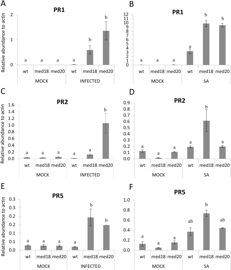
As the JA and SA signaling pathways often antagonize each other, we examined whether SA- associated defense genes were activated in med18 and med20 plants. We examined the expression of SA- associated PR genes, PR1, PR2 and PR5 with RT-qPCR and found that PR1 and the thaumatin-like PR5 gene was significantly induced in med18 and med20 roots relative to the WT after F. oxysporum infection. PR2 was also found to be induced in both med18 and med20, but the induction was not statistically significant in med18 (Fig 6). We examined these same genes in leaf tissue after treatment with SA to see if the med mutants had higher SA regulated gene expression in other tissues. These experiments showed that PR1 expression was approximately three fold higher in both mutants after SA treatment. PR5 was not significantly induced in either mutant, while PR2 was expressed higher in med18 leaves only (Fig 6). Therefore different patterns of PR gene induction are seen in the med18 and med20 leaves after SA induction as opposed to in roots after F. oxysporum infection. This is not surprising as infection with F. oxysporum would induce a response in gene expression based on a variety of elicitors and effectors as opposed to a single hormone treatment with SA. Overall we conclude that PR1 and PR5 are induced higher in med18 and med20 in response to F. oxysporum infection at early time points and may play a role in restricting pathogen colonization in the roots.
Fig 6. Real time qRT-PCR data of SA-associated genes in WT, med18 and med20 roots and leaves.
(A, C, E) PR1, PR2 and PR5 expression in roots of WT, med18 and med20 with and without F. oxysporum infection. (B, D, F) PR1, PR2 and PR5 expression in leaves of WT, med18 and med20 in response to 24 hours of mock or SA treatment. Results were obtained from three independent biological replicates of twenty plants per replicate. (a & b) indicates p-value < 0.05 using a one-way ANOVA, least significant difference test. Error bars represent standard deviation of the biological replicates.
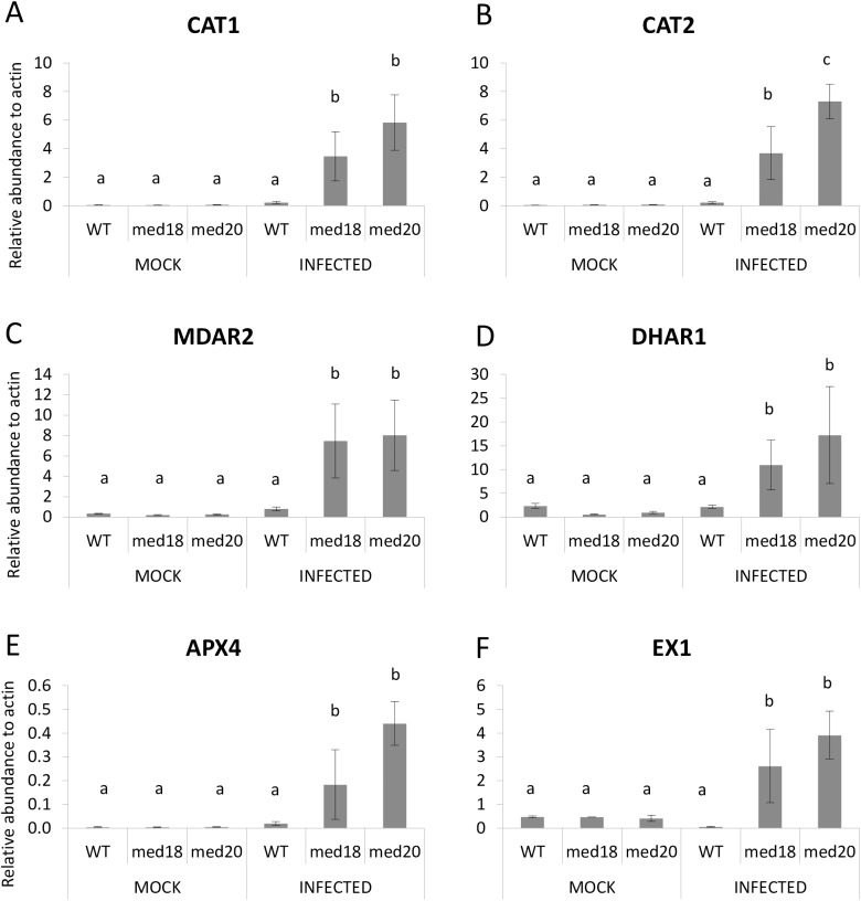
Activation of plant defenses by biotrophic fungal pathogens is often associated with increased SA-defenses, ROS production and the hypersensitive response in plants [47]. Response to ROS was also a significantly enriched GO term enriched in med20 versus the WT (S3 Table). To investigate and compare the involvement of ROS in the hemi-biotrophic infection of F. oxysporum, we conducted RT-qPCR expression analyses of genes known to have a role in ROS generation or metabolism. Genes found to be induced higher in med18 and med20 were the ROS scavenging CATALASE genes CAT1 and CAT2, as well as the oxidoreductases MONODEHYDROASCORBATE REDUCTASE2 (MDAR2), DEHYDROASCORBATE REDUCTASE1 (DHAR1) and ASCORBATE PEROXIDASE4 (APX4) (Fig 7; [48]). Also expressed higher was EXECUTER1 which plays an important role in singlet oxygen stress [49, 50].
Fig 7. ROS associated genes that are expressed higher in med18 and med20 roots in response to F. oxysporum infection.
(A-F) The expression of CAT1, CAT2, MDAR2, DHAR1, APX4 and EX1 was significantly higher in med18 and med20 roots than the WT after 24 hours F. oxysporum infection. Results were obtained from three independent biological replicates of twenty plants per replicate. (a, b & c) indicates p-value < 0.05 using a one-way ANOVA, least significant difference test. Error bars represent standard deviation of the biological replicates.
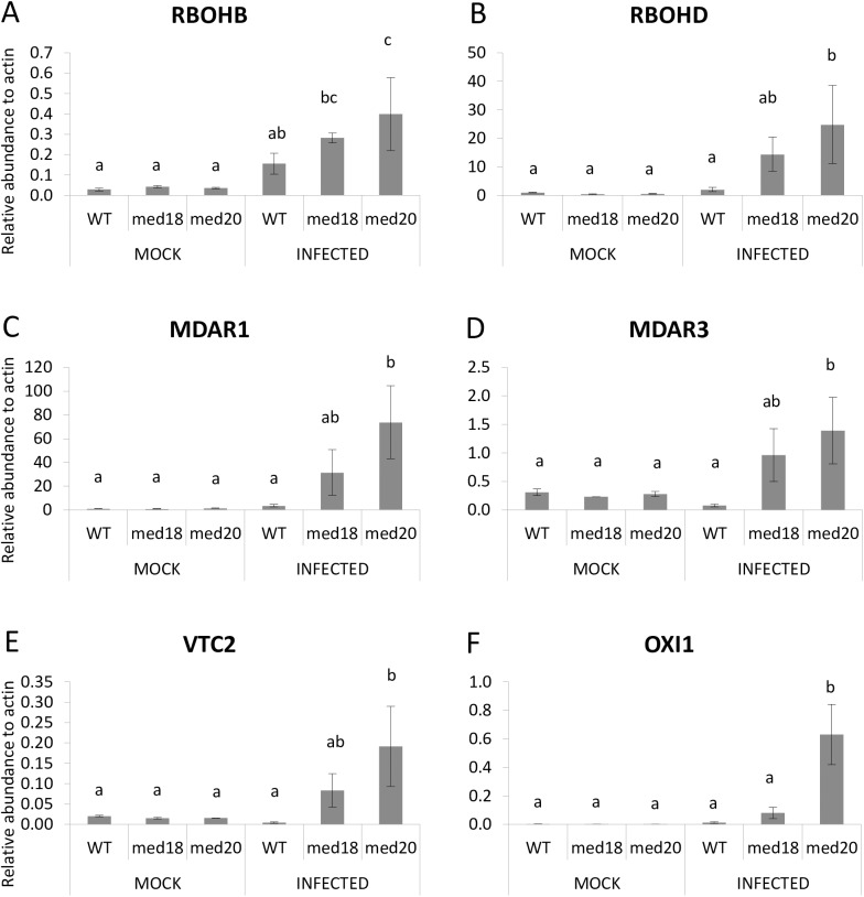
In addition, several genes were found to be significantly up-regulated only in med20, including RESPIRATORY BURST OXIDASE HOHMOLOGS, RBOHB and RBOHD, MONODEHYDROASCORBATE REDUCTASE1, MONODEHYDROASCORBATE REDUCTASE3, VITAMIN C DEFECTIVE2 and OXIDATIVE SIGNAL INDUCIBLE1 (Fig 8). The up-regulation of EX1 and ROS quenching genes such as the CATALASE and ascorbate reductase genes in both mutants suggest the possibility for increased ROS production in med18 and med20 roots in response to F. oxysporum.
Fig 8. ROS associated genes that are expressed higher in med20 roots in response to F. oxysporum infection.
(A-F) The expression of RBOHD, RBOHF, MDAR1, MDAR3, VTC2 and OXI1 was significantly higher in med20 roots after 24 hours F. oxysporum infection. Results were obtained from three independent biological replicates of twenty plants per replicate. (a, b & c) indicates p-value < 0.05 using a one-way ANOVA, least significant difference test. Error bars represent standard deviation of the biological replicates.
Discussion
Since the discovery of the Mediator complex in Arabidopsis [51], individual subunits of the complex have been shown to be involved in a wide range of biological processes in plants [25]. Through screening of twelve T-DNA insertion lines in Arabidopsis Mediator genes, we previously identified the MED8 and MED25 subunits to be required for susceptibility to F. oxysporum [26]. While the F. oxysporum resistance phenotype of the med25 mutant was linked to a defect in JA signaling, the med8 mutant demonstrated wild type levels of JA/ET defense marker gene expression [26]. To further investigate the role of Mediator subunits in F. oxysporum resistance, we inoculated mutants of the head domain subunits MED18 and MED20, which are predicted to be located adjacent to med8 in the Arabidopsis Mediator complex. Interestingly, the med18 and med20 mutants were found to be phenotypically similar to each other with almost complete resistance to F. oxysporum. The med8, med18, med20, med25 mutants and med8 med25 double mutant all display a delay in flowering time and enhanced resistance to F. oxysporum. It was previously hypothesized that a delay in flowering time might lead to enhanced resistance to F. oxysporum, as several mutants with delayed flowering time (e.g. myc2, arf2, fve-3), as well as Arabidopsis accessions with delayed flowering also have enhanced resistance [40]. However the link between flowering time and F. oxysporum resistance could be broken by removing the flowering repressor FLOWERING LOCUS C (FLC) in the fve-3 mutant or through vernalization in some natural accessions [40]. We found that the flowering time and F. oxysporum phenotypes are spatially separate in med18 using grafting. The flowering time phenotype was found to be regulated by the leaves as expected given that this is where the expression of FLOWERING LOCUS T (FT) is triggered [52], whereas F. oxysporum resistance occurs in the roots, resulting in failed root colonization in med18 roots.
Through RNAseq analysis we observed a significant reduction in JA-associated genes in med18 and med20, relative to the WT. This analysis identified genes affecting JA accumulation such as AOS, AOC3, LOX4, DONGLE, and a JA-Ile-hydroxylase as well as JA-signaling genes such as MYC2 and the JAZ genes. These results suggest that MED18 and MED20 are involved in regulating JA signaling in Arabidopsis. In accordance with previous findings on antagonistic interactions between JA- and SA-associated defense pathways, the expression of PR1 and PR5 was higher in med18 and med20 in response to F. oxysporum treatment. We also found up-regulation of singlet oxygen stress responsive genes, EX1, in med18 and med20 and OXI1 in med20 only, as well as up-regulation of ROS scavenging genes such as CAT1 and CAT2 and the ascorbate reductases in both mutants. OXI1 and EX1 have been recently shown to regulate singlet oxygen mediated cell death through independent pathways [50, 53]. Jasmonate has also been shown to play a role in singlet oxygen responses, which has been revealed through recent investigations with the flu and ch1 mutants (reviewed by [54]). It is possible that both JA signaling and singlet oxygen stress signaling is channelled through the Mediator complex via MED18 and MED20 or alternatively, mis-regulation of the JA pathway in med18 and med20 leads to defects in ROS production and tolerance. With both ROS producing and ROS scavenging genes being upregulated at the same timepoint, further work examining additional timepoints is needed to investigate the amplitude and types of ROS that are produced and whether altered ROS levels in med18 or med20 impact on F. oxysporum directly or instead play a role in defense signaling.
In addition to being more resistant to F. oxysporum, mutants of MED8, and MED18 have been shown to be susceptible to necrotrophic leaf pathogens such as Alternaria brassicicola and Botrytis cinerea [26, 39, 55]. Recently it was shown that MED18 is recruited by the histone acetyltransferase, HOOKLESS1, to the WRKY33 promoter and thereby increases WRKY33 expression [55]. WRKY33 has an important role in JA/SA crosstalk as well as redox homeostasis, [56]. Loss of WRKY33 results in activation of SA defense responses and down-regulation of JA-associated responses and therefore the JA/SA crosstalk we observed here could be associated with MED18’s recruitment to the WRKY33 promoter. WRKY33 was not differentially regulated in the RNAseq experiment at 24hours, but future work should examine whether changes in expression of this gene and other TFs with a role in cross talk are seen at earlier or later timepoints. MED18 has also been found to interact with TFs such as YIN YANG1, ABA INSENSITIVE4 and SUPPRESSOR OF FRIGIDA4 [39] and it would be important to test whether these TFs also affect F. oxysporum resistance. Alternatively, as mutations in med18 and med20 in the yeast Mediator complex affect the stabilization of RNA Pol II and TFIIB interactions [29] it is possible that disruption to this subdomain leads to a change in the binding surface which might subsequently affect these phenotypes through indirect mechanisms such as reduced RNA Pol II occupancy and altered histone modifications as has been demonstrated [39, 55].
Recently, the isolation of several mutants in an RNA binding KH domain protein termed SHINY1/ENHANCED STRESS RESPONSES1 (SHINY/ESR1) was identified through a forward genetic screen [57]. The esr1 mutants were found to have increased resistance to F. oxysporum as well as differential regulation in some but not all aspects of the JA pathway [57]. Previously SHINY1/ESR1 was shown to interact with FIERY2/RNA POLYMERASE II CARBOXYL TERMINAL DOMAIN PHOSPHATASE-LIKE 1 (FRY2/CPL1), a protein that de-phosphorylates the C terminal domain (CTD) of RNA Pol II [58, 59]. CPL1 has been shown to be essential for accurate miRNA processing and controls mRNA splicing and mRNA decay [60–62]. It has previously been shown that mutations in MED18 and MED20 also affect miRNA levels and a mutation in RNA Pol II leads to similar developmental phenotypes as med18 and med20 suggesting a connection between mutations in RNA binding proteins [57], the Mediator head domain and mutations in RNA Pol II itself [31]. This suggests that disruptions in this region of the RNA pol II holoenzyme result in similar developmental and biotic phenotypes in Arabidopsis.
While we detected interaction between MED18 and MED20 in reciprocal Yeast-2-Hybrid experiments, we were unable to identify a positive interaction with MED8. This was surprising as MED18 and MED20 form part of the movable jaw that connects through the C-terminal portion of MED8 in the yeast Mediator complex. However it has been shown that correct folding of yeast MED8, MED18 and MED20 as a trimer requires all three proteins [63], and therefore it is possible that all three proteins are required for proper interaction of the Arabidopsis proteins as well. Recent structural modelling of the yeast Mediator complex show MED18 and MED20 form a separate interface with RNA pol II as compared to MED8 [29]. MED18 and MED20 form an interface with RPB3 and RPB11 of RNA pol II, and the TFIIB B–ribbon domain, whereas MED8 forms interactions with the RPB4-RBP7 stalk of RNA pol II [29]. The Mediator complex has shown remarkable structural conservation across yeast and metazoan complexes [64, 65], and it is hypothesized that a similar structural organization occurs in the Arabidopsis complex. As in vivo protein pull down experiments with Arabidopsis Mediator subunits results in the entire complex being isolated [51], more detailed structural biology approaches are required to identify where MED18 and MED20 sit in the Arabidopsis Mediator complex and how disruption of either MED18 or MED20 subunits affects their interaction with the Arabidopsis RNA Pol II holoenzyme.
Overall we propose that MED18 and MED20 form a sub-domain within the Arabidopsis Mediator complex that regulates flowering time and pathogen defense. A reduction in JA signaling and enhanced SA- and ROS-associated gene expression was observed in both mutants which might contribute to the restriction of F. oxysporum growth during root infection. Further work should reveal how MED18 and MED20 dock in the Arabidopsis Mediator complex and provide further insights into the mechanistic process for producing the strong F. oxysporum resistance phenotype observed.
Supporting information
Photographs were taken two weeks after infection with F. oxysporum.
(PPTX)
Results were obtained from three independent biological replicates. ANOVA, LSD significant difference test (a, b, & c) indicates p-value < 0.05; error bars represent standard error of the biological replicates.
(PPTX)
(XLSX)
(XLSX)
(XLSX)
Data Availability
All relevant data are within the paper and its Supporting Information files. In addition the RNAseq Fastq files are available at the NCBI Sequence Read Archive under study number SRP092151.
Funding Statement
This work was supported by the Australian Research Council [http://www.arc.gov.au] grant no. DP110104354 to PMS, JMM, and KK. CD and SB were supported by grants from the Swedish Cancer Society (15 0537); the Swedish Research Council (621-2010-4969); the Swedish Governmental Agency for Innovation Systems, the Knut and Alice Wallenberg Foundation (2015-0056) and the Kempe Foundation. The funders had no role in study design, data collection and analysis, decision to publish, or preparation of the manuscript.
References
- 1.Havko NE, Major IT, Jewell JB, Attaran E, Browse J, Howe GA. Control of Carbon Assimilation and Partitioning by Jasmonate: An Accounting of Growth-Defense Tradeoffs. Plants (Basel, Switzerland). 2016;5(1). [DOI] [PMC free article] [PubMed] [Google Scholar]
- 2.Smakowska E, Kong J, Busch W, Belkhadir Y. Organ-specific regulation of growth-defense tradeoffs by plants. Curr Opin Plant Biol. 2016;29:129–37. doi: 10.1016/j.pbi.2015.12.005 [DOI] [PubMed] [Google Scholar]
- 3.Koornneef A, Pieterse CM. Cross talk in defense signaling. Plant Physiol. 2008;146(3):839–44. doi: 10.1104/pp.107.112029 [DOI] [PMC free article] [PubMed] [Google Scholar]
- 4.Tsuda K, Sato M, Stoddard T, Glazebrook J, Katagiri F. Network properties of robust immunity in plants. PLoS Genet. 2009;5(12):e1000772 doi: 10.1371/journal.pgen.1000772 [DOI] [PMC free article] [PubMed] [Google Scholar]
- 5.Windram O, Penfold CA, Denby KJ. Network modeling to understand plant immunity. Annu Rev Phytopathol. 2014;52:93–111. doi: 10.1146/annurev-phyto-102313-050103 [DOI] [PubMed] [Google Scholar]
- 6.Derksen H, Rampitsch C, Daayf F. Signaling cross-talk in plant disease resistance. Plant Sci. 2013;207:79–87. doi: 10.1016/j.plantsci.2013.03.004 [DOI] [PubMed] [Google Scholar]
- 7.Kazan K, Lyons R. Intervention of Phytohormone Pathways by Pathogen Effectors. Plant Cell. 2014;26(6):2285–309. doi: 10.1105/tpc.114.125419 [DOI] [PMC free article] [PubMed] [Google Scholar]
- 8.Katsir L, Schilmiller AL, Staswick PE, He SY, Howe GA. COI1 is a critical component of a receptor for jasmonate and the bacterial virulence factor coronatine. Proc Natl Acad Sci U S A. 2008;105(19):7100–5. doi: 10.1073/pnas.0802332105 [DOI] [PMC free article] [PubMed] [Google Scholar]
- 9.Cui F, Wu S, Sun W, Coaker G, Kunkel B, He P, et al. The Pseudomonas syringae type III effector AvrRpt2 promotes pathogen virulence via stimulating Arabidopsis auxin/indole acetic acid protein turnover. Plant Physiol. 2013;162(2):1018–29. doi: 10.1104/pp.113.219659 [DOI] [PMC free article] [PubMed] [Google Scholar]
- 10.Misas-Villamil JC, Kolodziejek I, Crabill E, Kaschani F, Niessen S, Shindo T, et al. Pseudomonas syringae pv. syringae uses proteasome inhibitor syringolin A to colonize from wound infection sites. PLoS Pathog. 2013;9(3):e1003281 doi: 10.1371/journal.ppat.1003281 [DOI] [PMC free article] [PubMed] [Google Scholar]
- 11.Zheng XY, Spivey NW, Zeng W, Liu PP, Fu ZQ, Klessig DF, et al. Coronatine promotes Pseudomonas syringae virulence in plants by activating a signaling cascade that inhibits salicylic acid accumulation. Cell Host Microbe. 2012;11(6):587–96. doi: 10.1016/j.chom.2012.04.014 [DOI] [PMC free article] [PubMed] [Google Scholar]
- 12.Miersch O, Bohlmann H, Wasternack C. Jasmonates and related compounds from Fusarium oxysporum. Phytochemistry. 1999;50(4):517–23. [Google Scholar]
- 13.Cole SJ, Yoon AJ, Faull KF, Diener AC. Host perception of jasmonates promotes infection by Fusarium oxysporum formae speciales that produce isoleucine- and leucine-conjugated jasmonates. Mol Plant Pathol. 2014;15(6):589–600. doi: 10.1111/mpp.12117 [DOI] [PMC free article] [PubMed] [Google Scholar]
- 14.Anderson JP, Badruzsaufari E, Schenk PM, Manners JM, Desmond OJ, Ehlert C, et al. Antagonistic interaction between abscisic acid and jasmonate-ethylene signaling pathways modulates defense gene expression and disease resistance in Arabidopsis. Plant Cell. 2004;16(12):3460–79. doi: 10.1105/tpc.104.025833 [DOI] [PMC free article] [PubMed] [Google Scholar]
- 15.Thatcher LF, Manners JM, Kazan K. Fusarium oxysporum hijacks COI1-mediated jasmonate signaling to promote disease development in Arabidopsis. Plant J. 2009;58(6):927–39. doi: 10.1111/j.1365-313X.2009.03831.x [DOI] [PubMed] [Google Scholar]
- 16.Trusov Y, Sewelam N, Rookes JE, Kunkel M, Nowak E, Schenk PM, et al. Heterotrimeric G proteins-mediated resistance to necrotrophic pathogens includes mechanisms independent of salicylic acid-, jasmonic acid/ethylene- and abscisic acid-mediated defense signaling. Plant J. 2009;58(1):69–81. doi: 10.1111/j.1365-313X.2008.03755.x [DOI] [PubMed] [Google Scholar]
- 17.Thatcher LF, Cevik V, Grant M, Zhai B, Jones JD, Manners JM, et al. Characterization of a JAZ7 activation-tagged Arabidopsis mutant with increased susceptibility to the fungal pathogen Fusarium oxysporum. J Exp Bot. 2016;67(8):2367–86. doi: 10.1093/jxb/erw040 [DOI] [PMC free article] [PubMed] [Google Scholar]
- 18.Edgar CI, McGrath KC, Dombrecht B, Manners JM, Maclean DC, Schenk PM, et al. Salicylic acid mediates resistance to the vascular wilt pathogen Fusarium oxysporum in the model host Arabidopsis thaliana. Australasian Plant Pathology. 2006;35(6):581–91. [Google Scholar]
- 19.Lyons R, Stiller J, Powell J, Rusu A, Manners JM, Kazan K. Fusarium oxysporum Triggers Tissue-Specific Transcriptional Reprogramming in Arabidopsis thaliana. PLoS One. 2015;10(4):e0121902 doi: 10.1371/journal.pone.0121902 [DOI] [PMC free article] [PubMed] [Google Scholar]
- 20.Kidd BN, Kadoo NY, Dombrecht B, Tekeoglu M, Gardiner DM, Thatcher LF, et al. Auxin signaling and transport promote susceptibility to the root-infecting fungal pathogen Fusarium oxysporum in Arabidopsis. Molecular Plant-Microbe Interactions. 2011;24(6):733–48. doi: 10.1094/MPMI-08-10-0194 [DOI] [PubMed] [Google Scholar]
- 21.Pantelides IS, Tjamos SE, Pappa S, Kargakis M, Paplomatas EJ. The ethylene receptor ETR1 is required for Fusarium oxysporum pathogenicity. Plant Pathology. 2013;62(6):1302–9. [Google Scholar]
- 22.Riechmann JL, Heard J, Martin G, Reuber L, Jiang C, Keddie J, et al. Arabidopsis transcription factors: genome-wide comparative analysis among eukaryotes. Science. 2000;290(5499):2105–10. [DOI] [PubMed] [Google Scholar]
- 23.McGrath KC, Dombrecht B, Manners JM, Schenk PM, Edgar CI, Maclean DJ, et al. Repressor- and activator-type ethylene response factors functioning in jasmonate signaling and disease resistance identified via a genome-wide screen of Arabidopsis transcription factor gene expression. Plant Physiol. 2005;139(2):949–59. doi: 10.1104/pp.105.068544 [DOI] [PMC free article] [PubMed] [Google Scholar]
- 24.Kidd BN, Cahill DM, Manners JM, Schenk PM, Kazan K. Diverse roles of the Mediator complex in plants. Seminars in Cell & Developmental Biology. 2011;22(7):741–8. [DOI] [PubMed] [Google Scholar]
- 25.Samanta S, Thakur JK. Importance of Mediator complex in the regulation and integration of diverse signaling pathways in plants. Front Plant Sci. 2015;6:757 doi: 10.3389/fpls.2015.00757 [DOI] [PMC free article] [PubMed] [Google Scholar]
- 26.Kidd BN, Edgar CI, Kumar KK, Aitken EA, Schenk PM, Manners JM, et al. The mediator complex subunit PFT1 is a key regulator of jasmonate-dependent defense in Arabidopsis. Plant Cell. 2009;21(8):2237–52. doi: 10.1105/tpc.109.066910 [DOI] [PMC free article] [PubMed] [Google Scholar]
- 27.Çevik V, Kidd BN, Zhang P, Hill C, Kiddle S, Denby KJ, et al. MEDIATOR25 Acts as an Integrative Hub for the Regulation of Jasmonate-Responsive Gene Expression in Arabidopsis. Plant Physiology. 2012;160(1):541–55. doi: 10.1104/pp.112.202697 [DOI] [PMC free article] [PubMed] [Google Scholar]
- 28.Lariviere L, Plaschka C, Seizl M, Wenzeck L, Kurth F, Cramer P. Structure of the Mediator head module. Nature. 2012;492(7429):448–51. doi: 10.1038/nature11670 [DOI] [PubMed] [Google Scholar]
- 29.Plaschka C, Lariviere L, Wenzeck L, Seizl M, Hemann M, Tegunov D, et al. Architecture of the RNA polymerase II-Mediator core initiation complex. Nature. 2015;518(7539):376–80. doi: 10.1038/nature14229 [DOI] [PubMed] [Google Scholar]
- 30.Campbell EJ, Schenk PM, Kazan K, Penninckx IA, Anderson JP, Maclean DJ, et al. Pathogen-responsive expression of a putative ATP-binding cassette transporter gene conferring resistance to the diterpenoid sclareol is regulated by multiple defense signaling pathways in Arabidopsis. Plant Physiol. 2003;133(3):1272–84. doi: 10.1104/pp.103.024182 [DOI] [PMC free article] [PubMed] [Google Scholar]
- 31.Kim YJ, Zheng B, Yu Y, Won SY, Mo B, Chen X. The role of Mediator in small and long noncoding RNA production in Arabidopsis thaliana. EMBO J. 2011;30(5):814–22. doi: 10.1038/emboj.2011.3 [DOI] [PMC free article] [PubMed] [Google Scholar]
- 32.Schumann U, Smith NA, Kazan K, Ayliffe M, Wang MB. Analysis of hairpin RNA transgene-induced gene silencing in Fusarium oxysporum. Silence. 2013;4(1):3 doi: 10.1186/1758-907X-4-3 [DOI] [PMC free article] [PubMed] [Google Scholar]
- 33.Jefferson RA, Kavanagh TA, Bevan MW. GUS fusions: beta-glucuronidase as a sensitive and versatile gene fusion marker in higher plants. EMBO J. 1987;6(13):3901–7. [DOI] [PMC free article] [PubMed] [Google Scholar]
- 34.Trapnell C, Roberts A, Goff L, Pertea G, Kim D, Kelley DR, et al. Differential gene and transcript expression analysis of RNA-seq experiments with TopHat and Cufflinks. Nat Protoc. 2012;7(3):562–78. doi: 10.1038/nprot.2012.016 [DOI] [PMC free article] [PubMed] [Google Scholar]
- 35.Pavlidis P, Noble WS. Matrix2png: a utility for visualizing matrix data. Bioinformatics. 2003;19(2):295–6. [DOI] [PubMed] [Google Scholar]
- 36.Ramakers C, Ruijter JM, Deprez RH, Moorman AF. Assumption-free analysis of quantitative real-time polymerase chain reaction (PCR) data. Neuroscience Letters. 2003;339(1):62–6. [DOI] [PubMed] [Google Scholar]
- 37.Yang Y, Ou B, Zhang J, Si W, Gu H, Qin G, et al. The Arabidopsis Mediator subunit MED16 regulates iron homeostasis by associating with EIN3/EIL1 through subunit MED25. Plant J. 2014;77(6):838–51. doi: 10.1111/tpj.12440 [DOI] [PubMed] [Google Scholar]
- 38.Zheng Z, Guan H, Leal F, Grey PH, Oppenheimer DG. Mediator subunit18 controls flowering time and floral organ identity in Arabidopsis. PLoS ONE. 2013;8(1):e53924 doi: 10.1371/journal.pone.0053924 [DOI] [PMC free article] [PubMed] [Google Scholar]
- 39.Lai Z, Schluttenhofer CM, Bhide K, Shreve J, Thimmapuram J, Lee SY, et al. MED18 interaction with distinct transcription factors regulates multiple plant functions. Nat Commun. 2014;5:3064 doi: 10.1038/ncomms4064 [DOI] [PubMed] [Google Scholar]
- 40.Lyons R, Rusu A, Stiller J, Powell J, Manners JM, Kazan K. Investigating the Association between Flowering Time and Defense in the Arabidopsis thaliana-Fusarium oxysporum Interaction. PLoS One. 2015;10(6):e0127699 doi: 10.1371/journal.pone.0127699 [DOI] [PMC free article] [PubMed] [Google Scholar]
- 41.Diener A. Visualizing and quantifying Fusarium oxysporum in the plant host. Mol Plant Microbe Interact. 2012;25(12):1531–41. doi: 10.1094/MPMI-02-12-0042-TA [DOI] [PubMed] [Google Scholar]
- 42.Du Z, Zhou X, Ling Y, Zhang Z, Su Z. agriGO: a GO analysis toolkit for the agricultural community. Nucleic Acids Res. 2010;38(Web Server issue):W64–70. doi: 10.1093/nar/gkq310 [DOI] [PMC free article] [PubMed] [Google Scholar]
- 43.Huffaker A, Ryan CA. Endogenous peptide defense signals in Arabidopsis differentially amplify signaling for the innate immune response. Proc Natl Acad Sci U S A. 2007;104(25):10732–6. doi: 10.1073/pnas.0703343104 [DOI] [PMC free article] [PubMed] [Google Scholar]
- 44.Gidda SK, Miersch O, Levitin A, Schmidt J, Wasternack C, Varin L. Biochemical and molecular characterization of a hydroxyjasmonate sulfotransferase from Arabidopsis thaliana. J Biol Chem. 2003;278(20):17895–900. doi: 10.1074/jbc.M211943200 [DOI] [PubMed] [Google Scholar]
- 45.Lorenzo O, Chico JM, Sanchez-Serrano JJ, Solano R. JASMONATE-INSENSITIVE1 encodes a MYC transcription factor essential to discriminate between different jasmonate-regulated defense responses in Arabidopsis. Plant Cell. 2004;16(7):1938–50. doi: 10.1105/tpc.022319 [DOI] [PMC free article] [PubMed] [Google Scholar]
- 46.Dombrecht B, Xue GP, Sprague SJ, Kirkegaard JA, Ross JJ, Reid JB, et al. MYC2 differentially modulates diverse jasmonate-dependent functions in Arabidopsis. Plant Cell. 2007;19(7):2225–45. doi: 10.1105/tpc.106.048017 [DOI] [PMC free article] [PubMed] [Google Scholar]
- 47.Torres MA, Jones JD, Dangl JL. Reactive oxygen species signaling in response to pathogens. Plant Physiol. 2006;141(2):373–8. doi: 10.1104/pp.106.079467 [DOI] [PMC free article] [PubMed] [Google Scholar]
- 48.Sewelam N, Kazan K, Thomas-Hall SR, Kidd BN, Manners JM, Schenk PM. Ethylene response factor 6 is a regulator of reactive oxygen species signaling in Arabidopsis. PLoS ONE. 2013;8(8):e70289 doi: 10.1371/journal.pone.0070289 [DOI] [PMC free article] [PubMed] [Google Scholar]
- 49.Wagner D, Przybyla D, Op den Camp R, Kim C, Landgraf F, Lee KP, et al. The genetic basis of singlet oxygen-induced stress responses of Arabidopsis thaliana. Science. 2004;306(5699):1183–5. doi: 10.1126/science.1103178 [DOI] [PubMed] [Google Scholar]
- 50.Lee KP, Kim C, Landgraf F, Apel K. EXECUTER1- and EXECUTER2-dependent transfer of stress-related signals from the plastid to the nucleus of Arabidopsis thaliana. Proc Natl Acad Sci U S A. 2007;104(24):10270–5. doi: 10.1073/pnas.0702061104 [DOI] [PMC free article] [PubMed] [Google Scholar]
- 51.Backstrom S, Elfving N, Nilsson R, Wingsle G, Bjorklund S. Purification of a plant mediator from Arabidopsis thaliana identifies PFT1 as the Med25 subunit. Mol Cell. 2007;26(5):717–29. doi: 10.1016/j.molcel.2007.05.007 [DOI] [PubMed] [Google Scholar]
- 52.Wigge PA. FT, a mobile developmental signal in plants. Curr Biol. 2011;21(9):R374–8. doi: 10.1016/j.cub.2011.03.038 [DOI] [PubMed] [Google Scholar]
- 53.Shumbe L, Chevalier A, Legeret B, Taconnat L, Monnet F, Havaux M. Singlet Oxygen-Induced Cell Death in Arabidopsis under High-Light Stress Is Controlled by OXI1 Kinase. Plant Physiol. 2016;170(3):1757–71. doi: 10.1104/pp.15.01546 [DOI] [PMC free article] [PubMed] [Google Scholar]
- 54.Laloi C, Havaux M. Key players of singlet oxygen-induced cell death in plants. Front Plant Sci. 2015;6:39 doi: 10.3389/fpls.2015.00039 [DOI] [PMC free article] [PubMed] [Google Scholar]
- 55.Liao CJ, Lai Z, Lee S, Yun DJ, Mengiste T. Arabidopsis HOOKLESS1 Regulates Responses to Pathogens and Abscisic Acid through Interaction with MED18 and Acetylation of WRKY33 and ABI5 Chromatin. Plant Cell. 2016;28(7):1662–81. doi: 10.1105/tpc.16.00105 [DOI] [PMC free article] [PubMed] [Google Scholar]
- 56.Birkenbihl RP, Diezel C, Somssich IE. Arabidopsis WRKY33 is a key transcriptional regulator of hormonal and metabolic responses toward Botrytis cinerea infection. Plant Physiol. 2012;159(1):266–85. doi: 10.1104/pp.111.192641 [DOI] [PMC free article] [PubMed] [Google Scholar]
- 57.Thatcher LF, Kamphuis LG, Hane JK, Onate-Sanchez L, Singh KB. The Arabidopsis KH-Domain RNA-Binding Protein ESR1 Functions in Components of Jasmonate Signalling, Unlinking Growth Restraint and Resistance to Stress. PLoS ONE. 2015;10(5):e0126978 doi: 10.1371/journal.pone.0126978 [DOI] [PMC free article] [PubMed] [Google Scholar]
- 58.Koiwa H, Barb AW, Xiong L, Li F, McCully MG, Lee BH, et al. C-terminal domain phosphatase-like family members (AtCPLs) differentially regulate Arabidopsis thaliana abiotic stress signaling, growth, and development. Proc Natl Acad Sci U S A. 2002;99(16):10893–8. doi: 10.1073/pnas.112276199 [DOI] [PMC free article] [PubMed] [Google Scholar]
- 59.Koiwa H, Hausmann S, Bang WY, Ueda A, Kondo N, Hiraguri A, et al. Arabidopsis C-terminal domain phosphatase-like 1 and 2 are essential Ser-5-specific C-terminal domain phosphatases. Proc Natl Acad Sci U S A. 2004;101(40):14539–44. doi: 10.1073/pnas.0403174101 [DOI] [PMC free article] [PubMed] [Google Scholar]
- 60.Manavella PA, Hagmann J, Ott F, Laubinger S, Franz M, Macek B, et al. Fast-forward genetics identifies plant CPL phosphatases as regulators of miRNA processing factor HYL1. Cell. 2012;151(4):859–70. doi: 10.1016/j.cell.2012.09.039 [DOI] [PubMed] [Google Scholar]
- 61.Chen T, Cui P, Xiong L. The RNA-binding protein HOS5 and serine/arginine-rich proteins RS40 and RS41 participate in miRNA biogenesis in Arabidopsis. Nucleic Acids Res. 2015;43(17):8283–98. doi: 10.1093/nar/gkv751 [DOI] [PMC free article] [PubMed] [Google Scholar]
- 62.Cui P, Chen T, Qin T, Ding F, Wang Z, Chen H, et al. The RNA Polymerase II C-Terminal Domain Phosphatase-Like Protein FIERY2/CPL1 Interacts with eIF4AIII and Is Essential for Nonsense-Mediated mRNA Decay in Arabidopsis. Plant Cell. 2016;28(3):770–85. doi: 10.1105/tpc.15.00771 [DOI] [PMC free article] [PubMed] [Google Scholar]
- 63.Shaikhibrahim Z, Rahaman H, Wittung-Stafshede P, Bjorklund S. Med8, Med18, and Med20 subunits of the Mediator head domain are interdependent upon each other for folding and complex formation. Proc Natl Acad Sci U S A. 2009;106(49):20728–33. doi: 10.1073/pnas.0907645106 [DOI] [PMC free article] [PubMed] [Google Scholar]
- 64.Asturias FJ, Jiang YW, Myers LC, Gustafsson CM, Kornberg RD. Conserved structures of mediator and RNA polymerase II holoenzyme. Science. 1999;283(5404):985–7. [DOI] [PubMed] [Google Scholar]
- 65.Tsai KL, Tomomori-Sato C, Sato S, Conaway RC, Conaway JW, Asturias FJ. Subunit architecture and functional modular rearrangements of the transcriptional mediator complex. Cell. 2014;157(6):1430–44. doi: 10.1016/j.cell.2014.05.015 [DOI] [PMC free article] [PubMed] [Google Scholar]
Associated Data
This section collects any data citations, data availability statements, or supplementary materials included in this article.
Supplementary Materials
Photographs were taken two weeks after infection with F. oxysporum.
(PPTX)
Results were obtained from three independent biological replicates. ANOVA, LSD significant difference test (a, b, & c) indicates p-value < 0.05; error bars represent standard error of the biological replicates.
(PPTX)
(XLSX)
(XLSX)
(XLSX)
Data Availability Statement
All relevant data are within the paper and its Supporting Information files. In addition the RNAseq Fastq files are available at the NCBI Sequence Read Archive under study number SRP092151.


