. 2017 Apr 25;6:e22058. doi: 10.7554/eLife.22058

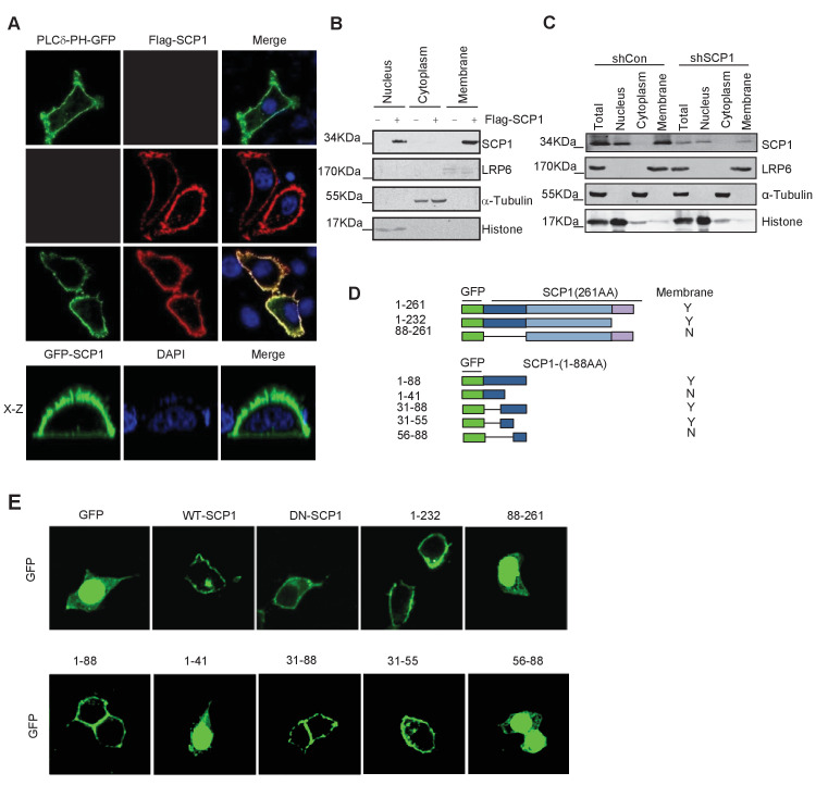
Figure 1. Membrane localization of SCP1.

(A) SCP1 was co-localized with PLCδ-PH on the cell membrane. FLAG-SCP1 was transfected with or without PLCδ-PH-GFP in HeLa. The subcellular localization of SCP1 and PLCδ-PH-GFP was analyzed using immunofluorescence assay, and both the horizontal section (X–Y) and vertical section (X–Z) were photographed. (B) and (C) Subcellular localization of SCP1 in cells. HEK-293T cells were transfected and the subcellular localizations of transfected SCP1 (B) or endogenous SCP1 (C) were analyzed using western blotting. (D) Cartoon of different deletion mutations of SCP1. Yes (Y) and no (N) represent SCP1 or truncated mutant membrane localizations, respectively. (E) HeLa cells were transfected with GFP-SCP1 or its mutants for 24 h and then analyzed for their subcellular localization using immunofluorescence assays.

DOI: http://dx.doi.org/10.7554/eLife.22058.003

Figure 1.

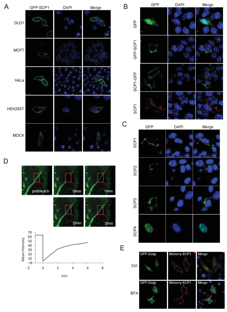
Figure 1—figure supplement 1. SCP1 is membrane localized.

Figure 1—figure supplement 1.

(A) SCP1 was localized on the cell membrane in various cells. SCP1 was expressed in DLD1, MCF7, HeLa, HEK293T, or MCDK cells and the subcellular localization of SCP1 was analyzed using immunofluorescence assay. (B) HeLa cells were transfected and the subcellular localization of SCP1 was analyzed using immunofluorescence assay. (C) SCP1, SCP2, and SCP3, but not SCP4, were localized on the plasma membrane. HeLa cells were transfected with GFP-SCP1/SCP2/SCP3/SCP4, respectively. (D) Membrane-localized SCP1 was photobleached (as showed in the red frame), and the fluorescence was recovered for 3 min as indicated. (E) The SCP1 membrane distribution was not affected by the disruption of the Golgi. HeLa cells were transfected with GFP-Golgi and mCherry-SCP1, respectively,and treated with DMSO or brefeldin A (BFA; 5 μg/ml) for 6 h.