Abstract
In the adult central nervous system (CNS) of higher vertebrates lesioned axons seemed unable to regenerate and reach their former target regions due to influences of the CNS microenvironment. Evidence from in vitro and biochemical experiments has demonstrated the presence of inhibitory substrate components in CNS tissue, in particular in white matter. These CNS components, which strongly inhibit neurite growth, were identified as minor membrane proteins of defined molecular mass (35 and 250 kDa) in oligodendrocyte membranes and CNS myelin. Oligodendrocyte development and myelin formation can be prevented by x-irradiation of newborn rats. Here we show that in myelin-free spinal cords cortico-spinal tract fibers transected at 2 weeks of age show reelongation of many millimeters within 2-3 weeks after the lesion. In normally myelinated controls, regenerative sprouts grew less than 1.7 mm caudal to the lesion.
Full text
PDF


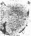
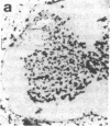
Images in this article
Selected References
These references are in PubMed. This may not be the complete list of references from this article.
- Bernstein D. R., Stelzner D. J. Plasticity of the corticospinal tract following midthoracic spinal injury in the postnatal rat. J Comp Neurol. 1983 Dec 20;221(4):382–400. doi: 10.1002/cne.902210403. [DOI] [PubMed] [Google Scholar]
- Black J. A., Waxman S. G., Ransom B. R., Feliciano M. D. A quantitative study of developing axons and glia following altered gliogenesis in rat optic nerve. Brain Res. 1986 Aug 13;380(1):122–135. doi: 10.1016/0006-8993(86)91436-8. [DOI] [PubMed] [Google Scholar]
- Borgens R. B., Blight A. R., Murphy D. J. Axonal regeneration in spinal cord injury: a perspective and new technique. J Comp Neurol. 1986 Aug 8;250(2):157–167. doi: 10.1002/cne.902500203. [DOI] [PubMed] [Google Scholar]
- Carbonetto S., Evans D., Cochard P. Nerve fiber growth in culture on tissue substrata from central and peripheral nervous systems. J Neurosci. 1987 Feb;7(2):610–620. doi: 10.1523/JNEUROSCI.07-02-00610.1987. [DOI] [PMC free article] [PubMed] [Google Scholar]
- Caroni P., Schwab M. E. Antibody against myelin-associated inhibitor of neurite growth neutralizes nonpermissive substrate properties of CNS white matter. Neuron. 1988 Mar;1(1):85–96. doi: 10.1016/0896-6273(88)90212-7. [DOI] [PubMed] [Google Scholar]
- Caroni P., Schwab M. E. Two membrane protein fractions from rat central myelin with inhibitory properties for neurite growth and fibroblast spreading. J Cell Biol. 1988 Apr;106(4):1281–1288. doi: 10.1083/jcb.106.4.1281. [DOI] [PMC free article] [PubMed] [Google Scholar]
- Casale E. J., Light A. R., Rustioni A. Direct projection of the corticospinal tract to the superficial laminae of the spinal cord in the rat. J Comp Neurol. 1988 Dec 8;278(2):275–286. doi: 10.1002/cne.902780210. [DOI] [PubMed] [Google Scholar]
- Crutcher K. A. Tissue sections from the mature rat brain and spinal cord as substrates for neurite outgrowth in vitro: extensive growth on gray matter but little growth on white matter. Exp Neurol. 1989 Apr;104(1):39–54. doi: 10.1016/0014-4886(89)90007-1. [DOI] [PubMed] [Google Scholar]
- David S., Aguayo A. J. Axonal elongation into peripheral nervous system "bridges" after central nervous system injury in adult rats. Science. 1981 Nov 20;214(4523):931–933. doi: 10.1126/science.6171034. [DOI] [PubMed] [Google Scholar]
- GILMORE S. A. The effects of x-irradiation on the spinal cords of neonatal rats. I. Neurological observations. J Neuropathol Exp Neurol. 1963 Apr;22:285–293. doi: 10.1097/00005072-196304000-00007. [DOI] [PubMed] [Google Scholar]
- GILMORE S. A. The effects of x-irradiation on the spinal cords of neonatal rats. II. Histological observations. J Neuropathol Exp Neurol. 1963 Apr;22:294–301. doi: 10.1097/00005072-196304000-00008. [DOI] [PubMed] [Google Scholar]
- Gribnau A. A., de Kort E. J., Dederen P. J., Nieuwenhuys R. On the development of the pyramidal tract in the rat. II. An anterograde tracer study of the outgrowth of the corticospinal fibers. Anat Embryol (Berl) 1986;175(1):101–110. doi: 10.1007/BF00315460. [DOI] [PubMed] [Google Scholar]
- Joosten E. A., Gribnau A. A., Dederen P. J. An anterograde tracer study of the developing corticospinal tract in the rat: three components. Brain Res. 1987 Nov;433(1):121–130. doi: 10.1016/0165-3806(87)90070-8. [DOI] [PubMed] [Google Scholar]
- Kalil K., Reh T. A light and electron microscopic study of regrowing pyramidal tract fibers. J Comp Neurol. 1982 Nov 1;211(3):265–275. doi: 10.1002/cne.902110305. [DOI] [PubMed] [Google Scholar]
- Matthews M. A., Duncan D. A quantitative study of morphological changes accompanying the initiation and progress of myelin production in the dorsal funiculus of the rat spinal cord. J Comp Neurol. 1971 May;142(1):1–22. doi: 10.1002/cne.901420102. [DOI] [PubMed] [Google Scholar]
- Mesulam M. M. Tetramethyl benzidine for horseradish peroxidase neurohistochemistry: a non-carcinogenic blue reaction product with superior sensitivity for visualizing neural afferents and efferents. J Histochem Cytochem. 1978 Feb;26(2):106–117. doi: 10.1177/26.2.24068. [DOI] [PubMed] [Google Scholar]
- Savio T., Schwab M. E. Rat CNS white matter, but not gray matter, is nonpermissive for neuronal cell adhesion and fiber outgrowth. J Neurosci. 1989 Apr;9(4):1126–1133. doi: 10.1523/JNEUROSCI.09-04-01126.1989. [DOI] [PMC free article] [PubMed] [Google Scholar]
- Schnell L., Schwab M. E. Axonal regeneration in the rat spinal cord produced by an antibody against myelin-associated neurite growth inhibitors. Nature. 1990 Jan 18;343(6255):269–272. doi: 10.1038/343269a0. [DOI] [PubMed] [Google Scholar]
- Schreyer D. J., Jones E. G. Growth and target finding by axons of the corticospinal tract in prenatal and postnatal rats. Neuroscience. 1982;7(8):1837–1853. doi: 10.1016/0306-4522(82)90001-x. [DOI] [PubMed] [Google Scholar]
- Schwab M. E., Caroni P. Oligodendrocytes and CNS myelin are nonpermissive substrates for neurite growth and fibroblast spreading in vitro. J Neurosci. 1988 Jul;8(7):2381–2393. doi: 10.1523/JNEUROSCI.08-07-02381.1988. [DOI] [PMC free article] [PubMed] [Google Scholar]
- Schwab M. E., Schnell L. Region-specific appearance of myelin constituents in the developing rat spinal cord. J Neurocytol. 1989 Apr;18(2):161–169. doi: 10.1007/BF01206659. [DOI] [PubMed] [Google Scholar]
- Schwab M. E., Thoenen H. Dissociated neurons regenerate into sciatic but not optic nerve explants in culture irrespective of neurotrophic factors. J Neurosci. 1985 Sep;5(9):2415–2423. doi: 10.1523/JNEUROSCI.05-09-02415.1985. [DOI] [PMC free article] [PubMed] [Google Scholar]
- Singh H., Pfeiffer S. E. Myelin-associated galactolipids in primary cultures from dissociated fetal rat brain: biosynthesis, accumulation, and cell surface expression. J Neurochem. 1985 Nov;45(5):1371–1381. doi: 10.1111/j.1471-4159.1985.tb07202.x. [DOI] [PubMed] [Google Scholar]
- Sommer I., Schachner M. Monoclonal antibodies (O1 to O4) to oligodendrocyte cell surfaces: an immunocytological study in the central nervous system. Dev Biol. 1981 Apr 30;83(2):311–327. doi: 10.1016/0012-1606(81)90477-2. [DOI] [PubMed] [Google Scholar]
- Tolbert D. L., Der T. Redirected growth of pyramidal tract axons following neonatal pyramidotomy in cats. J Comp Neurol. 1987 Jun 8;260(2):299–311. doi: 10.1002/cne.902600210. [DOI] [PubMed] [Google Scholar]
- Vidal-Sanz M., Bray G. M., Villegas-Pérez M. P., Thanos S., Aguayo A. J. Axonal regeneration and synapse formation in the superior colliculus by retinal ganglion cells in the adult rat. J Neurosci. 1987 Sep;7(9):2894–2909. doi: 10.1523/JNEUROSCI.07-09-02894.1987. [DOI] [PMC free article] [PubMed] [Google Scholar]


