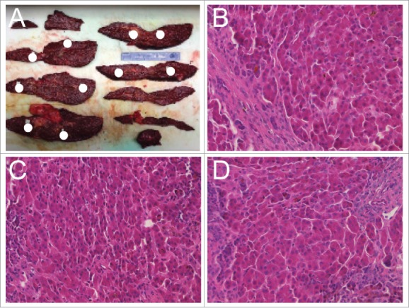
Figure 1.

Tissue dissection and sample pathologic characterization. (A) Liver dissection with coronal sections shown from case 2. Positions of nodules isolated for analysis throughout the liver are indicated with white circles. (B-D) Representative H&E staining for three nodules demonstrating overall similarity of regenerative nodules.
