Abstract
Wound healing complications associated with total knee arthroplasty present a considerable challenge to the orthopaedic surgeon. To ensure preservation of a functional joint, the management of periprosthetic soft-tissue defects around the knee requires rapid assessment, early and aggressive débridement, and durable, contoured coverage. Several reconstructive options are available to tailor soft-tissue coverage to the location, size, and depth of the wound. Special consideration should be given to the timing of the intervention, management of infection, and prosthesis salvage. The merits of each reconstructive option, including perforator, fasciocutaneous, muscular, and free microvascular flaps, should be weighed to select the most appropriate option. The proposed approach can guide surgeons in treating patients with these complex soft-tissue defects.
Keywords: flap, infection, knee arthroplasty, reconstruction, wound healing
Wound complications in patients who have undergone total knee arthroplasty (TKA) present a serious and common challenge for the orthopaedic surgeon. Infection, impaired wound healing, and soft-tissue compromise after TKA can result in devastating complications, including the need for removal of the prosthesis and/or loss of the limb.1 Annually, >600,000 primary TKA procedures are performed in the United States. The utilization of primary TKA is projected to increase substantially, to >1.5 million procedures per year by 2020.2 As the number of TKA procedures performed continues to increase, an associated increase in the overall number of complications is expected. In recent reports, the rate of infection or wound healing complications in patients who have undergone TKA ranges from 0.33% to 10.5%.1,3
Despite initial management, patients experiencing wound healing complications or infection are at increased risk of a subsequent deep wound infection and the need for additional surgical treatment within 2 years postoperatively, underscoring the negative effect of early wound healing complications on long-term outcomes.1,3,4 To ensure effective treatment of this increasing patient population, a renewed emphasis on accurate identification, classification, and treatment of wound complications after TKA is essential. The approach to the treatment of wound healing complications after TKA presented here was developed on the basis of the literature and the collaborative expertise of surgeons specializing in extremity reconstruction and joint arthroplasty. Optimal treatment of these patients requires careful evaluation of the surgical site; early, cooperative management by the arthroplasty and surgical reconstruction teams; and the use of a graded approach to the management of these complex wounds.
Risk Factors
Knowledge of the factors that place a patient at higher risk of surgical wound complications can guide management, allowing the surgeon to closely monitor at-risk patients and promptly detect any complications. Factors that increase the risk of wound complications after TKA can be systemic or local. Systemic risk factors include smoking, diabetes mellitus, increasing patient age, obesity, immunocompromised state, preexisting peripheral vascular disease, malnutrition, chronic renal insufficiency, and chemotherapy.4 Smoking, obesity, and malnutrition are particularly notable because they are associated with increased rates of infection and secondary surgery.4–7
Local risk factors include preexisting scars or dystrophic skin, prior surgery on the affected knee, and prior skin irradiation. In the immediate postoperative period, hematoma or superficial skin irradiation can increase the risk of serious wound complications.4 Existing incisions should be taken into account when the surgical approach is planned. Reusing preexisting longitudinal incisions is preferred. Because perfusion of the skin on the medial aspect of the knee is more robust than on the lateral side of the knee, the use of a lateral incision is recommended to avoid interruption of the primary dermal blood supply, which would place the lateral edges of the skin flap at greater risk of compromise.8
Other factors that influence the risk of wound complications include the choice of anticoagulation prophylaxis, the presence of prolonged postoperative wound drainage, early aggressive flexion of the knee, the use of a high-pressure tourniquet, and increased surgical times. Patients on anticoagulation therapy who undergo TKA are often administered higher perioperative doses of warfarin, which has been associated with substantial increases in the duration of wound drainage and the incidence of infection.9 Factors that influence tissue oxygenation have been linked to postoperative wound healing complications at the surgical site. The use of a high-pressure tourniquet decreases oxygenation and tissue perfusion at the wound site.10 Similarly, in a series of patients with notable flexion deformity who underwent TKA, an increased risk of necrosis of the skin edge at the incisional site in these patients was attributed to the severe ankylosis of the joint, which placed the skin flaps under increased tension when they were approximated over the anterior surface of the prosthesis.11 This finding emphasizes the importance of adequate, tension-free soft-tissue coverage over the prosthetic knee.
Assessment
In patients who have undergone TKA, prompt identification of delayed healing or infection is crucial to allow retention of the prosthesis. Consensus on standardization of periprosthetic wound evaluation has not been reached in the orthopaedic literature. Laing et al12 proposed a graded wound classification system; however, their system does not capture the complexity of soft-tissue defects after TKA because it does not address the presence of infection, the size of the wound, or characteristics of the tissue loss. Postoperative evaluation of patients who have undergone TKA should include serial assessments of the wound. If a delay in wound healing is observed, the surgeon should assess the area of soft-tissue loss and the depth of the wound, noting the presence and/or exposure of any bone, prosthesis, or cement in the wound. The surgeon should also note the presence or absence of erythema, purulent drainage, and a sinus tract.
Early identification of a periprosthetic infection is important and may influence the choice of initial treatment. Surface wound swabs of drainage are usually discouraged because they have high rates of contamination with skin microbiota and little correlation with deep infection.13 Joint aspiration around the prosthesis offers a better indication of deep infection than surface wound swabs provide. In the 6-week postoperative period, synovial white blood cell counts >27,800 cells/μL should raise a high index of suspicion for infection. In one study, the use of this parameter instead of the standard threshold of 3,000 cells/μL was associated with a positive predictive value of 94% and negative predictive value of 98%, resulting in a reduction in the number of surgical interventions that would have been performed in noninfected knees.14
Initial Intervention
After diagnosis of a wound healing complication has been reached, early and thorough débridement of any infected or devitalized tissue is crucial; all nonviable tissue must be excised. In patients with infection, the colonized soft tissue should be débrided in a similar manner. In patients with symptoms of infection, treatment consisting of irrigation, débridement, systemic antibiotic therapy, and immediate soft-tissue coverage has been associated with high rates of retention of the prosthesis.15–18 In patients with chronic deep periprosthetic infections, implant revision with long-term antibiotic therapy is recommended. Debate exists as to whether a single-stage revision or a two-stage revision with delayed reimplantation and the use of an antibiotic spacer or cement during the interval between stages leads to improved results.15 If a single-stage revision is chosen, flap coverage can be performed during the same procedure to augment blood flow to the region, increase local antibiotic delivery, and decrease bacterial proliferation. If a two-stage approach is chosen, flap coverage can be performed at the time of implant removal or can be delayed.19 Regardless of whether a single-stage or two-stage approach is chosen, coverage of the prosthesis depends on the often tenuous condition of the soft-tissue envelope after the wound has been débrided to healthy, bleeding tissue.
In patients who are at especially high risk of wound healing complications, prophylactic flap coverage has shown excellent outcomes in limited retrospective studies. Casey et al20 studied 23 patients who had prohibitive soft-tissue envelopes after TKA because of multiple scars, prior wound complications, or prior infections. These high-risk patients were treated with prophylactic flaps and were compared with 18 patients (19 knees) who underwent salvage flap procedures for infection, wound eschar, or wound dehiscence. All prostheses were retained in the group of patients who underwent prophylactic flap coverage, whereas 10 of the 19 prostheses in the salvage group required removal. Furthermore, 2 of the 19 limbs treated with salvage flap coverage ultimately required above-knee amputation.
Management Options
In patients with wound healing complications after TKA, the goal of soft-tissue reconstruction is to ensure definitive coverage of the prosthesis with thin, pliable, and durable tissue to facilitate joint function. An understanding of the arterial anatomy around the knee is crucial for appropriate planning. Perfusion of the skin surrounding the knee follows the angiosome concept, in which regions of skin and soft tissue are perfused by specific, longitudinally oriented source vessels.21 These perforator vessels originate deep to the fascia and have direct cutaneous, musculocutaneous, and septocutaneous branches.
Adjacent angiosomes are linked by so-called choke vessels, or intravascular anastomoses, which play an important role in perfusing the tissue around joints.21 This linkage allows for collateral blood supply to the soft tissue in the event of a wound healing complication. Because few muscles cross the knee joint, the soft tissues around the knee are supplied mostly by extramuscular vascular anastomoses among the genicular vessels.21 This peripatellar anastomotic ring is composed of the medial superior, medial inferior, lateral superior, lateral inferior, and descending genicular arteries and the anterior tibial recurrent artery.22 The medial-sided vessels provide a more dominant contribution, which should be taken into account during soft-tissue management. In patients with prior incisions and wound healing complications, perfusion to the soft tissue overlying the prosthesis may be compromised.
When planning reconstruction of soft-tissue defects, including defects occurring after TKA, the surgeon should consider the principle of the reconstructive ladder. This concept dictates that the simplest method of achieving wound closure (eg, primary closure) should be chosen when possible. However, the simplest method of closure with the most readily available tissue for reconstruction may not be optimal. Therefore, strict adherence to the principle of the reconstructive ladder is not advisable. Rather, the more modern “reconstructive elevator” principle is better suited to address wound defects after TKA. This concept allows ascension from the simplest to the most complex techniques, based on the specific characteristics of the wound and the soft-tissue coverage needed. It facilitates a comprehensive approach to treatment that focuses on early closure of the defect with stable soft-tissue coverage over the prosthesis while allowing for any future procedures the patient may require at the site of the wound.
Primary Closure and Healing by Secondary Intent
Primary closure is an option for the management of wounds with minimal necrosis or tissue loss if tension-free repair is possible after adequate débridement to healthy tissue. Primary closure is not advisable if the edges of the wound are not readily approximated or if closure would place skin flaps under tension. Wound healing by secondary intent occurs when the wound is allowed to granulate progressively from the edges with supportive dressing changes. This method is generally reserved for the management of small wounds over muscle or fascia that are not amenable to other treatment modalities because of the patient’s medical status. In most TKA patients, because of the increased risk of deep or periprosthetic infections associated with prolonged wound drainage, waiting for granulation of tissue to fill substantial defects is not advisable.
Negative-pressure Wound Therapy
Negative-pressure wound therapy (NPWT), originally developed to address chronic wounds refractory to nonsurgical management, has been explored more recently for the management of tissue defects associated with orthopaedic trauma and arthroplasty.23,24 NPWT provides a negative-pressure environment that continually removes fluid from the wound and provides constant, equal pressure across the entire exposed surface to promote epithelial migration from the edges toward the center of the wound. Immediate contraindications to the use of NPWT for the management of a wound after TKA include the need for placement of the NPWT device over necrotic tissue with eschar, exposed vessels, or exposed nerves.24 In other patients, NPWT remains an option for wound management after TKA and may be of value as a temporary measure for use before definitive management. However, it should not be used to delay definitive management or to manage wounds likely to require a prolonged time to heal. Prolonged NPWT is associated with increased rates of continual drainage and periprosthetic infection in addition to the previously mentioned risks associated with delayed wound coverage.24
Skin Grafts
Split-thickness skin grafts are a useful tool in the management of small defects that lack deep extension or evidence of infection. Skin grafts require a well-vascularized recipient site to oxygenate the grafted tissue. Both muscle and fascia are usually favorable recipient sites for graft survival. The role of skin grafts in the management of knee wounds is limited because skin grafts do not adequately eliminate dead space and because they limit future surgical procedures through the wound bed. Furthermore, split-thickness skin grafts have an associated risk of contracture inversely related to depth of the skin graft. Therefore, grafting across joint surfaces has the potential to limit long-term range of motion and negate some of the benefits of joint arthroplasty.
Flaps
Flaps can be classified according to the type of tissue to be transferred (eg, fasciocutaneous, muscle), the pattern of blood supply to the flap (eg, random, axial), the spatial relationship of the flap and the recipient site (eg, local, regional, distant), and the mechanism of transfer (eg, free, rotational, transposition). Flap selection for soft-tissue reconstruction after TKA is guided by the three-dimensional shape of the tissue defect and the location of the defect (Figure 1). Knowledge of the locations from which donor tissue can be harvested is important in determining the best option for wound coverage.
Figure 1.
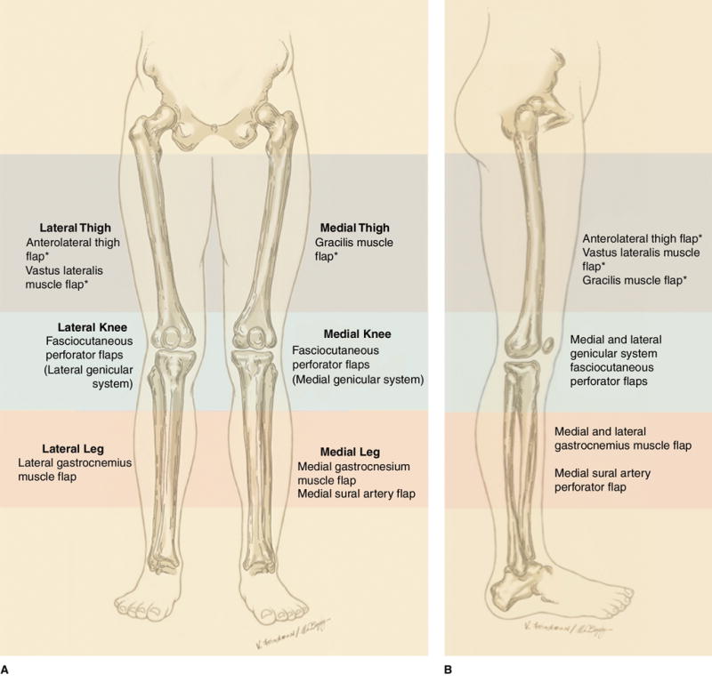
AP (A) and lateral (B) illustrations depicting standard flap donor sites for soft-tissue reconstruction of the knee after total knee arthroplasty. Asterisks indicate distally based flaps.
Local Flaps
Muscular and Musculocutaneous Local Flaps
Local muscular flaps are widely considered to be the workhorse flaps for coverage of soft-tissue defects surrounding the knee and have been discussed extensively in the literature.17,19,25 Muscle provides a well-vascularized flap with substantial bulk for elimination of dead space. Furthermore, muscular flaps may be advantageous in the management of periprosthetic infection because increased collagen deposition and greater inhibition and elimination of bacterial growth have been observed in chronically infected wounds covered by musculocutaneous flaps compared with those covered by fasciocutaneous flaps.26 These flaps can be especially useful in patients in whom additional surgical procedures on the knee are planned and have been associated with excellent outcomes in complex cases of infection or prosthetic exposure.17,19
For local coverage, the medial gastrocnemius muscle is most commonly used because of its reliability and ease of harvest. The medial head of the gastrocnemius is supplied by the medial sural artery and can be rotated to cover soft-tissue defects of the medial, anterior, and upper knee. The medial gastrocnemius flap ranges from 5 to 9 cm in width and from 13 to 20 cm in length. It is commonly used to cover wounds between 3 and 7 cm in width and with surface areas between 33 and 49 cm2.17,27 The flap can be easily rotated to cover medial and distal defects in the area of the tibial tubercle or patellar tendon. The arc of rotation can be further extended by 20% to 50% or 5 to 8 cm with dissection of the muscle origin and the pes anserinus19,25,27 (Figure 2). Division or excision of the superficial and deep fascia can extend the width and length of the muscle. This method may allow for coverage of a larger wound and facilitates healing of a skin graft to the underlying muscle (Figure 3). The lateral gastrocnemius muscle can be used to cover both lateral and anterior knee defects. Because of its smaller size (5 × 12 cm, on average) and obstructed anterior rotation caused by the fibula, the lateral gastrocnemius has a lesser arc of motion than the medial gastrocnemius and is used less frequently for coverage of a soft-tissue defect about the knee.17 In addition, careful dissection and decompression of the common peroneal nerve may be necessary; passage of the muscle deep to the nerve can prevent iatrogenic nerve compression (Figure 4). For the management of a larger wound centered over the patella and tibial tubercle, both the medial and lateral gastrocnemius muscles can be elevated and used for coverage of the wound.
Figure 2.
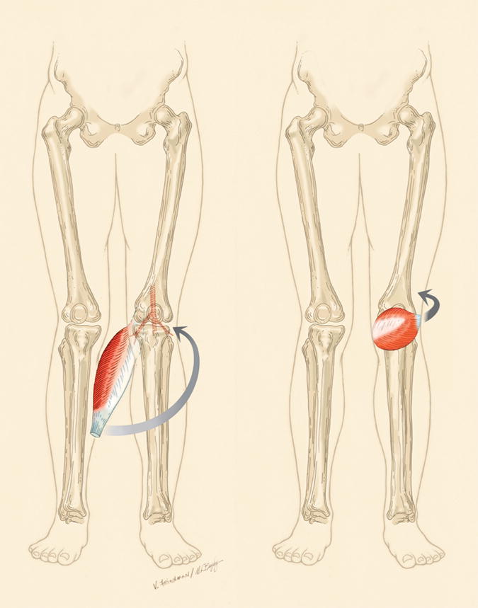
Illustrations depicting dissection of the medial gastrocnemius muscle and a small portion of the Achilles tendon away from the triceps surae complex. The muscle can be released up to its proximal blood supply to facilitate anterior rotation (arrows). Depending on the length and size of the muscle, the flap can be used for coverage of wounds as proximal as the superior pole of the patella or the distal quadriceps.
Figure 3.
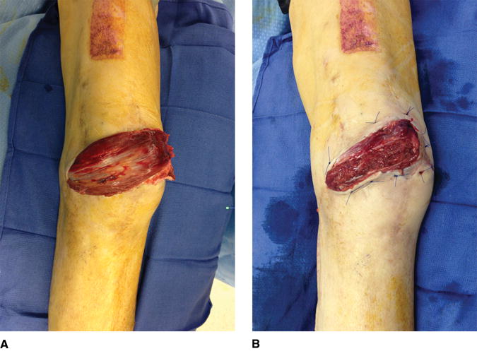
Intraoperative photographs demonstrating gastrocnemius flap coverage after revision total knee arthroplasty. The overlying fascia (A) is excised (B) to facilitate healing of a split-thickness skin graft (not pictured).
Figure 4.
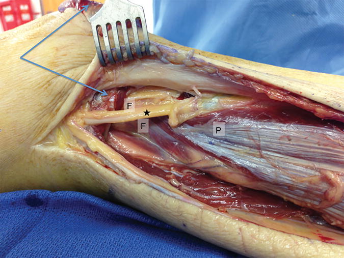
Intraoperative photograph depicting dissection before elevation of a lateral gastrocnemius flap. The common peroneal nerve is seen proximally (star). Decompression of the nerve as it passes around the fibular neck (F) into the peroneus longus interval (P) allows for passage of the lateral gastrocnemius deep to the nerve and into the recipient site on the anterior knee (blue arrows), which can prevent iatrogenic compression of the nerve.
In patients with larger or more proximal defects involving the anterior portion of the knee with deficiencies of the skin, anterior capsule, and quadriceps tendon, a gastrocnemius flap alone may be of insufficient size to provide coverage.17,19,28 These composite defects affecting the extensor mechanism can be managed with local transfer of the vastus lateralis muscle, in conjunction with the vastus medialis muscle and/or gastrocnemius muscle if necessary.28–30 The distally based vastus lateralis flap is based on the reverse flow in the anastomoses between the descending branch of the lateral circumflex femoral artery (LCFA) and the lateral superior genicular artery. The vastus lateralis muscle flap is 7 to 11 cm × 9 to 14 cm in size and can be used to cover defects of approximately 6 × 9 cm along the anterior proximal knee.30
Another locoregional muscle option for coverage of proximal and/or lateral knee wounds is the distally based pedicled gracilis flap, which is based on minor pedicles from the superficial femoral or popliteal artery that pass between the sartorius and adductor longus muscles31 (Figure 5). This flap carries a high risk of partial flap loss and is reserved for use in scenarios in which a pedicled gastrocnemius flap would be inadequate and when the patient is not a suitable candidate for a free flap.31
Figure 5.
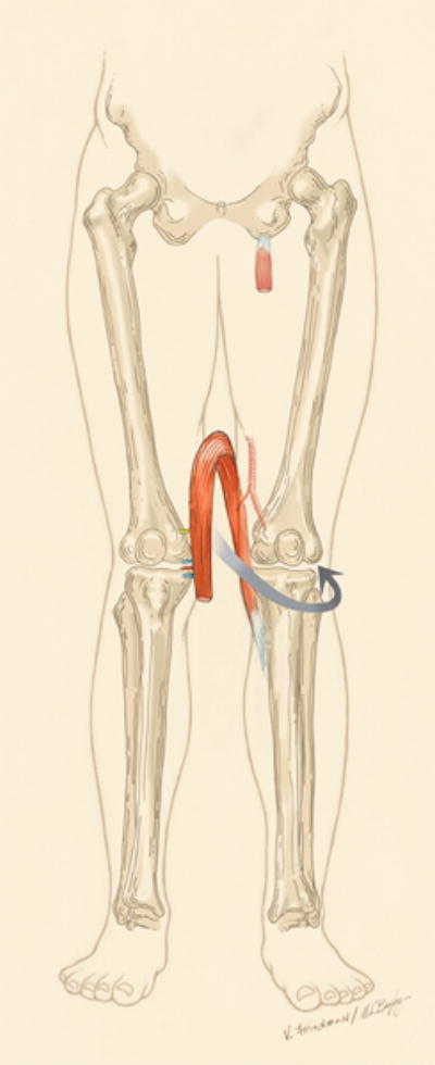
Illustration depicting a distally based gracilis flap, which can be useful for coverage of proximal wounds after total knee arthroplasty or as additional proximal coverage if a gastrocnemius flap is insufficient for wound coverage. The vascularity of the flap is based on intact flow at the distal extent of the muscle. The dominant arterial supply is ligated along with the branch of the obturator nerve that innervates the muscle.
Fasciocutaneous and Perforator Local Flaps
Fasciocutaneous flaps consist of skin, subcutaneous tissue, and underlying fascia. The vascular supply is derived from perforating arteries originating from longitudinally oriented source arteries that initially course under the deep fascia or in the intermuscular septum between adjacent muscles. These arteries provide multidirectional perforator vessels at the level of the deep fascia that form fascial plexuses, which perfuse the skin through a confluence of multiple vascular anastomoses at the subfascial, suprafascial, subcutaneous, and subdermal levels.32 Fasciocutaneous flaps are helpful in reconstructing soft-tissue defects around the knee because they are thin, pliable, and easy to contour to the shape of the defect. Because arterial perforators radiate in a stellate, multidirectional pattern throughout the fascial plexus, distally based flaps can be used (Figure 6). Historically, fasciocutaneous flaps have been used to manage incisional necrosis after TKA in patients without underlying infection or an exposed prosthesis. Flaps that are based proximally, along the axially oriented pattern of blood supply, preserve cutaneous innervation and have been advocated for coverage of areas of skin necrosis.17,32 Similarly, the use of unilateral or bilateral local fasciocutaneous flaps in a V-Y pattern allows soft-tissue advancement for closure of wounds 2 to 4 cm × 5 to 12 cm in size.33
Figure 6.
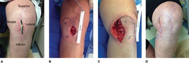
Photographs depicting the use of a fasciocutaneous rotational flap. A, Clinical photograph depicting a superficial infection and wound drainage after total knee arthroplasty in a 52-year-old woman. An attempt to irrigate, débride, and revise the primary closure was unsuccessful. B, Intraoperative photograph depicting markings on the patient’s skin to indicate the planned incisions for a distally based peninsular-type fasciocutaneous perforator flap based on the cutaneous branch of the descending genicular artery. C, Intraoperative photograph depicting the patient’s knee after the flap was elevated and transposed to close the midline knee wound. D, Postoperative clinical photograph obtained at 10 weeks depicting the patient’s knee. In many patients, the donor site can be closed, but in this patient, the flap was too large to allow for tension-free closure. Therefore, a small split-thickness skin graft (arrow) was used to close the donor site.
The use of perforator-based flaps has revolutionized the reconstruction of complex lower extremity wounds, including soft-tissue defects around the knee. Each clinically relevant perforator vessel supplies a so-called perforasome, or vascular territory of three-dimensional tissue. These territories are linked to adjacent territories both directly (through large vessels that communicate directly between perforators) and indirectly (in the form of recurrent flow through the subdermal plexus).34 Pedicled perforator flaps can be customized to the defect, minimize donor site morbidity, and allow coverage with tissue similar to that of the recipient site in adherence to the “like with like” principle of tissue replacement.
The perforator concept is extended in the use of propeller flaps, in which a local island of tissue is axially rotated 90° to 180° after dissection has been performed along the length of the pedicle. The design of these flaps and the orientation of the skin paddle take advantage of the axial direction of the linking vessels. A handheld Doppler ultrasonographic device is routinely used to locate perforators before the flap is harvested. Thigh perforator flaps have been increasingly used for coverage of large composite defects surrounding the knee. For the management of suprapatellar anterior or lateral defects, the distally based or reverse-flow anterolateral thigh (ALT) flap or the lateral supragenicular perforator flap can be used. The distal ALT flap is based on constant skin perfusion through retrograde blood flow between the descending branch of the LCFA and the lateral superior genicular artery and/or the deep femoral artery.35 The pivot point is 3 to 10 cm proximal to the superolateral patella, and the dominant perforator, which is commonly found in the mid thigh, is mobilized to its origin from the descending branch to yield a pedicle of 15 to 28 cm in length.35,36 A cuff of vastus lateralis muscle can be included to protect the pedicle, and flaps up to 10 × 16 cm in size can be used to cover wound defects ranging from 6 to 10 cm × 8 to 16 cm on the proximal anterior or lateral knee.35,36 The lateral supragenicular perforator flap is based on the lateral supragenicular perforator, which arises from the superior lateral genicular artery within 5 cm lateral and 7 cm proximal to the superolateral patella.37 The width of this flap is typically limited to 4 to 6 cm to allow primary closure of the donor site, whereas the length can be up to 18 cm.38
The distally based anteromedial thigh flap is useful for covering proximal anterior or medial knee wounds. This flap is based on a supragenicular perforator from the saphenous branch of the descending genicular artery that commonly emerges from the anterior margin of the sartorius muscle within 3 cm of the adductor tubercle.39 Flaps of 6 to 10 cm × 15 to 32 cm have been harvested to cover large tissue defects of 6 to 10 cm × 15 to 27 cm.39 Disadvantages of distally based or reverse-flow flaps are the increased risk of venous congestion and flap tip necrosis, the technical difficulty of perforator dissection, and the need to perform skin grafting at the donor site if primary closure is not possible.25 Proximally based perforator flaps based on the medial sural artery can be used to cover medial and distal knee defects with the advantage of less donor site morbidity than would result from the use of a gastrocnemius muscle flap. This flap can be 3 to 7 cm × 3 to 16 cm in size, have a rotation arc of 8 to 12 cm, and can be reliably harvested on the basis of a perforator located 8 cm distal to the popliteal crease on a line connecting the midpoint of the crease to the midpoint of the medial malleolus.40
Microvascular Free Flaps
Microvascular free tissue transfer consists of raising a donor flap from a site distant to the defect, isolating the vascular pedicle of the flap, and transferring the flap to a suitable recipient vascular pedicle by performing vascular anastomoses to restore blood flow. Free flap reconstruction has substantial utility in the salvage of complex knee wounds with large composite tissue defects, infection, and/or prosthesis exposure. Free flaps can be used when no suitable local options are present. Selection of the flap is based on assessment of the characteristics of the recipient site, the dimensions of the wound, associated morbidity of the donor site, the extent of the vascular pedicle, and the available recipient vessels around the knee. Microvascular tissue transfer requires considerable surgical expertise and is integral in the armamentarium of the reconstructive surgeon. Free flap reconstruction of knee defects can be complicated by vascular compromise, which may require further exploration in up to 20% of patients.41 A variety of recipient vessels can be used, and selection of appropriate recipient vessels is critical to success. Ideally, recipient vessels are well matched in diameter with the donor pedicle, are easily dissected, allow tension-free vascular anastomosis without kinking of the pedicle, and are located outside the zone of injury. The popliteal, geniculate, sural, deep femoral and superficial femoral arteries, and the descending branch of the LCFA have all been used, with the choice of artery depending on the location of the knee defect.42 Most recently, the descending genicular artery has emerged as a primary recipient option in the management of most knee defects because of its consistent location, the ease of dissection, the excellent size match for end-to-end anastomosis with most free flap donor pedicles, and the ability to avoid intraoperative position changes.43
Muscular and Musculocutaneous Free Flaps
The most common muscles used for free flap transfer to the knee are the latissimus dorsi and rectus abdominis muscles. In infected wounds with an exposed prosthesis, free muscle transfer augments the blood supply to the wound and improves the local immunologic environment. Muscular flaps also offer the ability to eliminate dead space. The latissimus dorsi free flap and the rectus abdominis free flap have both been used for coverage of soft-tissue defects with excellent limb salvage rates (>90%) in infected knees with an exposed prosthesis after TKA.16,18 The latissimus dorsi free flap can be harvested with a muscle dimension of 20 × 35 cm and with an overlying elliptic skin paddle of 9 × 25 cm that allows primary closure of the donor site. It is raised on a vascular pedicle containing the thoracodorsal artery, which allows for a length of 7 to 12 cm.18,44 The latissimus dorsi flap has a high rate of donor site complications, including wound dehiscence, seroma, and functional morbidity limiting overhead arm motion. The rectus abdominis muscle can be harvested with a transverse or vertical skin paddle. It is typically supplied by the deep inferior epigastric artery or the superior epigastric artery; however, when it is harvested as a free flap, the deep inferior epigastric artery is most commonly used. The flap can be 10 × 30 cm with a pedicle length of 5 to 7 cm.18,25 Because abdominal wall strength can be diminished after harvest of the rectus abdominis muscle, these flaps are not considered first-line treatment options.
Fasciocutaneous and Perforator Free Flaps
Muscles used in free tissue transfers are denervated during flap harvest, which can result in atrophy and fibrosis. To address these limitations and the donor site morbidity associated with muscle harvest, some authors have advocated the use of free adipocutaneous or fasciocutaneous flaps based on isolated perforators.16 The free ALT perforator flap is the most commonly used fasciocutaneous free flap for reconstruction of defects surrounding the knee. The pedicle of the ALT flap includes the descending branch of the LCFA. A chimeric flap incorporating vastus lateralis muscle or tensor fascia lata can be used if increased bulk is necessary to eliminate dead space.45 Compared with free myocutaneous flaps, fasciocutaneous or perforator free flaps offer the advantages of reduced donor site morbidity and improved contouring of the flap to the dimensions of the defect. Furthermore, perforator dissection increases the length of the pedicle, increasing the surgeon’s options in the choice of recipient vessel.
Summary
Soft-tissue coverage is crucial for prosthesis retention after TKA. Numerous options are available for the management of soft-tissue defects. An approach based on assessment of the characteristics of the patient and the wound can help guide treatment decisions. Early identification of a wound healing complication and prompt management with interdisciplinary cooperation among joint arthroplasty and reconstructive surgeons is essential to ensure optimal outcomes after TKA.
Acknowledgments
The authors thank Denis Nam, MD, MS, for his substantial contributions to this manuscript.
Dr. Osei acknowledges support by the Institute of Clinical and Translational Sciences Award program of the National Center for Advancing Translational Sciences at the National Institutes of Health (UL1 TR000448, KL2TR000450).
References
Evidence-based Medicine: Levels of evidence are described in the table of contents. In this article, references 10 and 23 are level I studies. Reference 6, 13, and 14 are level II studies. References 1, 3, 5, 9, and 12 are level III studies. References 7, 11, 16–20, 28–33, 35–39, and 41–45 are level IV studies. References 4, 8, 15, 24, and 25 are level V expert opinion.
References printed in bold type are those published within the past 5 years.
- 1.Galat DD, McGovern SC, Larson DR, Harrington JR, Hanssen AD, Clarke HD. Surgical treatment of early wound complications following primary total knee arthroplasty. J Bone Joint Surg Am. 2009;91(1):48–54. doi: 10.2106/JBJS.G.01371. [DOI] [PubMed] [Google Scholar]
- 2.Kurtz SM, Ong KL, Lau E, Bozic KJ. Impact of the economic downturn on total joint replacement demand in the United States: Updated projections to 2021. J Bone Joint Surg Am. 2014;96(8):624–630. doi: 10.2106/JBJS.M.00285. [DOI] [PubMed] [Google Scholar]
- 3.Gaine WJ, Ramamohan NA, Hussein NA, Hullin MG, McCreath SW. Wound infection in hip and knee arthroplasty. J Bone Joint Surg Br. 2000;82(4):561–565. doi: 10.1302/0301-620x.82b4.10305. [DOI] [PubMed] [Google Scholar]
- 4.Jones RE, Russell RD, Huo MH. Wound healing in total joint replacement. Bone Joint J. 2013;95-B(11 suppl A):144–147. doi: 10.1302/0301-620X.95B11.32836. [DOI] [PubMed] [Google Scholar]
- 5.Kapadia BH, Johnson AJ, Naziri Q, Mont MA, Delanois RE, Bonutti PM. Increased revision rates after total knee arthroplasty in patients who smoke. J Arthroplasty. 2012;27(9):1690–1695.e1. doi: 10.1016/j.arth.2012.03.057. [DOI] [PubMed] [Google Scholar]
- 6.Liabaud B, Patrick DA, Jr, Geller JA. Higher body mass index leads to longer operative time in total knee arthroplasty. J Arthroplasty. 2013;28(4):563–565. doi: 10.1016/j.arth.2012.07.037. [DOI] [PubMed] [Google Scholar]
- 7.Belmont PJ, Jr, Goodman GP, Waterman BR, Bader JO, Schoenfeld AJ. Thirty-day postoperative complications and mortality following total knee arthroplasty: Incidence and risk factors among a national sample of 15,321 patients. J Bone Joint Surg Am. 2014;96(1):20–26. doi: 10.2106/JBJS.M.00018. [DOI] [PubMed] [Google Scholar]
- 8.Vince KG, Abdeen A. Wound problems in total knee arthroplasty. Clin Orthop Relat Res. 2006;452:88–90. doi: 10.1097/01.blo.0000238821.71271.cc. [DOI] [PubMed] [Google Scholar]
- 9.Simpson PM, Brew CJ, Whitehouse SL, Crawford RW, Donnelly BJ. Complications of perioperative warfarin therapy in total knee arthroplasty. J Arthroplasty. 2014;29(2):320–324. doi: 10.1016/j.arth.2012.11.003. [DOI] [PubMed] [Google Scholar]
- 10.Zhang W, Li N, Chen S, Tan Y, Al-Aidaros M, Chen L. The effects of a tourniquet used in total knee arthroplasty: A meta-analysis. J Orthop Surg Res. 2014;9(1):13. doi: 10.1186/1749-799X-9-13. [DOI] [PMC free article] [PubMed] [Google Scholar]
- 11.Kim YH, Cho SH, Kim JS. Total knee arthroplasty in bony ankylosis in gross flexion. J Bone Joint Surg Br. 1999;81(2):296–300. doi: 10.1302/0301-620x.81b2.9183. [DOI] [PubMed] [Google Scholar]
- 12.Laing JH, Hancock K, Harrison DH. The exposed total knee replacement prosthesis: A new classification and treatment algorithm. Br J Plast Surg. 1992;45(1):66–69. doi: 10.1016/0007-1226(92)90120-m. [DOI] [PubMed] [Google Scholar]
- 13.Tetreault MW, Wetters NG, Aggarwal VK, et al. Should draining wounds and sinuses associated with hip and knee arthroplasties be cultured? J Arthroplasty. 2013;28(8 suppl):133–136. doi: 10.1016/j.arth.2013.04.057. [DOI] [PubMed] [Google Scholar]
- 14.Bedair H, Ting N, Jacovides C, et al. Diagnosis of early postoperative TKA infection using synovial fluid analysis. Clin Orthop Relat Res. 2011;469(1):34–40. doi: 10.1007/s11999-010-1433-2. [DOI] [PMC free article] [PubMed] [Google Scholar]
- 15.Moyad TF, Thornhill T, Estok D. Evaluation and management of the infected total hip and knee. Orthopedics. 2008;31(6):581–588. doi: 10.3928/01477447-20080601-22. quiz 589–590. [DOI] [PubMed] [Google Scholar]
- 16.Nahabedian MY, Orlando JC, Delanois RE, Mont MA, Hungerford DS. Salvage procedures for complex soft tissue defects of the knee. Clin Orthop Relat Res. 1998;356:119–124. doi: 10.1097/00003086-199811000-00017. [DOI] [PubMed] [Google Scholar]
- 17.Menderes A, Demirdover C, Yilmaz M, Vayvada H, Barutcu A. Reconstruction of soft tissue defects following total knee arthroplasty. Knee. 2002;9(3):215–219. doi: 10.1016/s0968-0160(02)00010-8. [DOI] [PubMed] [Google Scholar]
- 18.Cetrulo CL, Jr, Shiba T, Friel MT, et al. Management of exposed total knee prostheses with microvascular tissue transfer. Microsurgery. 2008;28(8):617–622. doi: 10.1002/micr.20578. [DOI] [PubMed] [Google Scholar]
- 19.Ries MD, Bozic KJ. Medial gastrocnemius flap coverage for treatment of skin necrosis after total knee arthroplasty. Clin Orthop Relat Res. 2006;446:186–192. doi: 10.1097/01.blo.0000218723.21720.51. [DOI] [PubMed] [Google Scholar]
- 20.Casey WJ, III, Rebecca AM, Krochmal DJ, et al. Prophylactic flap reconstruction of the knee prior to total knee arthroplasty in high-risk patients. Ann Plast Surg. 2011;66(4):381–387. doi: 10.1097/SAP.0b013e3181e37c04. [DOI] [PubMed] [Google Scholar]
- 21.Taylor GI, Pan WR. Angiosomes of the leg: Anatomic study and clinical implications. Plast Reconstr Surg. 1998;102(3):599–618. [PubMed] [Google Scholar]
- 22.Lazaro LE, Cross MB, Lorich DG. Vascular anatomy of the patella: Implications for total knee arthroplasty surgical approaches. Knee. 2014;21(3):655–660. doi: 10.1016/j.knee.2014.03.005. [DOI] [PubMed] [Google Scholar]
- 23.Howell RD, Hadley S, Strauss E, Pelham FR. Blister formation with negative pressure dressings after total knee arthroplasty. Curr Orthop Pract. 2011;22(2):176–179. [Google Scholar]
- 24.Harvin WH, Stannard JP. Negative-pressure wound therapy in acute traumatic and surgical wounds in orthopaedics. JBJS Rev. 2014;2(4):e4. doi: 10.2106/JBJS.RVW.M.00087. [DOI] [PubMed] [Google Scholar]
- 25.Andres LA, Casey WJ, Clarke HD. Techniques in soft tissue coverage around the knee. Tech Knee Surg. 2009;8:119–125. [Google Scholar]
- 26.Calderon W, Chang N, Mathes SJ. Comparison of the effect of bacterial inoculation in musculocutaneous and fasciocutaneous flaps. Plast Reconstr Surg. 1986;77(5):785–794. doi: 10.1097/00006534-198605000-00016. [DOI] [PubMed] [Google Scholar]
- 27.Veber M, Vaz G, Braye F, et al. Anatomical study of the medial gastrocnemius muscle flap: A quantitative assessment of the arc of rotation. Plast Reconstr Surg. 2011;128(1):181–187. doi: 10.1097/PRS.0b013e318217423f. [DOI] [PubMed] [Google Scholar]
- 28.Auregan JC, Bégué T, Tomeno B, Masquelet AC. Distally-based vastus lateralis muscle flap: A salvage alternative to address complex soft tissue defects around the knee. Orthop Traumatol Surg Res. 2010;96(2):180–184. doi: 10.1016/j.rcot.2010.02.013. [DOI] [PubMed] [Google Scholar]
- 29.Whiteside LA. Surgical technique: Vastus medialis and vastus lateralis as flap transfer for knee extensor mechanism deficiency. Clin Orthop Relat Res. 2013;471(1):221–230. doi: 10.1007/s11999-012-2532-z. [DOI] [PMC free article] [PubMed] [Google Scholar]
- 30.Sahasrabudhe P, Panse N, Baheti B, Jadhav A, Joshi N, Chandanwale A. Reconstruction of complex soft-tissue defects around the knee joint with distally based split vastus lateralis musculocutaneous flap: A new technique. J Plast Reconstr Aesthet Surg. 2015;68(1):35–39. doi: 10.1016/j.bjps.2014.09.034. [DOI] [PubMed] [Google Scholar]
- 31.Mitsala G, Varey AH, O’Neill JK, Chapman TW, Khan U. The distally pedicled gracilis flap for salvage of complex knee wounds. Injury. 2014;45(11):1776–1781. doi: 10.1016/j.injury.2014.06.019. [DOI] [PubMed] [Google Scholar]
- 32.Hallock GG. Salvage of total knee arthroplasty with local fasciocutaneous flaps. J Bone Joint Surg Am. 1990;72(8):1236–1239. [PubMed] [Google Scholar]
- 33.Papaioannou K, Lallos S, Mavrogenis A, Vasiliadis E, Savvidou O, Efstathopoulos N. Unilateral or bilateral V-Y fasciocutaneous flaps for the coverage of soft tissue defects following total knee arthroplasty. J Orthop Surg Res. 2010;5:82. doi: 10.1186/1749-799X-5-82. [DOI] [PMC free article] [PubMed] [Google Scholar]
- 34.Saint-Cyr M, Wong C, Schaverien M, Mojallal A, Rohrich RJ. The perforasome theory: Vascular anatomy and clinical implications. Plast Reconstr Surg. 2009;124(5):1529–1544. doi: 10.1097/PRS.0b013e3181b98a6c. [DOI] [PubMed] [Google Scholar]
- 35.Pan SC, Yu JC, Shieh SJ, Lee JW, Huang BM, Chiu HY. Distally based anterolateral thigh flap: An anatomic and clinical study. Plast Reconstr Surg. 2004;114(7):1768–1775. doi: 10.1097/01.prs.0000142416.91524.4c. [DOI] [PubMed] [Google Scholar]
- 36.Demirseren ME, Efendioglu K, Demiralp CO, Kilicarslan K, Akkaya H. Clinical experience with a reverse-flow anterolateral thigh perforator flap for the reconstruction of soft-tissue defects of the knee and proximal lower leg. J Plast Reconstr Aesthet Surg. 2011;64(12):1613–1620. doi: 10.1016/j.bjps.2011.06.047. [DOI] [PubMed] [Google Scholar]
- 37.Nguyen AT, Wong C, Mojallal A, Saint-Cyr M. Lateral supragenicular pedicle perforator flap: Clinical results and vascular anatomy. J Plast Reconstr Aesthet Surg. 2011;64(3):381–385. doi: 10.1016/j.bjps.2010.05.029. [DOI] [PubMed] [Google Scholar]
- 38.Wiedner M, Koch H, Scharnagl E. The superior lateral genicular artery flap for soft-tissue reconstruction around the knee: Clinical experience and review of the literature. Ann Plast Surg. 2011;66(4):388–392. doi: 10.1097/SAP.0b013e3181e37627. [DOI] [PubMed] [Google Scholar]
- 39.Lu LJ, Gong X, Cui JL, Liu B. The anteromedial thigh fasciocutaneous flap pedicled on the supragenicular septocutaneous perforator: Application in 11 patients. Ann Plast Surg. 2011;67(3):275–278. doi: 10.1097/SAP.0b013e3181f89151. [DOI] [PubMed] [Google Scholar]
- 40.Shim JS, Kim HH. A novel reconstruction technique for the knee and upper one third of lower leg. J Plast Reconstr Aesthet Surg. 2006;59(9):919–926. doi: 10.1016/j.bjps.2006.01.025. discussion 927. [DOI] [PubMed] [Google Scholar]
- 41.Louer CR, Garcia RM, Earle SA, Hollenbeck ST, Erdmann D, Levin LS. Free flap reconstruction of the knee: An outcome study of 34 cases. Ann Plast Surg. 2015;74(1):57–63. doi: 10.1097/SAP.0b013e31828d7558. [DOI] [PubMed] [Google Scholar]
- 42.Fang T, Zhang EW, Lineaweaver WC, Zhang F. Recipient vessels in the free flap reconstruction around the knee. Ann Plast Surg. 2013;71(4):429–433. doi: 10.1097/SAP.0b013e31824e5e6e. [DOI] [PubMed] [Google Scholar]
- 43.Venkatramani H, Sabapathy SR, Nayak S. Free-flap cover of complex defects around the knee using the descending genicular artery as the recipient pedicle. J Plast Reconstr Aesthet Surg. 2014;67(1):93–98. doi: 10.1016/j.bjps.2013.09.011. [DOI] [PubMed] [Google Scholar]
- 44.Hierner R, Reynders-Frederix P, Bellemans J, Stuyck J, Peeters W. Free myocutaneous latissimus dorsi flap transfer in total knee arthroplasty. J Plast Reconstr Aesthet Surg. 2009;62(12):1692–1700. doi: 10.1016/j.bjps.2008.07.038. [DOI] [PubMed] [Google Scholar]
- 45.Kuo YR, An PC, Kuo MH, Kueh NS, Yao SF, Jeng SF. Reconstruction of knee joint soft tissue and patellar tendon defects using a composite anterolateral thigh flap with vascularized fascia lata. J Plast Reconstr Aesthet Surg. 2008;61(2):195–199. doi: 10.1016/j.bjps.2006.06.012. [DOI] [PubMed] [Google Scholar]


