Abstract
Pilocytic astrocytoma (PA) is the most common pediatric brain tumor. A recurrent feature of PA is deregulation of the mitogen activated protein kinase (MAPK) pathway most often through KIAA1549-BRAF fusion, but also by other BRAF- or RAF1-gene fusions and point mutations (e.g. BRAFV600E). These features may serve as diagnostic and prognostic markers, and also facilitate development of targeted therapy. The aims of this study were to characterize the genetic alterations underlying the development of PA in six tumor cases, and evaluate methods for fusion oncogene detection. Using a combined analysis of RNA sequencing and copy number variation data we identified a new BRAF fusion involving the 5’ gene fusion partner GTF2I (7q11.23), not previously described in PA. The new GTF2I-BRAF 19–10 fusion was found in one case, while the other five cases harbored the frequent KIAA1549-BRAF 16–9 fusion gene. Similar to other BRAF fusions, the GTF2I-BRAF fusion retains an intact BRAF kinase domain while the inhibitory N-terminal domain is lost. Functional studies on GTF2I-BRAF showed elevated MAPK pathway activation compared to BRAFWT. Comparing fusion detection methods, we found Fluorescence in situ hybridization with BRAF break apart probe as the most sensitive method for detection of different BRAF rearrangements (GTF2I-BRAF and KIAA1549-BRAF). Our finding of a new BRAF fusion in PA further emphasis the important role of B-Raf in tumorigenesis of these tumor types. Moreover, the consistency and growing list of BRAF/RAF gene fusions suggests these rearrangements to be informative tumor markers in molecular diagnostics, which could guide future treatment strategies.
Introduction
Central nervous system (CNS) tumors are the second most common pediatric malignancies after acute lymphoblastic leukemia. Among all brain tumors, low-grade gliomas (LGG, World Health Organization (WHO) grade I and grade II) account for around 30–40% of cases [1]. The most common LGGs are the Pilocytic astrocytomas (PA, grade I) accounting for at least 17% of CNS neoplasms in children (0–14 years) [2]. The majority of pediatric PA occurs in the cerebellum (>40%), but can also be found in the supratentorial compartment, the optic pathway, hypothalamus, brainstem and spinal cord [3]. PA are histologically characterized by bipolar tumor cells, biphasic pattern, Rosenthal fibers and eosinophilic granular bodies but can exhibit varying histology and can show similarities to other high-grade astrocytomas, making the diagnosis somewhat challenging [4, 5]. PA has a favorable prognosis indicated by 20 years survival rate of 90% for low-grade astrocytomas [1]. Dissemination is uncommon, but may occur in newly diagnosed PAs [2]. Surgical resection is a first line therapy, and radiation and chemotherapy are applicable in case of inoperable or partly resected tumors. Despite good prognosis, recurrence of the tumor occurs in 10–20% of cases and the effects of tumor and current treatment strategies can cause severe psychosocial and physical dysfunction [6]. This emphasizes considerable need for reliable tumor markers to improve histological diagnosis of PA and ensure appropriate therapy, but also to guide and facilitate the development of personalized targeted therapy.
Until recently, the molecular mechanisms involved in development of PA were largely unknown. Through large genome wide DNA copy number variation (CNV) studies, gene fusions involving RAF paralogs were identified in PA [7–9]. These fusions are formed by tandem duplications or deletions on chromosome arms 7q.34 (involving BRAF) [7, 9–11] and 3p (involving the less common RAF1 gene) [8, 12]. Today, the KIAA1549-BRAF fusion, is the most prevalent genetic alteration in pediatric PA accounting for around 90% of cases [7]. Currently, there are several known KIAA1549-BRAF fusion junctions, where KIAA1549-BRAF 16–9 (∼60%); 15–9 (∼30%); 16-11(∼10%) fusions are the most prevalent ones, whereas KIAA1549-BRAF 18–10, 19–9, 16–10, 15–11, 17–10 fusions are more rare (< 1%) [7–9, 13, 14]. Also, other less frequent gene fusions found in PAs are FAM131-BRAF, SRGAP3-RAF1, RNF130-BRAF, CLCN6-BRAF, MKRN1-BRAF and GNAI1-BRAF [10, 12, 15], and the list of new RAF/BRAF fusions is continuously growing. The common feature for all reported BRAF/RAF fusions is the absence of inhibitory N-domain leading to constitutive active RAF kinase [7, 10, 12, 16]. In addition to gene fusions, point mutations in the MAPK pathway (e.g. BRAF, FGFR1, NF1, KRAS) can be found in PA, although rare [15]. Point mutations and rearrangements are reported to be mutually exclusive in this tumor type [15], which highlight a central role of the MAPK pathway in tumorigenesis.
The presence of the KIAA1549-BRAF fusion is associated with improved outcome in PA, and has been suggested as a prognostic marker [17]. However, it still remains generally accepted that patient age, location of the tumor, and extent of resection are the strongest prognostic indicators [18]. Since the KIAA1549-BRAF fusions are highly prevalent in pediatric PA, this feature can be used as a supportive diagnostic marker in cases where neuropathological distinction from other gliomas is difficult [19, 20]. The diagnostic and prognostic potential of KIAA1549-BRAF fusion in addition to ongoing development and evaluation of MAPK pathway targeted therapy requires reliable detection of all BRAF rearrangements for correct molecular subgrouping of tumors and patients treatment groups. To date, several different methods are used for molecular characterization of BRAF/RAF-rearrangements, e.g. quantitative PCR (qPCR), Fluorescence in situ hybridization (FISH), Copy number variation (CNV) microarray, and RNA sequencing, and there is no consensus regarding best practices for BRAF/RAF-fusion detection.
The aim of this study was to search for fusion oncogenes in a set of six pediatric PA and to evaluate methods for detection of BRAF aberrations. Through combined RNA sequencing and CNV detection we discovered a new GTF2I-BRAF 19–10 gene fusion in one PA case, which displayed MAPK activating properties. The four fusion-detection methods evaluated in this paper suggest the FISH break apart probe for BRAF to be the most suitable method for detection of different kinds BRAF rearrangement, irrespectively of its exon junction or fusion partner.
Material and methods
Patient data
Six PA tumors were collected from pediatric patients (1–18 years) that underwent surgical resection between years 2000–2003 at the department of Neurosurgery, Sahlgrenska University hospital, Gothenburg, Sweden. Tumor tissue was fresh-frozen at surgery or preserved in RNA-later (Thermo Fisher Scientific, www.thermofisher.com). Patients were followed up at the Children´s Cancer Centre, Queen Silvia Children's Hospital, Sahlgrenska University hospital (Table 1). Diagnosis was made by histological examination by a neuropathologist following the WHO criteria [5] (S1 Fig). The study was approved by the Regional Ethical Review Board in Gothenburg (approved 2013-05-22; approval number: Dnr 239–13). Written informed consent was obtained from the parents, caretakers, or guardians on behalf of the minors/children (<18 years old) enrolled in the study.
Table 1. Clinical data of the PA cases.
| Sample ID | Sex | Age (years) | Location | Extention of resection | Event | Other Treatment | Status | Follow-up (months) | Last Follow-up (date) |
|---|---|---|---|---|---|---|---|---|---|
| PA1 | M | 13 | C | GTR | Recurrence (local) | Re-op nov-01 | AND | 121 | aug-09 |
| PA2 | M | 9 | C | GTR | No | No | AND | 83 | aug-07 |
| PA3 | F | 17 | C | GTR | Pituitary prolactinoma | (Dopamine agonist for prolactinoma) | AND | 36 | nov-03 |
| PA4 | F | 12 | C | GTR | No | No | AND | 95 | jan-09 |
| PA5 | F | 8 | C | GTR | No | No | AND | 45 | nov-04 |
| PA6 | F | 4 | C | GTR | No | No | AND | 79 | jan-09 |
| PA6 | F | 4 | C | GTR | No | No | AND | 79 | jan-09 |
Sex: F, female; M, male; Age: Age at diagnosis (years). Location: C, cerebellum; Extention of resection = Partial (PR) or Gross Total resection (GTR). Event = Recurrence or No or Other. Other Treatment = description of other treatment in addition to resection. Re-op = reoperation. AND, Alive no evidence of disease; AWD, alive with disease; D, death.
DNA/RNA isolation and cDNA synthesis
Genomic DNA (gDNA) and total RNA were extracted from 25-30mg fresh frozen tumor tissue using the Tissue DNA Purification Kit and Simply RNA Tissue Kit respectively, and run on the Maxwell 16 instrument according to manufacturer’s protocol (Promega, Madison, WI, USA). Sample concentration and purity was measured with DS-11 Spectrophotometer (De Novix, http://www.denovix.com) and QuBit fluorometer (Thermo Fisher Scientific). The integrity of total RNA was assessed using a Bioanalyzer (Agilent Technologies, http://www.agilent.se), and RNA Integrity Numbers (RIN) from the six RNA samples were within the range of 6.7–8.6. A total amount of 750 ng total RNA was reverse transcribed into cDNA using SuperScript VILO cDNA Synthesis Kit according to manufacturer’s protocol (Thermo Fisher Scientific).
Quantitative PCR
Quantitative RT-PCR (qPCR) was performed in 384-well format using the ABI PRISM 7900HT instrument (Applied Biosystems by Life technologies, Thermo Fisher Scientific). TaqMan primers and probes for KIAA1549-BRAF fusions were custom-designed based on Tian et al. [21] and ordered directly from Life Technologies (S1 Table). Amplification reactions (10 μl) were carried out with 1 μl of 1:4 diluted template cDNA (approximately 20ng total RNA converted to cDNA), 1 x TaqMan Universal PCR Master Mix (Applied Biosystems), 1 x FAM-labeled Assay-on-Demand Gene expression Assay Mix (Applied Biosystems). Thermal cycling was initiated with a 2 minute incubation at 50°C, followed by a first denaturation step of 10 minutes at 95°C, and then by 40–50 cycles of 15 seconds at 95°C and 1 minute at 60°C. The qPCR amplification results were analyzed in the Sequence Detection System (SDS) software (Applied Biosystems). The KIAA1549-BRAF gene fusion status was determined as positive (quantitation cycle (Cq)<30) or negative (undetermined).
Competitive allele-specific TaqMan PCR
Competitive Allele-Specific TaqMan PCR (castPCR) was performed to detect and measure somatic mutation of BRAFV600E using the TaqMan Mutation Detection Assay (Hs00000111_mu, Applied Biosystems) for c.1799T>A in BRAF (RefSeq accession no: NM_004333.4). A “B-Raf V600E Genomic DNA Reference Standard” sample (Horizon Diagnostics, https://www.horizondiscovery.com) was used as positive control in a 1:2 dilution series of six samples (corresponding to an allelic frequency range: 0.78–50%), and a blood donor gDNA was used as a negative control. Mutation detection by castPCR was carried out in 10 μL reactions in 384-well format, each well comprising 20ng gDNA template, 1X Genotyping Master Mix, and 1X TaqMan Mutation Detection Assay (containing allele-specific forward primer, locus-specific TaqMan probe, locus-specific reverse primer, allele-specific MGB blocker), and run on an Applied Biosystems QuantStudio Real-Time PCR System using the following thermal cycling conditions: 95°C for 10 minutes; 5 cycles: 92°C for 15 seconds and 58°C for 1 minute; 40 cycles: 92°C for 15 seconds and 60°C for 1 minutes. All samples were run in triplicates. The mutation status and allele frequency (AF) was analyzed using Mutation Detector TM software (Applied Biosystems). Each sample was considered as positive (AF>0.1%) or negative (AF = 0%) for the BRAFV600E mutation.
RNA sequencing and data analysis
One μg of total RNA per sample was used as starting material for high-throughput paired-end 2 x 100 base pairs (bp) RNA sequencing run at an Illumina HiScanSQ instrument (https://www.illumina.com). The library preparation was performed according to manufacturer’s instruction using the TruSeq Stranded Total RNA Sample Preparation Kit (Protocol #15031048 rev C) with an integrated rRNA-depletion step using Ribo-Zero (Nordic biolabs, http://www.nordicbiolabs.se). The rRNA-depletion efficiency was checked by qPCR (with TaqMan) targeting 18S and GAPDH, by comparing the expression before and after removal of rRNA. The cDNA library was size selected, range 200–450 bp, using the PippinPrep instrument according to the manufacturer’s instructions (Sage Science, http://www.sagescience.com). The Illumina software pipeline was used to process image data into raw sequencing data. The quality of the raw sequence data was assessed using FastQC software. (http://www.bioinformatics.babraham.ac.uk/projects/fastqc/), generating a total of 200–300 million reads per sample. To search for fusion transcripts the FusionCatcher algorithm (version FusionCatcher_99.3e_ensembl v.77-May-2015) was run by default settings [22]. The associated ENSEMBL, UCSC, and RefSeq databases were automatically downloaded by FusionCatcher (https://code.google.com/p/fusioncatcher/). The output from FusionCatcher contained preliminary list and one final list of candidate fusion genes per sample. The final list output required two spanning and three supporting reads per fusion.
Copy number variation profiling
CNV genomic profiling of the six tumors was performed with CytoScan HD arrays (Affymetrix, Inc., Santa Clara, CA) according to manufacture’s protocol. The CytoScan HD array comprises more than 2.67 million copy number markers of which 750 000 are SNP probes and 1.9 million are non- polymorphic probes. Briefly, total genomic DNA (250 ng) was digested (NspI), ligated, PCR amplified, fragmented with DNase I, labeled with biotin and hybridized to a CytoScan HD array for 16–18 hours. The hybridized probes were washed using the GeneChip Fluidics Station 450, and marked with streptavidin-phycoerythrin. The arrays were scanned using a confocal laser scanner, GeneChip Scanner 3000 (Affymetrix, Inc.). Data analysis was performed with Affymetrix Chromosome Analysis Suite (ChAS) version 3.0 (Affymetrix, Inc.). CEL files were analyzed and converted to CYCHP result files by Single Sample Analysis and Normal Diploid Analysis in ChAS. Samples were viewed and inspected in ChAS browser. The calling threshold of CNVs for the CytoScan HD Array was set to the following: segment filter settings ≥ 200 kb with markercount ≥ 50. Manual screening was also performed through a number of parameters given by ChAS, such as smooth signal, weighted log2 ratio, and allele difference.
Bioinformatics
The stating of coding gene fusions was according to two criteria; 1) called gain/loss in CNV profiles by ChAS, and 2) presence of supporting reads in the RNA-seq data. Hence, the location of coding predicted fusions from the final list by FusionCatcher (S2 Table) were verified by inspection of CNV changes and breakpoints in the ChAS browser. Vice versa, genes located in CNV breakpoint regions from gain/loss segments called by ChAS were verified by their presence in FusionCatcher preliminary and final lists. In addition, for suspect CNV fusion junctions that could not be found in the FusionCatcher output lists, a 30 bp match sequence adjacent to the junction was utilized to screen through the RNA-seq data for supporting spanning reads verifying the breakpoint. The screening was performed using The BLAST-Like Alignment Tool (BLAT) [23] on the sequencing data in fasta format.
To identify all sequence reads around the fusion points, 600bp coding sequences for each potential exon-exon junction, 300 bases upstream (5´-end gene), and 300 bases downstream (3´-end gene) was outlined from mRNA reference sequences (S3 Table). Next, all reads were mapped to these coding sequences using BLAT with default settings. Spanning reads were defined as those spanning the exon-exon junction or fusion breakpoint. Split reads were required to clearly map to both sides of the exon-exon junction or fusion breakpoint with no spanning reads at the breakpoint. To avoid false positives it was required that at least 70 out of 75 bases should map coherently to the reference sequence, therefore the distance from map-start to map-end in the reference was set to maximum 80 bases. For spanning reads, a minimum number of bases at any side of the breakpoint was required to eliminate false positives; >2 for the GTF2I-BRAF and KIAA1549-BRAF fusions, and >5 for the DENND2A-GTF2IRD1 fusion. The calculation of relative expression was based on the total number of supporting reads normalized to the number of total raw data reads in each sample (S4 Table).
RT-PCR and Sanger sequencing
RT-PCR was carried out in 10 μl reactions with 20 ng cDNA and 10 μM of each primer (S1 Table) amplified with AmpliTaqGold Master Mix (Applied Biosystems) by Touch down 65–55 0C PCR using the following cycling conditions: 96°C for 10 minutes; 20 cycles: 94°C for 15 seconds, 65°C (reduced by 0,5°C per cycle from 65°C to 55°C) for 30 seconds, and 72°C for 30 seconds; 25 cycles: 94°C for 15 seconds, 55°C for 30 seconds, and 72°C for 30 seconds. PCR products were separated by electrophoresis on 2% agarose gel containing Gel Red, and photographed. RT-PCR products were sent to GATC Biotech for purification and Sanger sequencing using forward and reverse PCR primers, respectively (www.gatc-biotech.com).
Fluorescence in situ hybridization
Formalin fixed paraffin embedded tissue (FFPE) sections (2–5 μm) from all six PA cases, were used for interphase FISH analysis. Paraffin sections were pretreated in line with procedures recommended by Abbott, Vysis (Vysis Inc., Downers Grove IL), hybridized with a dual color BRAF Break Apart Probe (7q34) (Empire Genomics, Buffalo, NY), counterstained with 4´, 6´, -diamidino-2´-phenylindole dihydrochloride (DAPI), and photographed using a Zeiss Axioplan 2 Imaging fluorescence microscope. Two hundred interphase nuclei were counted by two independent reviewers. The interpretation of intact (normal), and split signals (fusion) was based on accepted international guidelines [24].
Transient transfection and Western blot
Human embryonic kidney cells (HEK293) were cultured in high glucose DMEM (Thermo Fisher Scientific) supplemented with 10% HyClone bovine growth serum (Thermo Fisher Scientific) at 37°C in 5% humidified CO2. Prior to transfection, 4x10⁵ of HEK293 cells were seeded in six well plates. Transient transfection of vector constructs pCMV6-Myc-DDK, pCMV6-BRAF-Myc-DDK (BRAFWT) (OriGene Technologies, Rockville, MD, USA), and pCMV6-GTF2I-BRAF-Myc-DDK (synthesized, subcloned and sequenced by Invitrogen GeneArt, Thermo Fisher Scientific) was performed using 2.5μg of each construct, 7.5μl Lipofectamine 3000, and 5μl P3000 reagentfollowing the manufacturer’s protocol (Thermo Fisher Scientific). After 72 hours cells were harvested and lysed using RIPA-buffer supplemented with phosphatase- and protease- inhibitors (Thermo Fisher Scientific). Protein lysates (30 μg) were resolved on 4–20% precast gels (Bio-Rad Laboratories) and transferred onto 0.45 μm PVDF membranes (Thermo Fisher Scientific). Western blot was performed using antibodies with ECL-detection (Supersignal West Maximum Fempto, Thermo Fisher Scientific) as follows: FLAG-DDK M2 mouse mAb (1:750, #F3165, Sigma Aldrich), GAPDH rabbit Ab (1:500, #sc-25778, Santa Cruz Biotechnology), phosphorylated-p44/42 MAPK (ERK1/2) (Thr202/Tyr204) rabbit mAb (1:500, #4370), p44/42 ERK (ERK1/2) rabbit mAb (1:1000, #4695) from Cell Signaling Technology, Amersham HRP-conjugated mouse or rabbit IgG (1:10 000, #NA931/NA934), GE Healthcare Life Sciences). Chemiluminescent signal from membranes were imaged using a LAS-400 imaging system (Fujifilm). Western blot was performed in triplicates for each sample and quantified using Image Studio Lite v 5.2.5 (www.licor.com/bio/products/software/image_studio_lite/). The pERK levels were calculated relative to the total ERK (tERK) protein expression, and normalized against GAPDH as loading control.
Immunohistochemistry
For routine pathological examination and assessment of tumor cell content, hematoxylin and eosin staining (HE) was performed. FFPE sections (5 μm) from all six cases were deparaffinized, rehydrated and antigen retrieved with citrate-based solution (low pH) (Vector Laboratories, Burlingame, CA, USA). Endogenous peroxidase activity was blocked with ready-to-use EnVision hydrogen peroxide (Dako) followed by incubation at 4°C overnight, with primary antibody against phosphorylated-44/42 MAPK (ERK1/2) (Thr202/Tyr204) rabbit mAb (1:400, #4370, Cell Signaling Technology, Danvers, MA). The antibody-antigen complex was visualized using the ready- to- use Dako EnVision FLEX HRP labeled polymer system and chromogen diaminobenzidine (DAB) staining, according to the manufacturer’s protocol (Dako, Glosterup, Denmark). Normal human brain cerebellum tissue sections from autopsy specimen were included as a reference tissue (NBP2-42613, Novus Biologicals, a Biotechne brand, www.novusbio.com).
Results
Characterization of PA cases and BRAF status
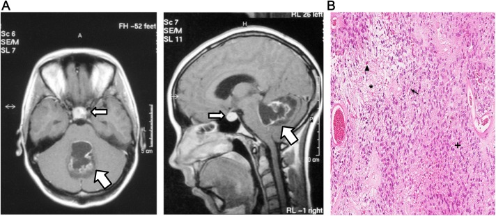
The Magnetic Resonance (MR) images showed classical features of PA in the cerebellum, and diagnosis was confirmed with histhopathological analysis; biphasic tumor areas with compact and loosen areas, Rosenthal fibers and eosinopfilic granular bodies (Fig 1, Table 1, and S1 Fig). Tumor cell content was estimated to be around 70% on average (>90% in tumor areas) in all six PA cases, based on Hematoxylin-Eosin-stained FFPE sections (S1 Fig). Each sample was investigated with RT-qPCR for presence of the three most common KIAA1549-BRAF mRNA fusion junctions (16–9, 15–9, and 16–11) [25] and BRAFV600E point mutation status (Table 2). The KIAA1549-BRAF 16–9 fusion was detected in five out of six cases (PA 1–2, PA 4–6). None of the samples was positive for BRAFV600E. Hence, a causative BRAF alteration could be identified in five out of six cases, whereas in case PA3 the genetic background was unknown. The cerebellar tumor in PA3 was partly cystic with irregular contrast enhancement. In addition to the PA tumor, a pituitary prolactinoma was revealed in the sella turcica, supported by the elevated S-prolactin level in the patient (Fig 1A). Co-occurrence of two or more brain tumors with different histological features is rare, although a few cases have been reported [26]. Moreover, in patients harboring pituitary tumors co-prevalence of other primary tumors is demonstrated to be significantly higher than expected in the general population [27]. Unfortunately, no material was available from the prolactinoma and hence only the cerebellar tumor (PA) from case PA3 was studied in the present paper. Patients underwent gross total resection of the Astrocytoma in the cerebellum, and all patients are alive with no evidence of disease. More information about event and other treatment can be found in Table 1.
Fig 1.
(A) Axial (left) and sagittal (right panel) MR images of PA3 tumor (T1-weighted, contrast enhanced) showing a cerebellar tumor (fat arrow), 4,5 x 3 x 3 cm, partly cystic with irregular contrast enhancement. In sella turcica, a 1.5 cm in diameter intensely contrast enhancing round tumor with a 0.5 cm cystic component is seen (slim arrow). The S-prolactin level was elevated, 1510 mIU/l (ref < 400), indicative of a pituitary prolactinoma. (B) Hematoxylin and Eosin staining of case PA3, demonstrates biphasic pattern with compact (+) and loose (*) areas, including Rosenthal fibers (arrow) and eosinophilic granular bodies (arrow head).
Table 2. Summary of results from five different methods for BRAF alteraration detection.
| castPCR | qPCR | RNA-seq | CNV (SNP array) | FISH | |||||||||
|---|---|---|---|---|---|---|---|---|---|---|---|---|---|
| Sample ID | BRAF mut | KIAA1549-BRAF fusion | Number of fusions from FusionCatcher | Detection of BRAF fusion | Rearrangements | BRAF split probe | |||||||
| V600E | Ex 16–9 | Ex 15–9 | Ex 16–11 | Pred fusions Prel. List | Pred fusions Final list | Coding fusions Final list | Prel. list | Final list | Gain/Loss | Breakpoints | Detection of rear. | % cells with rear. | |
| PA1 | Neg | Pos | Neg | Neg | 8159 | 17 | 1 | Y | N | 1 gain (7q34) | KIAA1549-BRAF | Y | 67% |
| PA2 | Neg | Pos | Neg | Neg | 8173 | 24 | 0 | Y | N | 1 gain (7q34) | KIAA1549-BRAF | Y | 76% |
| PA3 | Neg | Neg | Neg | Neg | 11527 | 34 | 2 | N | N | 2 gains (7q11.23 & 7q34) | GTF2I-BRAF & DENND2A-GTF2IRD1 | Y | 45% |
| PA4 | Neg | Pos | Neg | Neg | 4226 | 17 | 0 | N | N | 1 gain (7q34) | KIAA1549-BRAF | Y | 55% |
| PA5 | Neg | Pos | Neg | Neg | 9360 | 29 | 1 | Y | N | 1 gain (7q34) | KIAA1549-BRAF | Y | 55% |
| PA6 | Neg | Pos | Neg | Neg | 12726 | 47 | 0 | Y | N | Neg | Neg | Y | 40% |
Detection of BRAF-alterations with four methods: Competitive Allele-Specific TaqMan PCR (castPCR), quantitative PCR (qPCR), RNA sequencing (RNA-seq), Copy number variation (CNV) analysis with Single nucleotide polymorphism array (SNP-array), Fluorescense in situ hybridization (FISH). BRAF mut: BRAF point mutation; V600E = amino acid exchange; Neg = negative; Pos = Positive; Ex = Exon junction; Pred fusions = Number of Predicted fusions in Preliminary and Final list from FusionCatcher; Coding fusions: Fusions that will result in a new protein-coding fusion transcript; Detection of BRAF fusion = Presence of KIAA1549-BRAF or GTF2I-BRAF fusion in lists; Y = yes; N = No; Gain/Loss according to ChAS browser segment filter settings; BRAF split probe = BRAF break apart FISH result; Detection of rear. = Detection of rearrangement, Y = yes, n.d. = not determined; % of cells with rear. = frequency of rearrangement ("split probe"-positive cells when calculating 200 random interphase nuclei in the microscope, see methods for details).
Fusion detection by CNV and RNA-seq
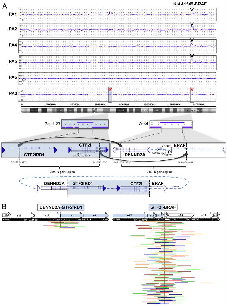
CNV screening with high-resolution SNP arrays caught the previously detected KIAA1549-BRAF 16–9 fusion in four out of five cases. One sample (PA6) showed a flat profile (no gains or losses on chromosome 7) (Fig 2A). Applying the FusionCatcher tool to RNA-seq data, none of the KIAA1549-BRAF 16–9 fusions could be identified in the final lists (Table 2 and S2 Table).
Fig 2.
(A) Copy number variation (CNV) genomic profiling with CytoScan HD SNP arrays. The weighted log2 ratio, smooth signal, and allele difference plot for chromosome 7 is shown for all six PA samples. Four out of five samples show the KIAA1549-BRAF duplication in 7q34. In case PA3 two novel duplications of approximately 250kb each were detected; one in 7q11.23 with breakpoint within GTF2IRD1 and GTF2I, and one in 7q34 with breakpoint within DENND2A and BRAF. The two duplicated regions give rise to two fusion junctions; DENND2A-GTF2IRD1 exons 14–2 and GTF2I-BRAF exon 19–10, probably through a circularization event followed by incorporation into the genome. The breakpoint positions are according GRCh37/Hg19 at UCSC Genome Browser (https://genome.ucsc.edu). Positions marked with star (*) are approximate by manual inspection in the ChAS software. (B) Supporting reads for the DENND2A-GTF2IRD1 14–2 and GTF2I-BRAF 19–10 fusion junctions in RNA sequencing data from case PA3. Spanning and split read pairs supporting the junction were extracted by BLAT and were aligned to 600bp of the predicted mRNA/cDNA sequence for each fusion. A schematic presentation of the mRNA junction is presented by the black box, showing exons (e), positions in cDNA, and GenBank accession numbers (DENND2A: NM_015689, GTF2IRD1: NM_016328, GTF2I: NM_032999), BRAF: NM_004333). Each row represent read pairs (or single reads) supporting a unique template. The RNA-seq data contained a total of 10 unique supporting read pairs/reads for DENND2A-GTF2IRD1 gene fusion and a total of 109 unique supporting read pairs/reads for the GTF2I-BRAF gene fusion. Only reads supporting the fusions are shown. Read pairs are in the same color. e = exon; i = intron.
Screening for novel fusions in the KIAA1549-BRAF-negative PA3 case was performed by a combined CNV and RNA-seq data analysis (see Material and Methods for details). RNA sequencing analysis with FusionCatcher predicted two coding fusions; DENND2A-GTF2IRD1 and GFAP-SPARC. Analyzing SNP array data of PA3, two CNV duplications of ∼250kb and ∼245kb each were identified; one involving GTF2I and GTF2IRD1 in 7q11.23 and a second involving DENND2A and BRAF in 7q34 (Fig 2A and Table 2). These CNV duplications verified the novel DENND2A-GTF2IRD1 14–2 fusion junction detected by RNA-seq, but also indicated a BRAF fusion formed by the same rearrangement event; BRAF-GTF2I. According to the CNV profile the two duplications were probably linked together into one fragment by a circularization event and somehow incorporated back into the genome (Fig 2A). No CNV gains or losses could be detected around the GFAP-SPARC fusion (17q21.31, 5q33.1) proposed by FusionCatcher, and since the junction could not be verified by RT-PCR this predicted fusion was excluded for further analysis.
In order to identify the precise fusion junction between GTF2I and BRAF, a 30 bp sequence from the 5’ junction site in BRAF (exon 10) was used to search for and extract all matching spanning reads present in the RNA-seq data (se Material and Methods for details). BLATing the extracted sequences revealed a novel gene junction between exon 19 in GTF2I and exon 10 in BRAF, supported by 109 reads or read pairs matching both GTF2I and BRAF (Fig 2B). The fusion also included a small 17 bp segment from BRAF intron 9, producing an in-frame fusion.
Confirmation of the novel fusion junctions
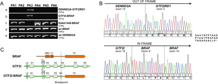
All fusion transcripts were verified by RT-PCR. Case PA3 was positive for PCR products from GTF2I-BRAF 19–10 (291bp) and DENND2A-GTF2IRD1 14–2 (222bp; Fig 3A). In accordance to results from qPCR, the PCR product of KIAA1549-BRAF 16–9 (249bp) could be detected in the five remaining PA cases. To verify and confirm the identity of the two novel fusion junctions, each transcript was sequenced by Sanger (Fig 3B). The DENND2A-GTF2IRD1 14–2 fusion, in which DENND2A is joined to six base pairs upstream of the ATG translation start in GTF2IRD1, produces an out-of-frame reading sequence with 41 new amino acids in the C-terminal of DENND2A. The truncated putative 815 aa DENND2A protein is similar in length as the DENND2A transcript version 3 (795 aa, NM_001318053). The GTF2I-BRAF 19–10 junction produces an in-frame reading sequence involving an inclusion of a 17 bp segment from BRAF intron 9. The putative GTF2I-BRAF fusion protein is 955 aa in length, containing the TFII-I DNA binding domains (R1-R3, BR and LZ) and nuclear localization (NLS) from GTF2I joined to the tyrosine kinase (TK) domain of BRAF (Fig 3C).
Fig 3.
(A) RT-PCR verification of gene fusions in six PA cases. Gene fusion PCR products of a) DENND2A-GTF2IRD1 14–2 (222 bp), b) GTF2I-BRAF 19–10 (291 bp), c) KIAA1549-BRAF 16–9 (249 bp), and wild type (wt) positive control products of d) BRAF and e) DENND2A. e = exon; i = intron. (B) Sanger sequencing of RT-PCR fusion junction products. Translation of codons is shown below the electropherograms. Upper panel: The DENND2A-GTF2IRD1 14–2 junction result in an out-of-frame truncated protein generating 41 new amino acids of the C-terminal of DENND2A. The ATG translation starting site in GTF2IRD1 is underlined. Lower panel: The GTF2I-BRAF 19–10 in-frame junction results in a putative protein involving an integrated sequence of 16bp from BRAF intron 9. New amino acids produced by the junctions are marked in bold. (C) Schematic illustration of the GTF2I and BRAF protein domains and localization of the fusion breakpoints. The TFII-I GTF2I protein (998 amino acids (aa), NP_127492) consists of six helix-loop-helix–like domains (R1-R6); the DNA binding domain basic region (BR); the nuclear localization signal (NLS); the leucine zipper domain (LZ). The BRAF (766 aa, NP_004324) consists of the Ras binding domain (RBD), the Phorbol ester/diacylglycerol binding zink finger domain (C1) and the tyrosine kinase (TK) domain. The breakpoint locus in GTF2I (position 575 aa) and BRAF (position 394 aa) is marked by a dashed vertical line. The GTF2I-BRAF putative fusion protein is 955 aa long and contains the TFII-I DNA binding domains (R1-R3, BR and LZ) and nuclear localization (NLS) from GTF2I joined to the tyrosine kinase (TK) domain of BRAF. Domains and positions are according to NextProt (http://www.nextprot.org, accessing date 2016-04-25).
Expression of fusions
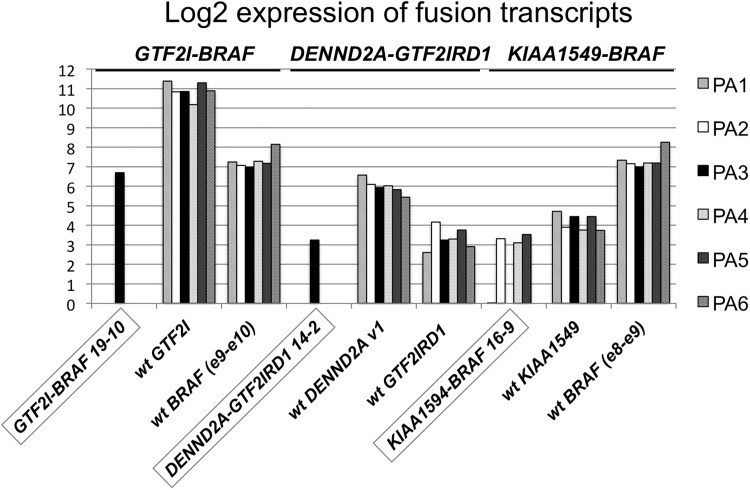
All sequence reads around fusion break points, both “spanning” reads and “split” read pairs, were extracted from the RNA-seq data. Expression of each fusion gene and corresponding wild type (wt) genes was calculated from the number of supporting reads, and normalized to the number of raw data reads (Fig 4 and S4 Table). In case PA3, 109 fragments supported the GTF2I-BRAF fusion (115 spanning reads, 9 split read pairs) and 11 reads supported the DENND2A-GTF2IRD1 fusion (11 spanning, 0 split read pairs) (Fig 2B and S4 Table). The three fusions showed a lower expression of fusion transcript than the 3’ wild type partner; GTF2I-BRAF versus GTF2I (18-fold), DENND2A-GTF2IRD1 versus DENND2A ver.1 (6-fold), KIAA1549-BRAF versus KIAA1549 (2-fold). The expression of the GTF2I-BRAF fusion (in PA3) indicates an elevated expression compared to KIAA1549-BRAF (13-fold, cases PA 1–2 and PA 4–5; Fig 4). Overall, the KIAA1549-BRAF expression was fairly low in all four positive cases with a maximum of 11 supporting reads in case PA5. Comparing the expression of GTF2I to KIAA1549 in all cases showed a 106-fold higher expression of GTF2I, indicating a stronger promoter (Fig 4). Although the PA6 case was positive for the KIAA1549-BRAF 16–9 fusion by qPCR (Table 2) and RT-PCR (Fig 3A), no supportive reads could be found in the RNA-seq data and this case also displayed a flat SNP-array profile (S4 Table and Fig 2A). An additional independent RT-PCR of sample PA6 was performed, and Sanger sequencing the PCR product confirmed the presence of the KIAA1549-BRAF 16–9 fusion (S2 Fig). Notably, the wild type BRAF expression was elevated by 2-fold in the PA6 case compared to the other five cases (Fig 4).
Fig 4. Expression analysis of fusion transcripts based on RNA-seq data.
Log2 RNA-seq expression data for three fusion transcripts; GTF2I-BRAF 19–10, DENND2A-GTF2IRD1 14–2, KIAA1549-BRAF 16–9 compared to wild type fusion partner genes in six PA cases. Expression data was calculated as total number of supporting reads normalized to the total number of raw reads in each sample. Exon-exon junction in genes are as follows; GTF2I-BRAF 19–10 (exon 19- exon 10), GTF2I (exon 19- exon 20), BRAF e9-e10 (exon 9- exon 10), DENND2A-GTF2IRD1 14–2 (exon 14- exon 2), KIAA1549-BRAF 16–9 (exon 16- exon 9), DENND2A v1 (transcript version 1, exon 14- exon 15), KIAA1549 (exon 16- exon 17), BRAF e8-e9 (exon 8- exon 9).
BRAF break apart FISH analysis
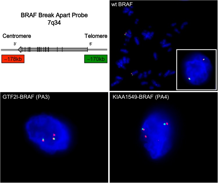
Out of the two novel fusions, the in-frame GTF2I-BRAF fusion was considered as the most plausible explanation factor for tumorigenesis in case PA3. Therefore, the new GTF2I-BRAF rearrangement together with the known KIAA1549-BRAF (16–9) fusion was further validated using BRAF Break Apart FISH assay on PA cases (Fig 5 and S3 Fig). First, the BRAF Break apart probes were verified to be located in 7q34 by metaphase FISH on normal blood cells (Fig 5). Normal and fusion-negative nuclei displayed two pairs of merged (yellow) or adjacent signals green/red (5’/3’), representing the two wild type BRAF alleles. Fusion-positive nuclei in tumor sections display the BRAF break apart pattern; two pairs of merged (yellow) or adjacent signals green/red (representing 5’/3’wt BRAF alleles), and one additional split red (3’) signal indicating a duplicated copy of the 3’ BRAF region. All six PA cases showed the break apart pattern, although the split 3’ signal could be close (e.g. PA2, PA5) or more distant (e.g. PA4) away from the normal allele in 7q34, probably depending on the integration site of the duplicated region (S3 Fig). Around 50% of cells in all PA cases were fusion-positive, i.e. showing split signal of 3’ BRAF (Table 2).
Fig 5. FISH analysis of BRAF fusions with BRAF break apart assay.
Upper left: Schematic presentation of the BRAF break apart assay, consisting of a 5’ 170 kb green probe and a 178 kb 3’ red probe in 7q34. Upper right (wt BRAF): Metaphase FISH of normal control and Interphase FISH of fusion-negative cell (right corner) showing two wild type BRAF alleles, displayed as a merged (yellow) or two adjacent green (5’)/red (3’) signals. Lower panels: Fusion-positive tumor cells (GTF2I-BRAF in PA3 and KIAA1549-BRAF in PA4) showing the BRAF split pattern; two normal BRAF alleles green /red signals, as well as one additional split BRAF red signal representing the duplicated 3’ region in the fusion gene. The same split signal pattern is seen for different types of BRAF fusions; GTF2I-BRAF in case PA3 and KIAA1549-BRAF in cases PA1-2 and PA4-6 (S3 Fig). Tissue sections were counterstained with DAPI (blue).
Activation of MAPK pathway
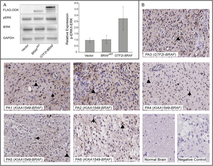
To elucidate expression and the MAPK activating potential of the GTF2I-BRAF fusion protein, HEK293 cells were transiently transfected with GTF2I-BRAF, BRAFWT, and empty vector constructs (pCMV6-Myc-DDK) respectively, and the level of pERK was measured with Western blot. Using anti- FLAG antibody the GTF2I-BRAF protein showed a band shift at the size of ~120 kDa, while the BRAFWT protein was detected at ~95 kDa. Cells expressing the GTF2I-BRAF fusion protein showed an elevated expression of pERK/tERK in comparison to cells transfected with BRAFWT and empty vector (Fig 6A). In addition, MAPK pathway activation was investigated in primary PA tissue sections with Immunohistochemistry. The pERK staining was increased in all six PA tumors compared to normal brain cerebellum regardless of the BRAF fusion type. pERK expression was predominantly found to be perinuclear and nuclear and to lesser extent cytoplasmic in tumor cells. Some blood vessels were also positive for pERK expression (Fig 6B). When omitting the primary antibody no staining was observed.
Fig 6. Activation of the MAPK pathway.
(A) Western Blot of protein lysates from HEK293 cells transiently transfected with pCMV6-Myc-DDK (empty vector), pCMV6-BRAF-Myc-DDK (BRAFWT) or pCMV6-GTF2I-BRAF-Myc-DDK (GTF2I-BRAF) were probed with antibodies against FLAG-DDK, phosphorylated ERK-Thr202/Tyr204 (pERK), total ERK (tERK) and GAPDH. Bars show relative mean pERK/tERK protein expression for each construct performed in triplicates (mean±SEM) after normalization to GAPDH. (B) Activation of the MAPK pathway in PA tumor tissue. FFPE sections from the six primary PA cases were immunostained with phosphorylated-ERK-Thr202/Tyr204 (pERK) antibody. Tumor tissue (PA1-6) showing perinuclear (arrow), nuclear (arrow head) and to lesser extent cytoplasmic (*) pERK staining. Normal human brain cerebellum reference tissue section from an autopsy specimen showing negative staining for pERK. Negative control with omitted primary antibody showing negative staining for pERK. Some endothelial cells were also positive for pERK. Original magnification x400.
Discussion
MAPK signaling pathway is the most important pathway for regulating cell growth, proliferation, apoptosis, and differentiation [28]. BRAF, a downstream target of RAS proteins, is a common target for activating mutations and fusions in diverse cancer forms including PA [29, 30]. The most frequent event in tumorigenesis of PA is BRAF gene rearrangements formed by duplication or deletions on chromosomal region 7q34, and KIAA1549-BRAF 16–9 is found in the majority (∼60%) of PA cases [7, 9–11]. In the present study we report for the first time a GTF2I-BRAF fusion in a pediatric brain tumor. The fusion was formed by rearrangement of two duplication events on 7q; one in 7q11.23 and one in 7q34. Partner genes in 7q34 (e.g. BRAF, KIAA1549) are transcribed in the telomere to centromere direction, and linear duplications/translocations may simply form fusions. In contrast, GTF2I (general transcription factor 2I) and neighboring genes in 7q11.23 are transcribed in opposite direction requiring an inversion in addition to the duplication and translocation to form a functional gene. The GTF2I intron 19 where the breakpoint occurred is also small compared to the common breakpoint introns of KIAA1549. These differences between KIAA1549 and GTF2I may explain why the former partner gene is much more common.
The new GTF2I-BRAF 19–10 fusion generates a putative protein containing the TFII-I DNA binding domains from GTF2I joined to the tyrosine kinase (TK) domain of BRAF. Interestingly, another GTF2I-BRAF fusion (exon 4–10) has been reported in one primary melanoma case [29], supporting a role of this fusion partnership in tumor development. Similar to other BRAF/RAF1 fusions in PA, the GTF2I-BRAF fusion protein lacks N-terminal inhibitory domain leading to constitutive active BRAF kinase and increased MEK/ERK signaling [7, 10, 12, 16]. In line with defined functional properties of BRAF fusions, the current study demonstrates GTF2I-BRAF 19-10-expressing HEK293 cells to exhibit increased pERK levels, thus confirming a role of GTF2I-BRAF in constitutive activation of the MAPK pathway. In addition, Immunohistochemistry show that the PA cases, harboring either KIAA1549-BRAF or GTF2I-BRAF, displays an elevated expression of pERK compared to normal brain tissue, further supporting an increase in MAP kinase activity mediated by BRAF fusions. Notably, the pERK was mainly located in the nucleus, which is essential for activation of transcription targets [31].
BRAF gene fusions are reported to involve many different 5’ partners, (e.g. KIAA1549, FAM131B, RNF130, CLCN6, MKRN1, GNAI1) although KIAA1549 is the most prevalent one [7–9, 12–15]. The possible role of BRAF fusions partners in PA tumorigenicity is largely unknown. Shin et al. [32] showed that the BRAF kinase domain alone, without fusion partner, was not able to induce tumors in mice indicating a certain role of fusion partners. The new GTF2I fusion partner has been implicated in regulation of several cellular processes involving growth factor induced signal transduction, proliferation, apoptosis, angiogenesis, TGFB signaling, and immune response [33]. Interestingly, GTF2I has also been shown to regulate nuclear translocation of ERK1/2 upon mitogenic signaling [31], and indicates an indirect role of GTF2I in activation of the MAPK pathway. Occurrence of additional GTF2I-fusions; GTF2I-BRAF 4–10 (primary melanoma) [29], GTF2I-NCOA2 (soft tissue angiofibroma development) [34] and GTF2I-RARA (acute promyelocytic leukemia) [35], further supports a oncogenic role. In the present study, the GTF2I promoter appears to be stronger than the KIAA1549 indicated by the elevated expression of both the GTF2I-BRAF fusion and the GTF2I transcript. However, no an altered disease progression could be observed in the index case (PA3) harboring the GTF2I-BRAF fusion. The patient underwent radical surgery of the Astrocytoma in the cerebellum in November 2003, and has not relapsed since then.
The two CNV gains on 7q also resulted in an additional rearrangement in the same tumor case; DENND2A-GTF2IRD1 14–2. DENND2A (DENN/MEDD domain 2A), located next to BRAF, encodes a guanine nucleotide exchange factor (GEF) that activates members of the Rab pathway. The small Rab GTPases regulate growth factor signaling and cell mobility through intracellular vesicle transport, and has important roles in migration and invasion of tumor cells [36]. Since the DENND2A-GTF2IRD1 putative fusion product will result in a truncated DENND2A protein, the consequence of this gene fusion is probably moderate and is not expected to play a role in PA tumorigenesis. Yet, inclusion of DENND2A in a 7q-rearrangement in PA has previously been reported by Cin and colleagues [10].
In recent years, RNA sequencing has become a prevalent method for fusion gene detection in cancer [37]. Indeed, transcriptomic sequencing has taken the fusion gene discovery into another level since it allows for detection of all (balanced or unbalanced) expressed rearrangements, including alternative splice variants resulting from a fusion event [37]. However, one main limitation of RNA sequencing is that it cannot detect rearrangement events involving non-transcribed regions. Also, since the dynamic range of expression is broad and tissue-specific, fusion genes expressed at low level can be difficult to detect. Current available bioinformatics algorithms for fusion discovery often report a very high number of false positive chimeras [38], or may be to stringent to detect the fusions that are actually present in the data [15]. This stresses the importance of evaluation and fine-tuning of bioinformatic pipeline for fusion gene discovery [39]. The new GTF2I-BRAF 19–10 fusion identified in present study would probably not have been discovered by RNA sequencing and CNV analysis alone. One issue that complicated the finding of this fusion gene with RNA-seq was the occurrence of pseudogenes for GTF2I on different chromosomal location, and it was excluded by the FusionCatcher algorithm. Moreover, the detection of GTF2I-BRAF fusion with CNV analysis was also problematic since the duplications generating the fusion were quite small, hence required modification of settings and thorough manual inspection. On the hand, the larger duplication producing the most prevalent KIAA1549-BRAF fusion was clearly detected by the SNP-arrays in most cases, but could not be captured by the FusionCatcher algorithm due to the low expression levels in the RNA-seq data. Our results are in line with Lin et al. [14] who demonstrated that KIAA1549-BRAF fusions is expressed at lower levels than BRAF, but at only slightly lower levels than the KIAA1549 promoter. In summary, occurrence of pseodogenes and low expression/few supporting reads [14, 15] complicates fusion detection by the RNA sequencing method.
Due to the high prevalence of BRAF rearrangements in pediatric PA and the documented better clinical outcome of KIAA1549-BRAF positive cases, the BRAF fusions have been suggested as a prognostic marker and supplement tool for better diagnostics [4, 17, 19, 20]. This emphasizes the need for reliable detection of variant BRAF rearrangements in clinical routine irrespectively of its fusion partner or exon-exon junction. The BRAF break apart FISH probe used in this paper to validate presence of BRAF fusions was able to detect both KIAA1549-BRAF and GTF2I-BRAF with the same sensitivity. The advantage of FISH method compared to qPCR, CNV analysis and RNA sequencing is that this method is a robust and informative diagnostic tool to indicate BRAF rearrangements independent of the fusion partner, fusion junction, size of duplication, or expression of the fusion gene. Moreover, BRAF break apart FISH probe method renders the possibility to assess the number of fusion-positive cells and is applicable for FFPE sections available in clinical routine. Disadvantages are that neither the fusion partner nor the fusion consequence (in-frame or out-of-frame) is detected. But complementary qPCR can be used to detect the most common KIAA1549-BRAF fusions in PA [21]. Nevertheless, the sensitivity and specificity of the break apart FISH method as a routine method needs further validation in larger PA cohorts.
The high occurrence and impact of BRAF rearrangements in pediatric gliomas, essentially PA, not only open the possibility for better diagnostics but also provides an opportunity for targeted therapy. Selective B-Raf inhibitors such as Vemurafinib and Sorafenib that have been developed for the deregulated B-Raf malignancy in other tumor types (e.g. melanoma), can be used for management of approximately 8% of all PA harboring BRAFV600E mutation [14, 40]. However, Raf inhibitors have been reported to generate a paradoxal activation of the MAPK pathway in the cells expressing wild type BRAF and BRAF fusions [41, 42]. Due to this fact, Phase II trial with Sorafenib for treatment of recurrent or progressive low-grade astrocytoma’s was discounted [43]. Instead, MAPK/ERK kinase (MEK) inhibitors are currently in clinical trials and can provide alternative therapy independent of BRAF status (ClinicalTrials.gov identifier NCT01386450; NCT01089101).
The knowledge about molecular genetics behind development of PA has increased tremendously in recent years. The new GTF2I-BRAF 19–10 fusion reported in this paper further emphasizes the central role of BRAF in tumorigenesis, and the MAPK pathway as a promising therapeutic target in PA. Moreover, the occurrence of an increasing number of BRAF fusion variants and possibility for MAPK pathway targeted therapy highlights the importance of a robust method for fast and cost-effective detection of BRAF deregulations to guide diagnosis, prognosis, and accurate targeted therapy.
Supporting information
(PDF)
(PDF)
(PDF)
(XLSX)
(XLSX)
(XLSX)
(XLSX)
Acknowledgments
We would like to thank the Genomics Core Facility and Bioinformatics resource unit at Gothenburg University for access to Illumina sequencing and qPCR systems and data management support. We are grateful to Anna Djos at Department of Clinical Genetics for assistance and guidance through SNP-array preparation and Carola Andersson at Clinical Molecular Pathology, Sahlgrenska University Hospital, for preparing histopathological material. This work was supported by the Swedish Children's Cancer Foundation (grant numbers PR2013-0010, FoAss10/008, NBCNSDoktBidr11/006, 2011–2013 to FA and NC2014-0036 to TT), the Assar Gabrielsson Foundation (grant numbers FB 15–75 2015 to TT, FB 13–68 2014 to AW); the Wilhelm and Martina Lundgren Research Foundation (year 2011–2013 to TT, AW and FA).
Abbreviations
- PA
Pilocytic astrocytoma
- MAPK
mitogen activated protein kinase
- CNV
copy number variation
- FISH
Fluorescence in situ hybridization
- RNA-seq
RNA sequencing
- LGG
low-grade gliomas
- WHO
World Health Organization
- NF1
Neurofibromatosis type 1
- qPCR
quantitative Reverse Transcriptase RT, -PCR; high-resolution
- SNP
Single Nucleotide Polymorphism arrays
- CGH
Comparative Genomic Hybridization
- SDS
Sequence Detection System; quantitation cycle
- castPCR
Competitive Allele-Specific TaqMan PCR
- ChAS
Affymetrix Chromosome Analysis Suite
- FFPE
formalin fixed paraffin embedded tissue
- DAPI
4´,6´,-diamidino-2´-phenylindole dihydrochloride
- DAB
diaminobenzidine
- pERK
phosphorylated ERK-Thr202/Tyr204)
- NSC
neural stem cells
- GEF
guanine nucleotide
- TK
tyrosine kinase
Data Availability
The novel cDNA and protein sequence of the GTF2I-BRAF 19-10 fusion gene has been deposited to GenBank (BankIt) at NCBI under Submission ID 2005648.
Funding Statement
This work was supported by the Swedish Children's Cancer Foundation (grant numbers PR2013-0010, FoAss10/008, NBCNSDoktBidr11/006, 2011-2013 to FA and NC2014-0036 to TT), the Swedish Assar Gabrielsson Foundation (grant numbers FB 15-75 2015 to TT, FB 13-68 2014 to AW); the Swedish Wilhelm and Martina Lundgren Research Foundation (year 2011-2013 to TT, AW and FA). The funders had no role in study design, data collection and analysis, decision to publish, or preparation of the manuscript.
References
- 1.Lannering B, Sandstrom PE, Holm S, Lundgren J, Pfeifer S, Samuelsson U, et al. Classification, incidence and survival analyses of children with CNS tumours diagnosed in Sweden 1984–2005. Acta Paediatr. 2009;98(10): 1620–7. doi: 10.1111/j.1651-2227.2009.01417.x [DOI] [PubMed] [Google Scholar]
- 2.Johnson KJ, Cullen J, Barnholtz-Sloan JS, Ostrom QT, Langer CE, Turner MC, et al. Childhood brain tumor epidemiology: a brain tumor epidemiology consortium review. Cancer Epidemiol Biomarkers Prev. 2014;23(12): 2716–36. doi: 10.1158/1055-9965.EPI-14-0207 [DOI] [PMC free article] [PubMed] [Google Scholar]
- 3.Burkhard C, Di Patre PL, Schuler D, Schuler G, Yasargil MG, Yonekawa Y, et al. A population-based study of the incidence and survival rates in patients with pilocytic astrocytoma. J Neurosurg. 2003;98(6): 1170–4. doi: 10.3171/jns.2003.98.6.1170 [DOI] [PubMed] [Google Scholar]
- 4.Gilles FH, Tavare CJ, Becker LE, Burger PC, Yates AJ, Pollack IF, et al. Pathologist interobserver variability of histologic features in childhood brain tumors: results from the CCG-945 study. Pediatr Dev Pathol. 2008;11(2): 108–17. doi: 10.2350/07-06-0303.1 [DOI] [PubMed] [Google Scholar]
- 5.Louis DN, Ohgaki H, Wiestler OD, Cavenee WK, Burger PC, Jouvet A, et al. The 2007 WHO classification of tumours of the central nervous system. Acta Neuropathol. 2007;114(2): 97–109. doi: 10.1007/s00401-007-0243-4 [DOI] [PMC free article] [PubMed] [Google Scholar]
- 6.Armstrong GT, Conklin HM, Huang S, Srivastava D, Sanford R, Ellison DW, et al. Survival and long-term health and cognitive outcomes after low-grade glioma. Neuro Oncol. 2011;13(2): 223–34. doi: 10.1093/neuonc/noq178 [DOI] [PMC free article] [PubMed] [Google Scholar]
- 7.Jones DT, Kocialkowski S, Liu L, Pearson DM, Backlund LM, Ichimura K, et al. Tandem duplication producing a novel oncogenic BRAF fusion gene defines the majority of pilocytic astrocytomas. Cancer Res. 2008;68(21): 8673–7. doi: 10.1158/0008-5472.CAN-08-2097 [DOI] [PMC free article] [PubMed] [Google Scholar]
- 8.Forshew T, Tatevossian RG, Lawson AR, Ma J, Neale G, Ogunkolade BW, et al. Activation of the ERK/MAPK pathway: a signature genetic defect in posterior fossa pilocytic astrocytomas. J Pathol. 2009;218(2): 172–81. doi: 10.1002/path.2558 [DOI] [PubMed] [Google Scholar]
- 9.Sievert AJ, Jackson EM, Gai X, Hakonarson H, Judkins AR, Resnick AC, et al. Duplication of 7q34 in pediatric low-grade astrocytomas detected by high-density single-nucleotide polymorphism-based genotype arrays results in a novel BRAF fusion gene. Brain Pathol. 2009;19(3): 449–58. doi: 10.1111/j.1750-3639.2008.00225.x [DOI] [PMC free article] [PubMed] [Google Scholar]
- 10.Cin H, Meyer C, Herr R, Janzarik WG, Lambert S, Jones DT, et al. Oncogenic FAM131B-BRAF fusion resulting from 7q34 deletion comprises an alternative mechanism of MAPK pathway activation in pilocytic astrocytoma. Acta Neuropathol. 2011;121(6): 763–74. doi: 10.1007/s00401-011-0817-z [DOI] [PubMed] [Google Scholar]
- 11.Roth JJ, Santi M, Pollock AN, Harding BN, Rorke-Adams LB, Tooke LS, et al. Chromosome band 7q34 deletions resulting in KIAA1549-BRAF and FAM131B-BRAF fusions in pediatric low-grade Gliomas. Brain Pathol. 2015;25(2): 182–92. doi: 10.1111/bpa.12167 [DOI] [PMC free article] [PubMed] [Google Scholar]
- 12.Jones DT, Kocialkowski S, Liu L, Pearson DM, Ichimura K, Collins VP. Oncogenic RAF1 rearrangement and a novel BRAF mutation as alternatives to KIAA1549:BRAF fusion in activating the MAPK pathway in pilocytic astrocytoma. Oncogene. 2009;28(20): 2119–23. doi: 10.1038/onc.2009.73 [DOI] [PMC free article] [PubMed] [Google Scholar]
- 13.Dahiya S, Yu J, Kaul A, Leonard JR, Gutmann DH. Novel BRAF Alteration in a Sporadic Pilocytic Astrocytoma. Case Rep Med. 2012;2012: 418672 doi: 10.1155/2012/418672 [DOI] [PMC free article] [PubMed] [Google Scholar]
- 14.Lin A, Rodriguez FJ, Karajannis MA, Williams SC, Legault G, Zagzag D, et al. BRAF alterations in primary glial and glioneuronal neoplasms of the central nervous system with identification of 2 novel KIAA1549:BRAF fusion variants. J Neuropathol Exp Neurol. 2012;71(1): 66–72. doi: 10.1097/NEN.0b013e31823f2cb0 [DOI] [PMC free article] [PubMed] [Google Scholar]
- 15.Jones DT, Hutter B, Jager N, Korshunov A, Kool M, Warnatz HJ, et al. Recurrent somatic alterations of FGFR1 and NTRK2 in pilocytic astrocytoma. Nat Genet. 2013;45(8): 927–32. doi: 10.1038/ng.2682 [DOI] [PMC free article] [PubMed] [Google Scholar]
- 16.Kaul A, Chen YH, Emnett RJ, Dahiya S, Gutmann DH. Pediatric glioma-associated KIAA1549:BRAF expression regulates neuroglial cell growth in a cell type-specific and mTOR-dependent manner. Genes Dev. 2012;26(23): 2561–6. doi: 10.1101/gad.200907.112 [DOI] [PMC free article] [PubMed] [Google Scholar]
- 17.Becker AP, Scapulatempo-Neto C, Carloni AC, Paulino A, Sheren J, Aisner DL, et al. KIAA1549: BRAF Gene Fusion and FGFR1 Hotspot Mutations Are Prognostic Factors in Pilocytic Astrocytomas. J Neuropathol Exp Neurol. 2015;74(7): 743–54. doi: 10.1097/NEN.0000000000000213 [DOI] [PMC free article] [PubMed] [Google Scholar]
- 18.Penman CL, Faulkner C, Lowis SP, Kurian KM. Current Understanding of BRAF Alterations in Diagnosis, Prognosis, and Therapeutic Targeting in Pediatric Low-Grade Gliomas. Front Oncol. 2015;5: 54 doi: 10.3389/fonc.2015.00054 [DOI] [PMC free article] [PubMed] [Google Scholar]
- 19.Jacob K, Albrecht S, Sollier C, Faury D, Sader E, Montpetit A, et al. Duplication of 7q34 is specific to juvenile pilocytic astrocytomas and a hallmark of cerebellar and optic pathway tumours. Br J Cancer. 2009;101(4): 722–33. doi: 10.1038/sj.bjc.6605179 [DOI] [PMC free article] [PubMed] [Google Scholar]
- 20.Lawson AR, Tatevossian RG, Phipps KP, Picker SR, Michalski A, Sheer D, et al. RAF gene fusions are specific to pilocytic astrocytoma in a broad paediatric brain tumour cohort. Acta Neuropathol. 2010;120(2): 271–3. doi: 10.1007/s00401-010-0693-y [DOI] [PubMed] [Google Scholar]
- 21.Tian Y, Rich BE, Vena N, Craig JM, Macconaill LE, Rajaram V, et al. Detection of KIAA1549-BRAF fusion transcripts in formalin-fixed paraffin-embedded pediatric low-grade gliomas. J Mol Diagn. 2011;13(6): 669–77. doi: 10.1016/j.jmoldx.2011.07.002 [DOI] [PMC free article] [PubMed] [Google Scholar]
- 22.Nicorici D, Satalan M, Edgren H, Kangaspeska S, Murumagi A, Kallioniemi O, et al. FusionCatcher—a tool for finding somatic fusion genes in paired-end RNA-sequencing data. bioRxiv. 2014.
- 23.Kent WJ. BLAT—The BLAST-like alignment tool. Genome Res. 2002;12(4): 656–64. doi: 10.1101/gr.229202 [DOI] [PMC free article] [PubMed] [Google Scholar]
- 24.(E.C.A) ECA. Permanent working group for cancer cytogenetics, solid tumours. FISH on histological sections of solid tumours: E.C.A. recommendations. Newsletter 2012.No. 29:: 28–30.
- 25.Jones DT, Gronych J, Lichter P, Witt O, Pfister SM. MAPK pathway activation in pilocytic astrocytoma. Cell Mol Life Sci. 2012;69(11): 1799–811. doi: 10.1007/s00018-011-0898-9 [DOI] [PMC free article] [PubMed] [Google Scholar]
- 26.Furtado SV, Venkatesh PK, Ghosal N, Hegde AS. Coexisting intracranial tumors with pituitary adenomas: genetic association or coincidence? J Cancer Res Ther. 2010;6(2): 221–3. doi: 10.4103/0973-1482.65246 [DOI] [PubMed] [Google Scholar]
- 27.Couldwell WT, Cannon-Albright LA. Co-prevalence of other tumors in patients harboring pituitary tumors. J Neurosurg. 2014;121(6): 1474–7. doi: 10.3171/2014.8.JNS132139 [DOI] [PubMed] [Google Scholar]
- 28.Peyssonnaux C, Eychene A. The Raf/MEK/ERK pathway: new concepts of activation. Biol Cell. 2001;93(1–2): 53–62. [DOI] [PubMed] [Google Scholar]
- 29.Ross JS, Wang K, Chmielecki J, Gay L, Johnson A, Chudnovsky J, et al. The distribution of BRAF gene fusions in solid tumors and response to targeted therapy. Int J Cancer. 2016;138(4): 881–90. doi: 10.1002/ijc.29825 [DOI] [PMC free article] [PubMed] [Google Scholar]
- 30.Davies H, Bignell GR, Cox C, Stephens P, Edkins S, Clegg S, et al. Mutations of the BRAF gene in human cancer. Nature. 2002;417(6892): 949–54. doi: 10.1038/nature00766 [DOI] [PubMed] [Google Scholar]
- 31.Hakre S, Tussie-Luna MI, Ashworth T, Novina CD, Settleman J, Sharp PA, et al. Opposing functions of TFII-I spliced isoforms in growth factor-induced gene expression. Mol Cell. 2006;24(2): 301–8. doi: 10.1016/j.molcel.2006.09.005 [DOI] [PubMed] [Google Scholar]
- 32.Shin CH, Grossmann AH, Holmen SL, Robinson JP. The BRAF kinase domain promotes the development of gliomas in vivo. Genes Cancer. 2015;6(1–2): 9–18. doi: 10.18632/genesandcancer.48 [DOI] [PMC free article] [PubMed] [Google Scholar]
- 33.Roy AL. Biochemistry and biology of the inducible multifunctional transcription factor TFII-I: 10 years later. Gene. 2012;492(1): 32–41. doi: 10.1016/j.gene.2011.10.030 [DOI] [PMC free article] [PubMed] [Google Scholar]
- 34.Arbajian E, Magnusson L, Mertens F, Domanski HA, Vult von Steyern F, Nord KH. A novel GTF2I/NCOA2 fusion gene emphasizes the role of NCOA2 in soft tissue angiofibroma development. Genes Chromosomes Cancer. 2013;52(3): 330–1. doi: 10.1002/gcc.22033 [DOI] [PubMed] [Google Scholar]
- 35.Li J, Zhong HY, Zhang Y, Xiao L, Bai LH, Liu SF, et al. GTF2I-RARA is a novel fusion transcript in a t(7;17) variant of acute promyelocytic leukaemia with clinical resistance to retinoic acid. Br J Haematol. 2015;168(6): 904–8. doi: 10.1111/bjh.13157 [DOI] [PubMed] [Google Scholar]
- 36.Porther N, Barbieri MA. The role of endocytic Rab GTPases in regulation of growth factor signaling and the migration and invasion of tumor cells. Small GTPases. 2015;6(3): 135–44. doi: 10.1080/21541248.2015.1050152 [DOI] [PMC free article] [PubMed] [Google Scholar]
- 37.Wang Q, Xia J, Jia P, Pao W, Zhao Z. Application of next generation sequencing to human gene fusion detection: computational tools, features and perspectives. Brief Bioinform. 2013;14(4): 506–19. doi: 10.1093/bib/bbs044 [DOI] [PMC free article] [PubMed] [Google Scholar]
- 38.Carrara M, Beccuti M, Cavallo F, Donatelli S, Lazzarato F, Cordero F, et al. State of art fusion-finder algorithms are suitable to detect transcription-induced chimeras in normal tissues? BMC Bioinformatics. 2013;14 Suppl 7: S2. [DOI] [PMC free article] [PubMed] [Google Scholar]
- 39.Liu S, Tsai WH, Ding Y, Chen R, Fang Z, Huo Z, et al. Comprehensive evaluation of fusion transcript detection algorithms and a meta-caller to combine top performing methods in paired-end RNA-seq data. Nucleic Acids Res. 2016;44(5): e47 doi: 10.1093/nar/gkv1234 [DOI] [PMC free article] [PubMed] [Google Scholar]
- 40.Schindler G, Capper D, Meyer J, Janzarik W, Omran H, Herold-Mende C, et al. Analysis of BRAF V600E mutation in 1,320 nervous system tumors reveals high mutation frequencies in pleomorphic xanthoastrocytoma, ganglioglioma and extra-cerebellar pilocytic astrocytoma. Acta Neuropathol. 2011;121(3): 397–405. doi: 10.1007/s00401-011-0802-6 [DOI] [PubMed] [Google Scholar]
- 41.Poulikakos PI, Zhang C, Bollag G, Shokat KM, Rosen N. RAF inhibitors transactivate RAF dimers and ERK signalling in cells with wild-type BRAF. Nature. 2010;464(7287): 427–30. doi: 10.1038/nature08902 [DOI] [PMC free article] [PubMed] [Google Scholar]
- 42.Sievert AJ, Lang SS, Boucher KL, Madsen PJ, Slaunwhite E, Choudhari N, et al. Paradoxical activation and RAF inhibitor resistance of BRAF protein kinase fusions characterizing pediatric astrocytomas. Proc Natl Acad Sci U S A. 2013;110(15): 5957–62. doi: 10.1073/pnas.1219232110 [DOI] [PMC free article] [PubMed] [Google Scholar]
- 43.Karajannis MA, Legault G, Fisher MJ, Milla SS, Cohen KJ, Wisoff JH, et al. Phase II study of sorafenib in children with recurrent or progressive low-grade astrocytomas. Neuro Oncol. 2014;16(10): 1408–16. doi: 10.1093/neuonc/nou059 [DOI] [PMC free article] [PubMed] [Google Scholar]
Associated Data
This section collects any data citations, data availability statements, or supplementary materials included in this article.
Supplementary Materials
(PDF)
(PDF)
(PDF)
(XLSX)
(XLSX)
(XLSX)
(XLSX)
Data Availability Statement
The novel cDNA and protein sequence of the GTF2I-BRAF 19-10 fusion gene has been deposited to GenBank (BankIt) at NCBI under Submission ID 2005648.


