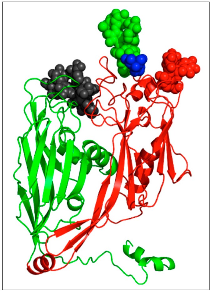
Figure 3.
Structure of the revised PBCV-1 Vp54 monomer. The two jelly-roll domains are colored in green and red, respectively. The glycans located on the surface are shown as a space-filling representation of their atoms and are colored according to the residue they are attached to (Asn-280: green, Asn-302: black, Asn-399: red, Asn-406: blue). Taken from De Castro et al. [49] with permission.

