Abstract
During chronic liver injuries, progenitor cells expand in a process called ductular reaction, which also entails the appearance of inflammatory cellular infiltrate and epithelial cell activation. The progenitor cell population during such inflammatory reactions has mostly been investigated using single surface markers, either by histological analysis or by flow cytometry-based techniques. However, novel surface markers identified various functionally distinct subsets within the liver progenitor/stem cell compartment. The method presented here describes the isolation and detailed flow cytometry analysis of progenitor subsets using novel surface marker combinations. Moreover, it demonstrates how the various progenitor cell subsets can be isolated with high purity using automated magnetic and FACS sorting-based methods. Importantly, novel and simplified enzymatic dissociation of the liver allows for the isolation of these rare cell populations with a high viability that is superior in comparison to other existing methods. This is especially relevant for further studying progenitor cells in vitro or for isolating high-quality RNA to analyze the gene expression profile.
Keywords: Developmental Biology, Issue 120, liver, progenitor cells, gp38, CD133, cell sorting, flow cytometry analyses
Introduction
Liver regeneration is mostly associated with the self-renewal capacity of hepatocytes. Nevertheless, chronic liver injuries occur with progenitor cell activation and expansion, which have been associated with their ability to differentiate into hepatocytes and cholangiocytes1,2,3,4. This is especially relevant because, during chronic injuries, hepatocyte proliferation is not effective. Despite multiple genetic tracing studies targeting progenitor cells, their role in liver regeneration remains controversial5,6,7,8. Moreover, the activation of progenitor cells has been linked to increased fibrotic response in the liver, which raises questions about their exact role during injuries9,10.
The heterogeneous nature of the progenitor cell compartment has long been suggested by gene expression studies that isolated progenitor cells expressing a single surface marker using microdissection or cell sorting-based methods1,11. Indeed, recently, a novel surface marker combination using gp38 (podoplanin) unequivocally linked previous single markers of progenitor cells to various subsets12. Importantly, these subsets not only differed in their surface marker expression but also exhibited functional alterations during injuries12.
Multiple animal models have been utilized to investigate progenitor cell activation and liver regeneration. It seems that the various injury types promote the activation of differing subsets of progenitor cells12. This might explain the phenotypic divergence of the ductular reaction observed in humans4. Thus, the complex phenotypic and functional analyses of progenitor cells are pivotal to understand their role in injuries and the true significance of the ductular reaction in liver diseases.
Besides surface marker combinations, the crucial differences in cell isolation protocols further complicate the conclusions based on previous studies2. A substantial amount of studies addressed the role of progenitor cells that greatly differ in their isolation protocol (e.g., liver dissociation (enzyme combination and duration of the process), density medium, and centrifugation speed)2. An optimized isolation technique, providing better viability for rare cell populations and reflective of subset composition, has been developed and published recently12. The aim of this article is to provide a more detailed protocol of this liver cell isolation procedure and the subset analysis to allow for the proper reproduction of the technique. Additionally, the protocol includes a comparison with the previous isolation method to demonstrate the differences compared to the new protocol.
Protocol
All experimental procedures were conducted with the approval of the ethics and animal care committees of Homburg University Medical Center.
1. Preparation of Materials and Buffers
Freshly prepare all buffers required for liver digestion using sterile components and a laminar hood to avoid bacterial contamination.
Prepare the collection buffer (CB) by mixing 49.5 mL of RPMI medium and 0.5 mL of fetal bovine serum (FBS; low endotoxin, heat inactivated) to achieve a 1% (v/v) solution. Store the solution on ice until further usage. NOTE: Approximately 25 mL of CB is necessary to digest one whole liver.
Prepare the digestion buffer (DB) by using the following ingredients: RPMI medium, 1% (v/v) FBS (low endotoxin, heat inactivated), collagenase P (0.2 mg/mL), DNase-I (0.1 mg/mL), and dispase (0.8 mg/mL). NOTE: Approximately 25 mL of DB is necessary to digest one whole liver. Pre-warm the DB in the 37 °C water bath before use.
Reconstitute the enzymes upon arrival in Hanks' balanced salt solution (HBSS; collagense P and dispase) or in DNase-I buffer (50% (v/v) glycerol, 1 mM MgCl2, and 20 mM Tris-HCl; pH 7.5), aliquot it, and store it at -20 °C. Store the DNase-I buffer at 4 °C and use it within two months.
2. Preparation of Liver Single-cell Suspension
Euthanize untreated wild-type mice by cervical dislocation in accordance with the local ethics and animal care committees.
Place the mice on a dissection board and wet the fur with 70% ethanol. Using scissors, open the abdomen with a midline incision of the skin, followed by a Y-incision towards the limbs. Open the peritoneum up to the sternum using the scissors. In order to uncover the liver, displace the intestine gently to the right side using a cotton swab.
With the help of scissors and forceps, remove the liver lobes, leaving the gall bladder behind, and avoid contamination with connective tissue. Weigh the liver and place it on ice in a Petri dish containing HBSS.
Place the liver lobes on a dry Petri dish and cut the liver tissue into homogeneous cubes approximately 2 mm a side by using a scalpel. Transfer the pieces into a 15-mL conical centrifuge tube.
Add 2.5 mL of DB to the 15-mL conical centrifuge tube containing the liver pieces and place it in a 37 °C water bath to start the digestion process (a healthy liver takes 60-70 min and a cirrhotic liver takes 80-90 min). NOTE: If one entire liver is being digested, the liver should be separated into two 15-mL conical centrifuge tubes to ensure good cell viability.
Prepare a new 15-mL conical centrifuge tube to collect released liver cells. Place a polyamide 100-µm filter mesh on the top of the tube and wet the mesh with 800 µL of CB. Place the conical centrifuge tube on ice.
Mix the samples in the 37 °C water bath after 5 and 10 min in order to support the digestion process by shaking the 15-mL conical centrifuge tube containing the liver pieces.
15 min after starting the digestion, gently mix the liver pieces using a 1,000-µL pipette with a cut tip that enables the liver pieces to pass through easily. Place the tubes back into the water bath and allow the pieces to settle for 2 min.
Remove the supernatant containing the disseminated cells (typically 2x 700 µL) and add it to the tube prepared in step 2.6. Replace the removed supernatant with DB (2x 700 µL) and place it back into the 37 °C water bath.
Repeat the procedure described in step 2.8 (typically at 30, 40, 50, 55, and 60 min) until approximately 60-70 min have passed since the start of the digestion. From 40 min onwards, the remaining liver pieces should be small enough to pass through an uncut 1,000-µL pipette tip. NOTE: Healthy liver is digested fully within 60-70 min, while fibrotic liver typically needs 80-90 min. By this time point, the liver tissue should not be visible in the conical 15-mL centrifuge tube containing the liver pieces, and all released cells should be transferred into the tube prepared in step 2.6.
At the end of the digestion process, collect the cells and centrifuge them for 8 min at 180 x g and 4 °C (using reduced acceleration/4 and deceleration/2).
Resuspend the cell pellet in 1 mL of ammonium-chloride-potassium (ACK) lysis buffer and incubate it for 1 min at room temperature in order to lyse the red blood cells. Stop the reaction by adding 5 mL of CB, and then centrifuge the cells for 8 min at 180 x g and 4 °C (using reduced acceleration/4 and deceleration/2). Resuspend the cells in 4 mL of CB and store them on ice. Count the cells, as described in step 3. NOTE: The cell pellet is loose, and therefore the supernatant is pipetted away instead of being decanted.
3. Determination of the Cell Count Using Flow Cytometry
NOTE: For determining the cell counts, an automated cell counter or, ideally, the flow cytometry-based cell quantification described below is suggested instead of the classical Neubauer chamber-based method. The liver single-cell suspension described in step 2 contains parenchymal and non-parenchymal cells (NPC) with greatly differing sizes and granularities. The proper exclusion of cellular debris together with the gating-on forward scatter, side scatter (FSC-SSC) characteristic of NPCs when using flow cytometry ensures the success of the described protocol12.
Prepare an aliquot of the liver cell suspension (20 µL) and add 174 µL of phosphate-buffered saline (PBS) and 6 µL of propidium iodine (PI; end concentration 0.375 µg/mL). Add counting beads that allow for the quantification of the cells.
Gate on SSC-A-FSC-A to avoid debris and further exclude doublets using FSC-H and FCS-A. NOTE: It is important to follow the gating strategy depicted in Figure 2.
Gate out PI-positive dead cells, measure 30 µL from your samples, and record the events. Follow the manufacturer's guideline to calculate the cell count. NOTE: Follow steps 4 for flow cytometry measurement, steps 5 and 6 for magnetic based progenitor cell isolation, and steps 7 for flow cytometry sort of progenitor subpopulations.
4. Staining of the Liver Single-cell Suspension for the Fow Cytometry Analysis of Progenitor Subsets
| Antibody | Clone | Host/Isotype | Stock Concentration [mg/mL] | Dilution |
| CD64 | X54-5/7.1 | Mouse IgG1, κ | 0.5 | 1:100 |
| CD16/32 | 93 | Rat IgG2a, λ | 0.5 | 1:100 |
| CD45 | 30-F11 | Rat IgG2b, κ | 0.2 | 1:200 |
| CD31 | MEC13.3 | Rat IgG2a, κ | 0.5 | 1:200 |
| ASGPR1 | Polyclonal Goat IgG | 0.2 | 1:100 | |
| Podoplanin | 1/8/2001 | Syrian Hamster IgG | 0.2 | 1:1,400 |
| Podoplanin | 1/8/2001 | Syrian Hamster IgG | 0.5 | 1:1,400 |
| CD133 | Mb9-3G8 | Rat IgG1 | 0.03 | 3 µL |
| CD133 | 315-2C11 | Rat IgG2a, λ | 0.5 | 1:100 |
| CD34 | RAM34 | Rat IgG2a, κ | 0.5 | 1:100 |
| CD90.2 | 53-2,1 | Rat IgG2, κ | 0.5 | 1:800 |
| CD157 | BP-3 | Mouse IgG2b, κ | 0.2 | 1:600 |
| EpCAM | G8.8 | Rat IgG2a, κ | 0.2 | 1:100 |
| Sca-1 | D7 | Rat IgG2a, κ | 0.03 | 10 µL |
| Mouse IgG2b, κ | MPC-11 | 0.2 | ||
| Rat IgG1 | RTK2071 | 0.2 | ||
| Rat IgG2b, κ | RTK4530 | 0.2 | ||
| Rat IgG2a, κ | RTK2758 | 0.5 | ||
| Rat IgG2a, κ | RTK2758 | 0.2 | ||
| Syrian Hamster IgG | SHG-1 | 0.2 | ||
| Syrian Hamster IgG | SHG-1 | 0.5 | ||
| Normal Goat IgG Control | Polyclonal Goat IgG | 1 | ||
| Donkey anti-Goat IgG | Donkey IgG | 2 | 1:800 | |
| Streptavidin | 1 | 1:400 |
Table 1.
| Antibody | 1 | 2 | 3 | 4 | 5 |
| CD45 APC/Cy7 | Rat IgG2b, κ 0.5 µL | + | + | + | + |
| CD31 Biotin | + | Rat IgG2a, κ 0.5 µL | + | + | + |
| ASGPR1 purified | + | + | Normal Goat IgG Control 0.2 µL | + | + |
| Podoplanin APC | + | + | + | Syrian Hamster IgG 1 µL of a 1:14 Dilution | + |
| CD133 PE | + | + | + | + | Rat IgG1 0.45 µL |
| Donkey anti-Goat Alexa Fluor 488 | + | + | + | + | + |
| Streptavidin Alexa Fluor 405 | + | + | + | + | + |
Table 2.
Place an aliquot of the cell suspension from step 2.11 into a reaction tube (1.5 mL) containing 2.5 x 10⁵ cells, determined as described in step 3.
Centrifuge the cells for 3 min at 300 x g and 4 °C using a microcentrifuge. NOTE: At this point, a vacuum pump can be utilized to carefully remove the supernatant.
Resuspend the cell pellet in Fc-block mix and incubate it for 5 min on ice. Prepare Fc-block mix with a total volume of 50 µL per stain. Add 10 µL of FcR-blocking reagent, 1 µL of purified anti-CD64 (1:100; 0.5 µg/stain), and 40 µL of staining buffer (ST buffer: HBSS, 1% (v/v) FBS, and 0.01% sodium azide) per stain. NOTE: Sodium azide is highly toxic. Use appropriate personal protective equipment, such as nitrile gloves, a laboratory coat, and a face mask.
Add 50 µL of staining mix to the cells (the total volume of cells is now 100 µL) and incubate the cells with the antibody mix, protected from light and for 20 min on ice.
Prepare the antibody mix with a total volume of 50 µL per stain. For 50 µL of ST buffer, add the following conjugated antibodies for basic staining: CD45 (0.5 µL; 1:200; 0.1 µg/stain), CD31 (0.5 µL; 1:200; 0.25 µg/stain), ASGPR1 (1 µL; 1:100; 0.2 µg/stain), podoplanin (1 µL of a 1:14 dilution; 1:1,400 end dilution; 0.014 µg/stain), and CD133 (3 µL; 0.09 µg/stain). NOTE: Table 1 summarizes the antibody clones and dilutions utilized for basic stains and for additional surface markers of the various subsets. Prepare extra cell suspensions for the antibody mix containing the corresponding isotype controls and/or fluorescence minus one controls (FMOs), as depicted in Table 2.
Add 400 µL of ST buffer to each sample and centrifuge the cells for 4 min at 300 x g and 4 °C.Discard the supernatant and resuspend the cells in 100 µL of secondary antibody mix containing 0.125 µL of donkey anti-goat IgG (1:800; 0.25 µg/stain) and 0.25 µL of fluorescent-conjugated streptavidin (1:400; 0.25 µg/stain) in 100 µL of ST buffer.
Incubate the cells while protected from light and with the antibody mix for 20 min on ice. Add 400 µL of ST buffer to each sample and centrifuge the cells for 4 min at 300 x g and 4 °C. Discard the supernatant and resuspend the cells in 300 µL of ST buffer containing 0.25 µg/mL PI. NOTE: The cell viability of progenitor cells decreases with the time; therefore, it is suggested to measure fresh samples as soon as possible.
5. Magnetic Microbead-based Enrichment of Progenitor Cells
Transfer an aliquot of the liver single-cell suspension containing 1.5-2 x 10⁶ cells, based on the cell count determined as described in step 3, into a new 15-mL conical centrifuge tube.
Centrifuge the cells for 8 min at 180 x g and 4 °C (using reduced acceleration/4 and deceleration/2). NOTE: Typically, one healthy C57Bl/6 mouse liver (or 1 g of liver tissue) will need to be separated into three 15-mL conical centrifuge tube.
Resuspend the cells in 400 µL of HBSS 0.5% (v/v) bovine serum albumin (BSA) and add 40 µL of anti-CD31 microbeads and 30 µL of anti-CD45 microbeads. Incubate the cells for 15 min at 4 °C. NOTE: Do not exceed the incubation time, as the purity of the separation will be significantly reduced.
Add 5 mL of HBSS 0.5% (v/v) BSA to the cells and centrifuge them for 8 min at 180 x g and 4 °C (using reduced acceleration/4 and deceleration/2).
Resuspend the cell pellet in 100 µL of dead cell removal beads and incubate them at room temperature for 15 min. In the meantime, prepare an LS separation column and calibrate it with 3 mL of HBSS 0.5% (v/v) BSA. NOTE: Do not exceed the incubation time, as the purity of the separation will be significantly reduced.
Add 900 µL of HBSS 0.5% (v/v) BSA to the cells, filter them using 100-µm polyamide filter mesh, and load them on the LS separation column. NOTE: Load one entire liver onto one LS separation column.
Wash the column three times with 3 mL of HBSS 0.5% (v/v) BSA and collect the flow-through.
Centrifuge the flow-through for 8 min at 180 x g and 4°C (using reduced acceleration/4 and deceleration/2).
Resuspend the cell pellet either in rinsing buffer (RB) (PBS and 2 mM ethylenediaminetetraacetic acid (EDTA)) containing 0.5% (v/v) BSA or ST buffer, depending upon the further experimental procedure.
6. Magnetic Microbead-based Automated Cell Purification of Progenitor Cell Subsets Combined from Multiple Livers
NOTE: Since progenitor cell subsets represent rare cell populations, combining cells from multiple livers is often needed to achieve sufficient cell numbers for further experiments. As an example, CD133+ and gp38+ cell separation is described below.
- Isolation of CD133+ progenitors
- After step 5.9, count the cells, as described in step 3, and resuspend up to 106 cells (typically pooling 4-6 healthy liver samples) in 100 µL of RB.
- Add anti-CD64 1:100 and anti-CD16/32 1:100 to the cells and incubate them on ice for 5 min. Add 1:100 anti-CD133 biotinylated antibody to the cells and incubate them on ice for a further 10 min.
- Add 5 mL of RB and centrifuge for 8 min at 180 x g and 4 °C (using reduced acceleration/4 and deceleration/2).
- Resuspend the cells in 400 µL of RB and add 10 µL of anti-biotin microbeads. Incubate the sample for 15 min at 4 °C. Do not exceed the incubation time, as this reduces the purity of the samples.
- Add 5 mL of RB and centrifuge for 8 min at 180 x g and 4 °C (using reduced acceleration/4 and deceleration/2). Resuspend the pellet in 1 mL of RB.
- Isolation of gp38+ progenitors
- After step 5.9, count the cells, as described in step 3, and resuspend up to 106 cells (typically pooling 4-6 healthy liver samples) in 100 µL of RB.
- Add anti-CD64 1:100 and anti-CD16/32 1:100 to the cells and incubate them on ice for 5 min. Next, add 1:100 anti-gp38 biotinylated antibody to the cells and incubate them on ice for a further 10 min.
- Add 5 mL of RB and centrifuge for 8 min at 180 x g and 4 °C (using reduced acceleration/4 and deceleration/2).
- Resuspend the cells in 400 µL of RB and add 5 µL of anti-biotin microbeads. Incubate the sample for 15 min at 4 °C. Do not exceed the incubation time, as this reduces the purity of the samples.
- Add 5 mL of RB and centrifuge for 8 min at 180 x g and 4 °C (using reduced acceleration/4 and deceleration/2).
- Resuspend the cell pellet in 1 mL of RB.
- Common steps after step 6.1 or 6.2
- Place the 15-mL conical centrifuge tube containing the magnetically labeled cell suspensions on a chill 5 rack with two additional empty tubes for collecting the positive and negative fractions. Place the chill rack on the separation platform of the separator.
- Use the positive selection program (Possel_d2) with an extensive washing step between each sample (rinse option). NOTE: Using column-based manual separation of cells instead of the automatic separator will reduce the purity of the sample.
- Collect the positive fraction and centrifuge for 8 min at 180 x g and 4 °C (using reduced acceleration/4 and deceleration/2).
- Resuspend the cell pellet either in RB or ST buffer, depending upon the further experimental procedure.
- For a purity check, take an aliquot of the cells and stain them as described in step 4.
7. Flow Cytometry Cell Sorting
NOTE: A high purity sort of any progenitor cell subset could be achieved with the protocol described below. The overall yield of cells is much lower than that described in step 6 and is best for gene expression analysis.
| Parameter | Setting |
| Nozzle Size | 85 µm |
| Frequency | 46.00 - 46.20 |
| Amplitude | 38.30 - 55.20 |
| Phase | 0 |
| Drop Delay | 28.68 - 28.84 |
| Attenuation | Off |
| First Drop | 284 - 297 |
| Target Gap | 9.-14 |
| Pressure | 45 psi |
Table 3.
Acquire cells from the magnetic-based enrichment (described in step 5); count them, as described in step 3; and stain them for progenitor markers (CD133, gp38), CD31, ASGPR1 and CD45, as explained in step 4.
Prepare sorting medium (SM): phenol-red free Dulbecco's Modified Eagle's Medium (DMEM) containing 0.5% (v/v) BSA and 0.01% (v/v) sodium azide. Resuspend the stained cells in SM, transfer them into a polypropylene round-bottom tube, and place them on ice. NOTE: Sodium azide is highly toxic. Use appropriate personal protective equipment, such as nitrile gloves, a laboratory coat, and a face mask. Do not use sodium azide if the sorted cells are used for functional assays.
Use the appropriate software for the type of sorter used for the cell isolation. Open the software and follow the manufacturer's instructions to set up the sorting parameters and machine specifications. NOTE: The machine specifications are summarized in Table 3. While machine specifications could be different at various institutions, it is important to note that the reduction of sorting pressure and a larger nozzle size is necessary (45 psi and 85-µm nozzle) to increase the viability of the progenitor cell population and the quality of the RNA prepared from the sorted cells. It is important to use enriched liver progenitor cells for setting the compensation. Other liver or leukocyte populations have different auto-fluorescence and result in a suboptimal resolution of the stromal population and in a false gating strategy.
Calibrate the machine and place a 5-mL polypropylene round-bottom tube containing 350 µL of SM in the sort collection device. Start sample acquisition and sort. Collect 1,000 events of the subpopulation and stop the sample flow.
Take the tube out of the sorting device and measure the sorted cells on the flow cytometer. Identify the percentage of the sorted cell population present among the living cells. This percentage gives the purity of the sorted cells.
If sort purity exceeds 95%, place a 5-mL polypropylene round-bottom tube containing 350 µL of RLT lysis buffer in the sort collection device.
Start the sort and collect 7,000-10,000 events per stromal subpopulation.
Transfer the RLT lysis buffer containing the sorted cells in a DNase- and RNase-free reaction tube and store the samples at -80 °C until further analysis.
Representative Results
The procedure presented here for the digestion of the liver using a novel mixture of enzymes results in a single-cell suspension containing parenchymal and non-parenchymal liver cells (Figures 1 and 2a). After the ACK-lysing of red blood cells, the direct flow cytometry analysis of the single-cell suspension is possible (Figures 1 and 2). The gating strategy involves the exclusion of doublets and dead cells (Figure 2a). The cells that are negative for CD45, CD31, and ASGPR1 are gated. This population includes the liver progenitor cells and can be grouped based upon their gp38 and CD133 expression (Figure 2a). The gp38+CD133+ and the gp38-CD133+ cells represent the most abundant cell populations among CD45-CD31-ASGPR1- cells in the healthy adult mouse liver (Figure 2a, b). These subsets differ in the expression of additional surface markers previously associated with progenitor cells, such as Epcam, Sca-1, and CD34 (Figure 2c).
Progenitor cells could be depleted from hematopoietic and parenchymal cells, such as hepatocytes and liver sinusoidal endothelial cells (LSECs) (Figures 1 and 3), and progenitor cells could be further purified with magnetic microbead-based isolation (Figure 3a, b) or with high-purity sorting (Figures 1 and 4). The magnetic microbead-based isolation of CD133+ or gp38+ cells results in over 90% purity (Figure 3a, b) regarding any contaminating CD45, CD31, or ASGPR1+ cells, and the cells remain highly viable (Figure 3a, b).
The major obstacle in progenitor cell biology is that the cells are representing rare cell populations, even in the case of inflammation. On the other hand, the variety of isolation techniques available can differ in the cellular composition and viability of isolated liver cells2. This is especially relevant for the choice of enzyme mixture utilized for tissue dissociation. Here, collagenase P instead of collagenase D was used for the liver1,2,8. Importantly, the expression level of CD133 on progenitor cells and the yield of isolated cell populations were greatly reduced in the presence of collagenase D (Figure 5a, b). In addition to this, our mixture contained dispase, which is highly suitable for gentle disaggregation and subculturing of various cell types13. Collagenase P together with dispase represents an ideal combination for cell dissociation of the liver for progenitor cell analysis. This combination was also superior to trypsin or pronase-based digestion of the liver (data not shown). It is equally important to note that density centrifugation, such as Percoll-based enrichment2,14, was not necessary for the progenitor analyses presented in this protocol.
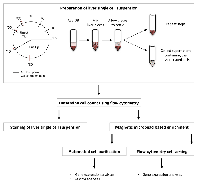 Figure 1: Workflow of the described protocol.
Please click here to view a larger version of this figure.
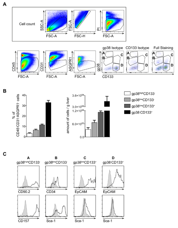 Figure 2: Novel classification of liver progenitor subsets. A single-cell suspension was prepared from healthy livers and subsequently stained with a panel of surface markers: CD45, CD31, CD133 and gp38 (A). For dead cell exclusion, propidium iodide (PI) was utilized. Representative dot plots with the gating strategy are depicted. The gates used for flow cytometry cell count determination are also marked. (B) The percentages and the absolute number of cells present per g of liver tissue of the various stromal cell subsets among the CD45-negative cells in wild-type untreated animals are shown. (C) The histograms display distinguishing marker characteristics of stromal cell subsets in a healthy liver. High expression of CD90.2 and the presence of CD157 are specific for A, while CD34 is specific for subpopulation B. Sca-1 is present in B, C, D and Epcam is only present in population C and D. Mean ± SEM; the data (A-C) represent 2-3 independent experiments with n = 3-4 per experiment. The data were compared using an unpaired, two-tailed T test * P<0.05, ** P<0.005, *** P<0.0001. Please click here to view a larger version of this figure.
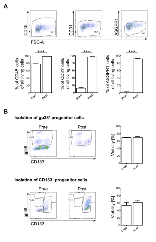 Figure 3: Magnetic microbead-based enrichment and automated cell purification. (A) Magnetic microbead-based enrichment of CD45-, CD31- and ASGPR1- cells are depicted before and after enrichment. (B) Representative dot plots of the automated microbead-based cell isolations are depicted for CD133+ and gp38+ cells, on the left. On the right, the viability of the cell populations before and after enrichment are shown as the percentage of propidium iodide (PI)-negative cells. Mean ± SEM; the data (A-B) represent 3 independent experiments with n = 3 per experiment. The data were compared using an unpaired, two-tailed T test * P<0.05, ** P<0.005, *** P<0.0001. Please click here to view a larger version of this figure.
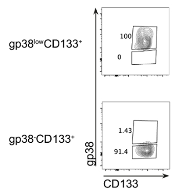 Figure 4: Flow cytometry sort of progenitor subsets with high purity. Dot plots depict the purity of the cell populations in the sorted samples (post-sort). The data represent 3 independent experiments. Please click here to view a larger version of this figure.
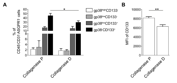 Figure 5: Comparison of digestion enzymes. The single-cell suspension was prepared using collagenase P (as described in step 2) or collagenase D (1 mg/mL + DNase-I 0.1 mg/mL) from healthy livers and was subsequently stained with a panel of surface markers: CD45, CD31, CD133, and gp38 (A). The percentages of cells present per g of liver tissue of the various stromal cell subsets among the CD45-negative cells in wild-type untreated animals are shown. (B) The median fluorescence intensity of CD133 is depicted for the CD133+ cell population. Mean ± SEM; the data (A-B) represent 2 independent experiments with n = 3 per experiment. The data were compared using an unpaired, two-tailed T test * P<0.05, ** P<0.005, *** P<0.0001. Please click here to view a larger version of this figure.
Discussion
Liver inflammation and injury of different origins trigger regenerative processes in the liver that are accompanied by progenitor cell expansion and activation2,3. These liver progenitor cells possess stem cell characteristics and likely play a significant role in the pathomechanism of various liver diseases.
The heterogeneity of liver progenitor cells has long been suggested. The re-evaluation of liver progenitor subsets using a novel surface marker combination of CD133 and gp38 could identify subsets that carry different surface markers and express a unique set of inflammation-related genes during liver injury12. The method described here allows for the direct flow cytometry analysis of rare cells and their direct comparison in various liver injury models. This is especially relevant, as various injuries trigger the activation of multiple progenitor subsets that could be reflective of the cellular heterogeneity observed in ductular reactions3,4,11,12. Notably, a small fraction of murine liver tissue (0.2 g) is enough to isolate sufficient cells for the flow cytometry analysis of progenitors using the described method.
Flow cytometry sorting providing high purity populations of the subsets is necessary to explore their unique gene expression profiles. Previous reports described flow cytometry-based progenitor cell isolation15,16. Nevertheless, the protocol presented here provides an optimized method that ensures the high purity and viability of these cells. Notably, in vitro culturing and expansion techniques described previously can be nicely combined with the protocol presented here15,16.
Many types of flow cytometry sorters are available at various institutions, and their particular instrumentations might be slightly different. Nevertheless, the general principles for cell sorting are presented in the described protocol. Generally, a lower pressure and bigger nozzle size are absolutely necessary, independent of the machine types and specifications. Moreover, the utilization of an appropriate sorting medium can significantly increase the viability and the RNA quality of the sorted cells, which is an important factor, particularly for gene expression studies. The sorting of progenitor cells, however, greatly reduces cell viability, and the yield of cells after sorting is relatively low (for high purity sort of 7-10,000 events/subpopulation, 4-5 healthy livers need to be pooled). This could be a limiting factor for further in vitro analyses. We suggest magnetic microbead-based isolation for in vitro assays, because of its higher yield and cell viability, and flow cytometry sorting for gene expression analysis, especially when more specified subset analyses and isolations are needed.
Based on the fact that the viability of cells is greatly reduced after flow cytometry sorting, an automated magnetic microbead-based isolation of progenitor cells has been developed and presented here. It allows for the isolation of a larger number of progenitor cells with high purity (combining multiple livers). Furthermore, the viability of the cells is maintained, since the time necessary for the isolation process is greatly reduced. While lower numbers of progenitor cells can be expanded in vitro1,2,15,16, it is recommended to consider how these in vitro manipulations could alter the cellular features of stem/progenitor cells described for other mesenchymal and stromal cell populations17,18.
The major limitation of the presented magnetic microbead-based isolation is that the CD133+ and the gp38+ cell populations represent heterogeneous cells. Additional surface markers might be necessary to identify smaller cell populations.
The understanding of how hepatic stem cells and progenitors relate to each other is far from complete, despite excellent studies by Lola Reid and others1,8,19. Thus, the careful consideration of techniques resulting in higher yields and viability, such as the one suggested here, could help to extend the analyses of progenitor subsets and the understanding of their interconnected relationships.
Overall, we have described the detailed isolation and analysis of liver progenitor subsets that were recently distinguished12. Moreover, by employing a novel enzyme combination, rare progenitors could be analyzed by direct flow cytometry measurements. Additionally, the protocol demonstrates how to enrich progenitor cells with a high purity, using either cell sorting or an automated microbead-based technique.
Troubleshooting:
The percentages of cell debris and dying cells are high
If, during digestion (step 2), the pipetting of the liver samples is too harsh, the percentage of dying cells in the single-cell suspension increases. If the liver is cut into larger pieces than suggested, the digestion is less efficient and results in more cell death during preparation.
After completing the procedure in step 5, the purity of the samples is not sufficient
If the percentages of cell debris and dying cells in the single-cell suspension prepared in step 2 are too high, the microbeads bind unspecifically, and therefore, the purity of the isolation is greatly reduced.
Low cell number after microbead-based purification
For the success of the described protocol, it is important that the ratio of cells to microbeads is optimal, as suggested in the protocol. Utilizing more microbeads reduces the purity and decreases the yield and viability of the isolated cells. If surface markers other than those presented in the protocol are used for progenitor subset isolation, the optimal ratio of cells to microbeads must be determined.
Magnetic microbead based isolation of gp38+CD133- population
Follow steps in section 5. In step 5.3 add additionally 10 µL anti-CD133+ microbeads, then follow steps 5, 6.2 and 6.3 as described.
Calculating absolute cell numbers
The liver weight is used to calculate how many cells of a certain subset are present per gram liver tissue.
(% of the subpopulation in living cells (based on flow cytometry) / 100) x total living cell number isolated from the liver piece = Z
(1/ weight of liver piece) x Z = cell number/g liver tissue
Other cell populations that can be isolated with this method
It is possible to isolate the following liver cells using this digestion protocol: CD45+ cells (or subpopulations of hematopoietic cells, e.g. Kupffer cells, dendritic cells, T cells, NK cells, etc.) and LSECs. The digestion protocol is not suitable for isolation of hepatocytes or hepatic stellate cells. These cells are present at relatively low cell numbers and they show reduced viability compared to other available methods.
Disclosures
The authors have no competing financial interests.
Acknowledgments
This work was supported by the Alexander von Humboldt Foundation Sofja Kovalevskaja Award to VLK.
References
- Dolle L, et al. Successful isolation of liver progenitor cells by aldehyde dehydrogenase activity in naive mice. Hepatology. 2012;55(2):540–552. doi: 10.1002/hep.24693. [DOI] [PubMed] [Google Scholar]
- Dolle L, et al. The quest for liver progenitor cells: a practical point of view. J Hepatol. 2010;52(1):117–129. doi: 10.1016/j.jhep.2009.10.009. [DOI] [PubMed] [Google Scholar]
- Dorrell C, et al. Surface markers for the murine oval cell response. Hepatology. 2008;48(4):1282–1291. doi: 10.1002/hep.22468. [DOI] [PMC free article] [PubMed] [Google Scholar]
- Gouw AS, Clouston AD, Theise ND. Ductular reactions in human liver: diversity at the interface. Hepatology. 2011;54(5):1853–1863. doi: 10.1002/hep.24613. [DOI] [PubMed] [Google Scholar]
- Tarlow BD, Finegold MJ, Grompe M. Clonal tracing of Sox9+ liver progenitors in mouse oval cell injury. Hepatology. 2014;60(1):278–289. doi: 10.1002/hep.27084. [DOI] [PMC free article] [PubMed] [Google Scholar]
- Shin S, et al. Foxl1-Cre-marked adult hepatic progenitors have clonogenic and bilineage differentiation potential. Genes Dev. 2011;25(11):1185–1192. doi: 10.1101/gad.2027811. [DOI] [PMC free article] [PubMed] [Google Scholar]
- Furuyama K, et al. Continuous cell supply from a Sox9-expressing progenitor zone in adult liver, exocrine pancreas and intestine. Nat Genet. 2011;43(1):34–41. doi: 10.1038/ng.722. [DOI] [PubMed] [Google Scholar]
- Font-Burgada J, et al. Hybrid Periportal Hepatocytes Regenerate the Injured Liver without Giving Rise to Cancer. Cell. 2015;162(4):766–779. doi: 10.1016/j.cell.2015.07.026. [DOI] [PMC free article] [PubMed] [Google Scholar]
- Kuramitsu K, et al. Failure of fibrotic liver regeneration in mice is linked to a severe fibrogenic response driven by hepatic progenitor cell activation. Am J Pathol. 2013;183(1):182–194. doi: 10.1016/j.ajpath.2013.03.018. [DOI] [PMC free article] [PubMed] [Google Scholar]
- Pritchard MT, Nagy LE. Hepatic fibrosis is enhanced and accompanied by robust oval cell activation after chronic carbon tetrachloride administration to Egr-1-deficient mice. Am J Pathol. 2010;176(6):2743–2752. doi: 10.2353/ajpath.2010.091186. [DOI] [PMC free article] [PubMed] [Google Scholar]
- Spee B, et al. Characterisation of the liver progenitor cell niche in liver diseases: potential involvement of Wnt and Notch signalling. Gut. 2010;59(2):247–257. doi: 10.1136/gut.2009.188367. [DOI] [PubMed] [Google Scholar]
- Eckert C, et al. Podoplanin discriminates distinct stromal cell populations and a novel progenitor subset in the liver. Am J Physiol Gastrointest Liver Physiol. 2016;310(1):G1–G12. doi: 10.1152/ajpgi.00344.2015. [DOI] [PMC free article] [PubMed] [Google Scholar]
- Epting CL, et al. Stem cell antigen-1 is necessary for cell-cycle withdrawal and myoblast differentiation in C2C12 cells. J Cell Sci. 2004;117(Pt 25):6185–6195. doi: 10.1242/jcs.01548. [DOI] [PubMed] [Google Scholar]
- Tirnitz-Parker JE, Tonkin JN, Knight B, Olynyk JK, Yeoh GC. Isolation, culture and immortalisation of hepatic oval cells from adult mice fed a choline-deficient, ethionine-supplemented diet. Int J Biochem Cell Biol. 2007;39(12):2226–2239. doi: 10.1016/j.biocel.2007.06.008. [DOI] [PubMed] [Google Scholar]
- Rountree CB, et al. A CD133-expressing murine liver oval cell population with bilineage potential. Stem Cells. 2007;25(10):2419–2429. doi: 10.1634/stemcells.2007-0176. [DOI] [PubMed] [Google Scholar]
- Rountree CB, Ding W, Dang H, Vankirk C, Crooks GM. Isolation of CD133+ liver stem cells for clonal expansion. J Vis Exp. 2011. [DOI] [PMC free article] [PubMed]
- Sidney LE, McIntosh OD, Hopkinson A. Phenotypic Change and Induction of Cytokeratin Expression During In Vitro Culture of Corneal Stromal Cells. Invest Ophthalmol Vis Sci. 2015;56(12):7225–7235. doi: 10.1167/iovs.15-17810. [DOI] [PubMed] [Google Scholar]
- Hass R, Kasper C, Bohm S, Jacobs R. Different populations and sources of human mesenchymal stem cells (MSC): A comparison of adult and neonatal tissue-derived MSC. Cell Commun Signal. 2011;9:12. doi: 10.1186/1478-811X-9-12. [DOI] [PMC free article] [PubMed] [Google Scholar]
- Schmelzer E, et al. Human hepatic stem cells from fetal and postnatal donors. J Exp Med. 2007;204(8):1973–1987. doi: 10.1084/jem.20061603. [DOI] [PMC free article] [PubMed] [Google Scholar]


