

Abstract
Thoracic spinal psammomatous meningioma is a rare subtype of meningioma. Among diverse types of mesenchymal differentiation, osseous metaplasia is found to be still rarer. We are presenting a new case of thoracic psammomatous spinal meningioma with osseous metaplasia in a middle aged female which that gives a sense of cancellous bone in the spinal canal. To conclude, meningiomas with osseous metaplasia are very rare tumors that complicate the surgical removal in certain cases. Ossification, if predicted prior to operation with computed tomography reconstruction, makes planning of removal easier. In our case, maintained cerebrospinal fluid spaces despite hard consistency of tumor made its removal easier once cerebrospinal fluid was drained. We have submitted this article because it is very rare and curable pathology and preoperative diagnosis helps in prevention of neurological injury during its excision.
Keywords: Ossified meningioma, psammomatous meningioma, osseous metaplasia
Introduction
Meningioma together with schwannomas represents majority of intradural extramedullary spinal tumors. Presence of psammomatous bodies, gritty foci of calcification are frequently found in spinal meningioma. However, extensive or entire ossification is not that common and when encountered, it complicates removal in certain cases, and thus affects postoperative course and prognosis. Meningioma with metaplasia is a rare subtype of meningioma. By definition, the mesenchymal differentiation includes osseous, cartilaginous, lipomatous, myxoid, or xanthomatous changes in meningiomas.[1,2]
To our knowledge, there are less than 25 cases reporting ossified psammomatous spinal meningiomas.[3] Aim of this article is to present a new case of ossified spinal meningioma that looks like an oval stone in thoracic spinal meningioma.
Case Report
A 60-year-old female presented with the following complaints:
Progressive weakness bilateral lower limb, left more than right for the last one year
Numbness starting just above umbilicus lowers down for the last one year
Urinary incontinence for the last two months
Inability to walk for the last twenty days.
Neurological examination revealed paraparesis, knee and ankle jerk exaggerated, plantar's bilateral upwards, ankle clonus present and tone in lower limb increased. Thoracic magnetic resonance imaging showed a hypointense lesion on T1 and T2 image at the level of T7 and T8 and causing compression of the cord anterolaterally on the left side with widening of posterior cerebrospinal fluid column with positive supra-and infra- meniscal sign above MR signals suggesting a calcified meningioma [Figures 1 and 2].
Figure 1.

MRI transverse section showing meningioma at D7 compressing cord anterolaterally
Figure 2.
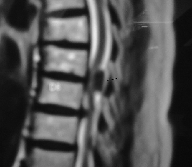
Saggital section MRI dorsolumbar spine showing meningioma at D7 compressing cord anteriorly
Complete T7 and T8 laminectomy was done. Overlying dura was thick and firm. Midline dural opening was made and dura dissected free from tumor on both sides with difficulty, as it was adherent to the tumor. Around it, tumor was easy to dissect from its ventral aspect, as it was not adherent and hard ventrally. Dura affliction was more on the right side than on the left. Unlike rest of the tumor, the dural attachment was soft to firm and richly vascular [Figure 3]. Tumor was removed in toto and cauterization of dural affiliction was done [Figure 4]. Postoperatively, patient had immediate improvement with disappearance of band-like tightening sensation of supraumbilical area in the abdomen.
Figure 3.

Intraoperative picture showing dural exposure with center hard gritty part of meningioma
Figure 4.

Intraoperative picture after excision of meningioma and dural repair
Patient was in follow-up regularly every month for eight months postoperatively. Clinically, patient improved from grade ‘0’ muscle power in lower limbs to grade ‘4+’ and was catheter free after six months.
Pathological findings
The excised tumor mass was subjected to histopathological examination, which on gross inspection revealed a tumor measuring 1.5 × 1.2 × 0.6 cm with attached dura. The tumor was composed predominantly of psammoma bodies and immature bony trabeculae with interspersed whorls and lobules of meningothelial cells containing ovoid nuclei, inconspicuous nucleoli, and moderate amount of eosinophilic cytoplasm.[4,5,6,7,8] There was no evidence of mitotic activity or necrosis [Figures 5 and 6]. Adjoining dural collagen was infiltrated by tumor. Final impression was psammomatous meningioma with osseous metaplasia, WHO grade I intradural, T7 vertebral region.
Figure 5.

Tissue diagnosis showing ossification with psammoma bodies and osseous metaplasia at magnification ×100
Figure 6.

Tissue diagnosis showing ossification with psammoma bodies at magnification ×40
Discussion
Ossification in spinal meningioma is a rare event. First case of ossified meningioma was reported by Roger in 1928.[9] Less than 25 cases are reported till date, and most of the cases are in thoracic region, as in our case. Even after reports of certain new cases, there are no definitive figures of its incidence, which is roughly estimated to be 0.7-5.5% of all spinal tumors.[5,6,10,11]
Because of its rigid nature, removal of tumor is associated with complications affecting surgical outcome. But in our case, oval shape of the tumor and its nonadherence on the spinal cord, made its removal easier, once the superior and inferior poles of the tumor identified and its dissection led to drainage of CSF from the spaces. Different long-term outcomes of ossified meningioma surgery have been reported. Roux et al. reported 3 ossified meningioma of 54 spinal meningioma with total removal with good outcome in 2 of these. One removal was subtotal and required a second surgery and radiotherapy.[12]
Conclusion
In conclusion, meningiomas with osseous metaplasia are very rare tumors which complicate surgery in certain cases. Ossification predicted prior to surgery with computed tomography reconstruction makes planning of removal of even hard tumor easier. In our case, maintained CSF spaces made removal easier, once CSF is drained; even without prior computed tomography reconstructive images.
Financial support and sponsorship
Nil.
Conflicts of interest
There are no conflicts of interest.
Acknowledgment
We are thankful to the medical superintendent of C.S.S.H. Hospital attached to Subharti Medical College, Meerut for granting us the permission to publish this material. We declare that this is our work, except where acknowledged specifically as the published or unpublished work of others. We are also thankful to the patient and his attendant for cooperation during preparation of this material.
References
- 1.Louis DN, Scheithauer BW, Budka H. Meningiomas. In: Kleihues P, Cavenee WK, editors. World Health Organization Classification of Tumours, Pathology and Genetics of Tumours of the Nervous System. Lyon: IARC Press; 2000. pp. 176–84. [Google Scholar]
- 2.Perry A, Louis DN, Scheithauer BW. Meningiomas. In: Louis DN, Ohgaki H, Wiestler OD, editors. World Health Organization Classification of Tumors of the Central Nervous System. Lyon: IARC Press; 2007. pp. 164–72. [Google Scholar]
- 3.Liu CL, Lai PL, Jung SM, Liao CC. Thoracic ossified meningioma and osteoporotic burst fracture: Treatment with combined vertebroplasty and laminectomy without instrumentation: Case report. J Neurosurg Spine. 2006;4:256–9. doi: 10.3171/spi.2006.4.3.256. [DOI] [PubMed] [Google Scholar]
- 4.Naderi S, Yilmaz M, Canda T, Acar U. Ossified thoracic spinal meningioma in childhood: A case report and review of the literature. Clin Neurol Neurosurg. 2001;103:247–9. doi: 10.1016/s0303-8467(01)00157-3. [DOI] [PubMed] [Google Scholar]
- 5.Kato K, Chernov M, Urino T, Kasuya H, Kubo O, Isehi H, et al. Ossified frontosphenoorbital meningioma en plaque, mimicking extensive hyperostosis. Minim Invasive Neurosurg. 2008;51:237–9. doi: 10.1055/s-2008-1080906. [DOI] [PubMed] [Google Scholar]
- 6.Kaufman AB, Dunsmore RH. Clinicopathological considerations in spinal meningeal calcification and ossification. Neurology. 1971;21:1243–8. doi: 10.1212/wnl.21.12.1243. [DOI] [PubMed] [Google Scholar]
- 7.Pepler WJ. Alkaline phosphatase in the meninges and meningiomas. Nature. 1960;186:979. doi: 10.1038/186979a0. [DOI] [PubMed] [Google Scholar]
- 8.Bosnjak R, Derham C, Popović M, Ravnik J. Spontaneous intracranial meningioma bleeding: Clinicopathological features and outcome. J Neurosurg. 2005;103:473–84. doi: 10.3171/jns.2005.103.3.0473. [DOI] [PubMed] [Google Scholar]
- 9.Rogers L. A spinal meningioma containing bone. Br J Surg. 1928;15:675–7. [Google Scholar]
- 10.Colón GP, Ross DA, Hoff JT. Sequential outer table craniotomy a in hyperossified meningioma. Technical note. J Neurosurg. 1998;88:346–8. doi: 10.3171/jns.1998.88.2.0346. [DOI] [PubMed] [Google Scholar]
- 11.Mathuriya SN, Vasishta RK, Khandelwal N, Pathak A, Sharma BS, Khosla VK. Calcified falx meningioma. Neurol India. 2000;48:285–7. [PubMed] [Google Scholar]
- 12.Roux FX, Nataf F, Pinaudeau M. Intraspinal meningiomas: Review of 54 cases with discussion of poor prognosis factors and modern therapeutic management. Surg Neurol. 1996;46:458–64. doi: 10.1016/s0090-3019(96)00199-1. [DOI] [PubMed] [Google Scholar]