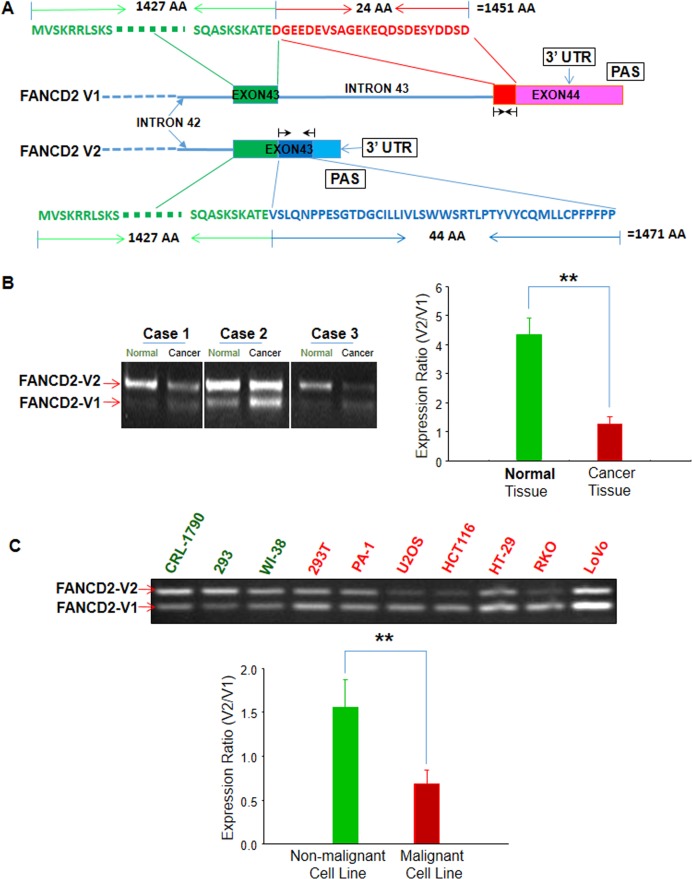
Figure 1. Two variants of FANCD2: namely “FANCD2-V1 (V1)” for the long-known FANCD2 and “FANCD2-V2 (V2)” for the overlooked one.
V2 is relatively highly expressed in benign cells or tissues as compared to V1. (A) FANCD2 gene contains two potential polyadenylation signaling motifs, which result in two proteinic variants: V1 and V2 that have a large common amino terminal (1427 AAs) (green-colored) and a 24 or 44 unique AA at the C-terminal respectively (red/V1 or blue/V2) (Black arrowheads indicate the RT-PCR primers used for detecting V1 or V2 mRNA expression.). (B) RT-PCR shows V1 or V2 relative expressions in malignant or matched non-malignant lung tissues. V2 is relatively high expressed in non-malignant tissues as compared to V1 (t-test, **p < 0.01). (C) RT-PCR shows V1 or V2 relative expressions in malignant or non-malignant human cell lines. V1 expression is relatively higher than V2 in malignant cells or transformed cells (PA-1, U2OS, HCT116, RKO, HT-29, LoVo and 293T) compared to non-malignant cells tested (CRL-1790, 293 and WI38) (t-test, **: p < 0.01). (All bar graphs were plotted from ImageJ quantification of RT-PCR results.)

