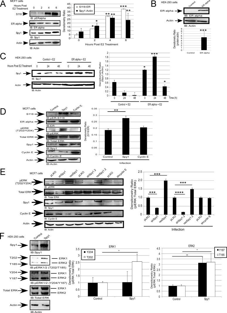
Figure 1. Spy1 is upregulated downstream of the estrogen receptor.
(A) MCF7 cells were treated with 50 nM of estradiol (E2) or vehicle control (DMSO) over the indicated time course. Representative blot (left), densitometry averages Spy1 and S118 (right). (B–C) Hek-293 cells were transfected with pEGFP-C1-ERα. B-Representative blot confirming expression. C-Treatment with 50 nM of E2 over the indicated time course. Representative blot (left), densitometry averages for Spy1 (right). (D) MCF7 cells infected with control, Spy1, or Cyclin E1.(E) Cells were infected pLKO, 2 constructs of shSpy1.1, shSpy1.2, shCyclin E, or rescue vectors, followed by SDS-PAGE and IB. Representative blot (left), densitometry averages for relative ER-S118 (right). (F) Representative blot (upper panel) showing phosphorylation status of ERK1 and ERK2 using phosphor-specific antibodies for pERK threonine or tyrosine sites. Lower panel depicts quantification of both sites for either ERK1 (left) or ERK2 (right). Error bars reflect SE between at least 3 separate experiments. Student's t-test was performed; *p < 0.05, **p < 0.01, ***p < 0.001.

