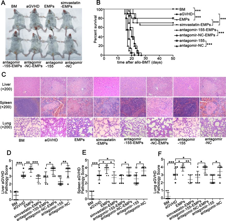
Figure 5. Suppression of miR-155 in EMPs by antagomir-155 ameliorates the exacerbated aGVHD induced by high concentration of EMPs.
After being subjected to lethal total body irradiation, BALB/c mice were transplanted intravenously with 1 × 10⁷ BM cells and 2 × 10⁷ spleen cells isolated from C57BL/6 donors to establish aGVHD model. EMPs (60 μg) isolated from mouse TNF-α-stimulated MAECs, TNF-α-stimulated MAECs protected by 2.5 μmol/L simvastatin, TNF-α-stimulated MAECs transfected by antagomir-155, TNF-α-stimulated MAECs transfected by antagomir-NC were given intravenously to aGVHD mice on day0 and +7d, retrospectively. Antagomir-155 and antagomir-NC of 25 mg/kg was given intravenously on +7d followed by 5 mg/kg twice weekly up to +21d. (A) Representative photograph of clinical severity of aGVHD in each group. (B) Survival of recipients was compared using the Mantel-Cox log-rank test. Shown is the mean ± SD from three combined independent experiments. ***P < 0.001. (C) Liver, spleen and lung were harvested at +21d and representative histological analysis of hematoxylin-eosin (H&E) staining was shown (original magnification ×200). (D–F) H&E-stained slides of liver (D), spleen (E) and lung (F) were scored for histopathologic damage and lymphocyte infiltration. Values are means ± SD. *P < 0.05, *P < 0.01, ***P < 0.001.

