Abstract
Synovial chondromatosis affecting the glenohumeral joint is rare. Treatment primarily consists of arthroscopic loose body removal and synovectomy. Shoulder arthroplasty has been mentioned in the literature as a treatment option for patients with coexisting arthritis, although the results have been underreported. The case of an 84-year-old man with long standing synovial chondromatosis of the shoulder resulting in severe degenerative disease is presented. The patient was treated with a reverse total shoulder arthroplasty, loose body removal, and a complete synovectomy. Three and six month follow up results have shown a decrease in the visual analogue scale for pain, improved range of motion, and no radiographic evidence of disease recurrence. Reverse total shoulder arthroplasty is a viable treatment option for synovial chondromatosis in patients with coexisting glenohumeral arthritis demonstrating good short term outcomes.
Keywords: Arthroplasty, Chondromatosis, Reverse, Shoulder, Treatment
Introduction
Synovial chondromatosis is a disease process affecting diarthroidal joints, characterized by metaplastic chondroid tissue within a proliferative synovial lining (1). The exact prevalence is unknown given to the rarity of the disease, but cases have been described globally (1-4). Young adults aged 30-50 are most often described as being affected, with a male to female predilection of 2:1 (3). Large joints are more commonly affected, with the knee being the most common site followed by the hip (4). Rarely does it affect the shoulder, although case series have been reported (1-7).
Synovial chondromatosis has been classified as occurring from primary or secondary sources. The primary form is thought to occur spontaneously from metaplastic changes within ectopic foci of cartilage in the synovium (1). Secondary disease occurs in the setting of underlying joint pathology (8). Although synovial chondromatosis is considered a relatively benign process, the intra-articular loose bodies formed can ultimately lead to joint destruction and degeneration. Sarcomatous transformation has also been reported in the literature occurring at 5% (9).
Treatment primarily consists of open or arthroscopic removal of loose bodies and synovectomy. Outcomes reported have generally been favorable with a low recurrence rate(6, 7, 9). There is a paucity of literature describing the results or recurrence of patients undergoing arthroplasty for severe degenerative joint disease caused by synovial chondromatosis. In this paper we present the case of a patient who underwent a reverse total shoulder arthroplasty for synovial chondromatosis and achieved good short term results with no evidence of recurrence. The authors have obtained the patient’s informed written consent for print and electronic publication of the case report.
Case presentation
An 84-year-old right hand dominant male presented with the chief complaint of right shoulder pain and loss of motion that has progressively worsened over the past ten years. He reports no previous history or incident of trauma to that shoulder. The pain has limited his ability to perform activities of daily living, and he does note a progressive loss of strength. A self-reported visual analogue scale (VAS) for pain was described as 7/10. Conservative treatment consisting of steroid injections and physical therapy was initiated in the past with limited success.
On physical exam, there was no gross deformity, effusion, or erythema noted to the right shoulder. Mild atrophy was noted to the deltoid musculature when compared to the contralateral side. Range of motion (ROM) was noted to be 0-80 degrees forward flexion, 0-60 degrees of abduction, and internal rotation to the level of the hip with palpable crepitus throughout. No palpable masses were present. A firm mechanical block to motion was noted at terminal flexion and abduction. Motor testing revealed 3/5 strength to the rotator cuff musculature when compared to contralateral extremity.
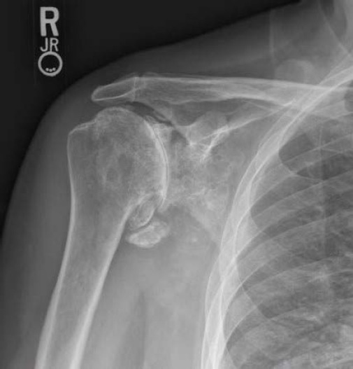
Plain radiographs revealed severe degenerative changes to the glenohumeral joint including subchondral sclerosis as well as multiple radiodense loose bodies [Figure 1]. Magnetic resonance imaging (MRI) confirmed the radiographic findings and also demonstrated high grade rotator cuff tedinopathy with atrophy and fatty infiltration to the rotator cuff musculature. The decision was made to proceed with a reverse total shoulder arthroplasty given the severe degenerative changes present as well as the tearing and atrophy noted to the rotator cuff.
Figure 1.

Right shoulder radiograph demonstrating severe glenohumeral degenerative changes with multiple radiodense loose bodies.
Operative procedure
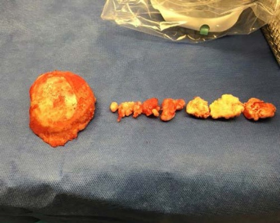
A standard deltopectoral approach was used to access the glenohumeral joint. Upon making the capsulotomy, several loose bodies were encountered within the synovial tissue and subsequently removed. Inspection of the humeral head revealed severe loss of articular cartilage. Following excision of the humeral head, multiple loose bodies were noted in the surrounding synovium of the posterior as well as anteroinferior portion of the glenohumeral joint [Figure 2]. The loose bodies had a “shiny popcorn” appearance to them, consistent with synovial chondromatosis. An extensive synovectomy was performed and all visible loose bodies were removed. All specimens were sent to the pathology department for evaluation. The reverse total shoulder arthroplasty was then completed in a standard fashion. Post-operative radiographs taken in the recovery room revealed residual calcification within the axillary recess that we were unable to appreciate intraoperatively.
Figure 2.

From left to right, the excised humeral head and several intra-articular loose bodies. The humeral head demonstrates gross loss of articular cartilage.
Gross examination revealed the loose bodies consisted of osseous nodular tissue covered in tan-grey cartilage. Histologic evaluation revealed osteochondromatous bodies with degenerative changes and focal synovial chondrometaplasia, confirming the diagnosis of synovial chondromatosis.
Post operative course
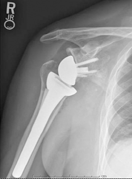
Post operatively the patient was placed into a sling and initially started on pendulum exercises. Physical therapy was initiated at week two with protection of external rotation until week six post surgery. At the three month follow up appointment the patient reported a VAS score of 1/10. The patient denied any episodes of instability or mechanical blockage with range of motion. Range of motion was noted to be 140 degrees of forward flexion, 100 degrees of abduction, and internal rotation was noted to level of L5-S1 junction. Radiographs were taken in the office and revealed well fixed components with no evidence of disease recurrence at three months and six months post-operatively [Figure 3].
Figure 3.

Radiograph taken at the sixth-month post-operative visit demonstrating a stable reverse total shoulder arthroplasty with no evidence of disease recurrence.
Discussion
Synovial chondromatosis of the shoulder is rare disease that has been primarily described in small series and case reports (4, 6, 7, 9, 10). Currently, the etiology of synovial chondromatosis is unknown. Jaffe was the first to describe the disease process in 1958 as intrasynovial loose bodies composed of metaplastic chondroid tissue (1). Milgram further defined the origin of loose bodies into those arising from primary synovial proliferation or those arising from secondary sources such as osteophytes or osteochondral fractures (3). Three stages of primary synovial chondromatosis have been described by Milgram consisting of an early, transitional, and a late stage (3). The early stage is composed of active synovium without evidence of loose bodies. The transitional stage contains both active synovium as well as loose bodies while the late stage contains only loose bodies. Villacin et al developed a histologic criterion to differentiate primary and secondary forms which have been shown to correlate with clinical outcomes (8). Patients suffering from primary disease had a 60% recurrence rate whereas those with secondary disease had no recurrence (8). Our patient most likely suffered from end stage arthritis due to long standing untreated synovial chondromatosis. Although degenerative changes were present to suggest this as the source of the intra-articular loose bodies, histologic evaluation did show focal synovial chondrometaplasia. Based on the presence of intra-articular loose bodies with active synovium, this patient demonstrated to be in the transitional stage of the disease (4).
Symptoms in patients with synovial chondromatosis often include insidious pain, swelling, and decreased ROM, closely resembling other disease processes (3). Joint effusion, crepitus, and palpable loose bodies are common physical exam findings. Plain radiographs classically reveal multiple calcified intra-articular loose bodies, although these findings have been shown to be absent in up to 30% of cases (5). Due to the vague symptomatology and rarity of the disease, diagnosis is often delayed or missed (7). No correlation has been established regarding the duration of symptoms and pathologic stage of the disease (5).
Primarily affecting young adults, the goal of treatment is prevention of articular cartilage damage. In the shoulder this is accomplished by arthroscopic or open removal of loose bodies with or without synovectomy (2-4, 6, 7, 9). Despite good overall clinical results, recurrence rates remain variable (6, 7, 9). Urbach et al looked at ten year follow up data on a subset of five patients diagnosed with synovial chondromatosis of the shoulder that underwent arthroscopic removal of loose bodies and partial synovectomy. Two of the five patients had recurrence of the disease despite good clinical outcomes with no progression of arthritis on radiographs (9). Maurice et al had a recurrence rate in knees of 11.5% and found no difference between removal of loose bodies alone or combined with synovectomy. They attributed the recurrence rates to patients that had an early stage of the disease with active synovium (2). Late recurrence at an average of 18 months following arthroscopic synovectomy in the shoulder has been shown in two additional case studies (6, 7). Shoulder arthroplasty for end stage arthritis associated with synovial chondromatosis has been mentioned as a treatment option for certain patients, but outcomes have never been reported (6, 7, 9). Ackerman et al reported the results of eleven patients who underwent total knee or total hip arthroplasty for synovial chondromatosis. They showed improved pain, functional scores, and ROM, although ROM was decreased compared to patients undergoing arthroplasty for primary osteoarthritis. Two of the patients had recurrence of the disease despite total synovectomy requiring a subsequent synovectomy (10). Our patient underwent reverse total shoulder arthroplasty given the existing severe degenerative changes to the glenohumeral joint and concomitant rotator cuff arthropathy. The decision was made intraoperatively to perform a complete synovectomy with removal of all visible loose bodies. While recurrence tends to be low following surgical intervention, the histologic finding of active synovium in our patient could certainly increase the risk.
To our knowledge, there is no literature published on arthroplasty of the shoulder for synovial chondromatosis. Little is known about the outcomes or recurrence rates of synovial chondromatosis following shoulder arthroplasty other than what is extrapolated from total hip and knee studies (10). Recurrence could certainly have consequences on pain, ROM, and component longevity. This case study demonstrates that reverse total shoulder arthroplasty is a viable option for synovial chondromatosis with underlying degenerative joint disease. Our patient showed improved VAS pain scores as well as improvement in overall ROM and functionality with short term follow up. Long term follow up is warranted in this patient given the reports of late recurrence.
References
- 1.Jaffe HL. Synovial chondromatosis and other benign articular tumors. In: Jaffe HL, editor. Tumors and tumorous conditions of the bones and joints. Philadelphia: Lea & Febiger; 1958. pp. 558–67. [Google Scholar]
- 2.Maurice H, Crone M, Watt I. Synovial chondromatosis. J Bone Joint Surg. 1988;70(5):807–11. doi: 10.1302/0301-620X.70B5.3192585. [DOI] [PubMed] [Google Scholar]
- 3.Milgram JW. Synovial osteochondromatosis: a histopathological study of thirty cases. J Bone Joint Surg Am. 1977;59(6):792–801. [PubMed] [Google Scholar]
- 4.McFarland EG, Neira CA. Synovial chondromatosis of the shoulder associated with osteoarthritis: conservative treatment in two cases and review of the literature. Am J Orthop (Belle Mead NJ) 2000;29(10):785–7. [PubMed] [Google Scholar]
- 5.Hermann G, Klein M, Abdelwahab I, Kenan S. Synovial chondrosarcoma arising in synovial chondromatosis of the right hip. Skeletal Radiol. 1997;26(6):366–9. doi: 10.1007/s002560050249. [DOI] [PubMed] [Google Scholar]
- 6.Jeon IH, Ihn JC, Kyung HS. Recurrence of synovial chondromatosis of the glenohumeral joint after arthroscopic treatment. Arthroscopy. 2004;20(5):524–7. doi: 10.1016/j.arthro.2004.03.014. [DOI] [PubMed] [Google Scholar]
- 7.Covall DJ, Fowble CD. Arthroscopic treatment of synovial chondromatosis of the shoulder and biceps tendon sheath. Arthroscopy. 1993;9(5):602–4. doi: 10.1016/s0749-8063(05)80414-1. [DOI] [PubMed] [Google Scholar]
- 8.Villacin AB, Bringham LN, Bullough PG. Primary and secondary synovial chondrometaplasia: histopathologic and clinicoradiologic differences. Hum Pathol. 1979;10(4):439–51. doi: 10.1016/s0046-8177(79)80050-7. [DOI] [PubMed] [Google Scholar]
- 9.Urbach D, McGuigan FX, John M, Neumann W, Ender SA. Long-term results after arthroscopic treatment of synovial chondromatosis of the shoulder. Arthroscopy. 2008;24(3):318–23. doi: 10.1016/j.arthro.2007.08.034. [DOI] [PubMed] [Google Scholar]
- 10.Ackerman D, Lett P, Galat DD, Jr, Parvizi J, Stuart MJ. Results of total hip and total knee arthroplasties in patients with synovial chondromatosis. J Arthroplasty. 2008;23(3):395–400. doi: 10.1016/j.arth.2007.06.014. [DOI] [PubMed] [Google Scholar]


