Abstract
Background:
Management of acetabular fractures in the senior population can be one of the most challenging injuries to manage. Furthermore, treating surgeons have a paucity of information to guide the treatment in this patient population.
The purpose of this study was to determine:
(1) demographic and epidemiologic data, (2) mortality rates for nonoperative compared to operative management at different time points, (3) common fracture configurations, and (4) fracture fixation strategies in senior patients treated with acetabular fractures.
Methods:
Retrospective review of prospectively gathered data at a Level I trauma center over a five-year period. 1123 acetabular fractures were identified. 156 of them were for patients over the age of 65 (average age of 78).
Results:
Falls and motor vehicle accidents accounted for the two most common mechanisms of injury. 82% of patients had significant medical comorbidities. 51 patients (33%) died within one year, in which 75% of them died within 90 days of their acetabular fracture. 84% of the deceased patients, i.e. from the group of 51 patients, had non-operative treatment. For patients treated with traction alone, there was a 79% one-year mortality and 50% mortality rate within 90 days. Within the entire cohort, 70% had either an associated both-column (ABC) or anterior column/posterior hemitransverse (AC/PHT) fracture pattern. Fifty-seven patients (36.5%) underwent open reduction and internal fixation using standard reduction techniques and surgical implants via two main surgical exposures of ilioinguinal (69%) and Kocher-Langenbeck (29%).
Conclusion:
Geriatric patients with acetabular fractures are uncommon accounting for only 14% of all acetabular fractures. Patients who undergo surgery show lower mortality rates. ABC and AC/PHT fracture patterns are the two most common fracture patterns. Routine fixation constructs and implants can be used to manage these challenging fractures. Most patients are unable to return to their homes and instead require skilled nursing facility during their convalescence.
Keywords: Acetabular fractures, Geriatric patient, Outcomes, Senior
Introduction
The ideal treatment strategy for the geriatric patient with an acetabular fracture, whether due to high-energy trauma or a manifestation of osteoporosis and low-energy trauma, has not yet been completely characterized. There are few studies in the literature detailing the epidemiology of this subset of trauma patients. The treatment of geriatric patients is especially challenging given their increased likelihood of medical comorbidities and high probability of osteopenic or osteoporotic bone. To ensure appropriate treatment strategies are developed and utilized, it is important to have a thorough understanding of the underlying epidemiology of the senior patients with an acetabular fracture.
We sought to answer four specific questions: (1) what are the demographic and epidemiological data relating to this patient population, (2) what is the mortality rate in patients treated in a nonoperative manner and patients treated operatively at various time points, (3) what are common fracture configurations, and (4) can routine fixation strategies utilized in younger patients be utilized in geriatric patients with acetabular fractures?
Materials and Methods
After obtaining Institutional Review Board approval, we used a prospective fracture database including 1,123 acetabular fractures that were treated over a five-year period at Harborview Medical Center (Seattle, Wa). In order to highlight recent treatment strategies, we selected a subset of patients aged 65 years or older. This age limit was selected as a general indication of individuals entering their senior years – an age affiliated with the United States of America social security benefits. This subset analysis yielded 156 patients who had either operative or non-operative treatment for an acetabular fracture. This final cohort was reviewed for demographic data, mechanism of injury, pre-injury status, fracture type, treatment details, and survival at one year post-injury. Operation details were recorded for patients who had undergone open reduction internal fixation (ORIF). Patient mortality was identified by two means: medical chart data indicating patient death or search of the Social Security Death Index.
Radiographs and CT scans were reviewed from the date of injury and from the most recent follow-up to determine fracture configuration and fixation construct used on operatively managed patients. Data points were analyzed utilizing SPSS 17.0 (SPSS Inc., Chicago, IL).
IRB: This study was approved by the University of Washington Institutional Review Board.
Results
Demographic Data
The average age of the study population was 77.5 years (65 – 97). The average ages of patients treated operatively and nonoperatively were 75.5 (±8) and 78.6 (±7.4) (P= 0.018). There were 113 male and 43 female patients. Pre-injury status demonstrated an active population with 80% being community ambulators. For those patients treated with ORIF and/or exam-under-anesthesia (EUA), there was an average delay of 3.8 days (ranged from 1 to 20 days) from admission to time of surgery. The average hospital stay was 13.8 days (± 9.7) for patients who underwent surgical intervention and 11.1 (± 10.1) for those who had non-operative management (P=0.11). Post-hospital disposition showed that 25 (16%) were discharged to home and 120 (77%) were discharged to skilled nursing facility. Patient comorbidities were common with 128 (82%) of the group having an average of two medical comorbidities (ranged from 0 to 8). The most common comorbidities were 41 cases of hypertension (32%) and 33 cases of diabetes (26%). In the operative group, 43 of the patients (76%) had comorbidities with an average of 1.6 comorbidities (±1.3) per patient. In the nonoperative group, 84 of the patients (86%) had comorbidities with an average of 2.2 (±1.6) comorbidities per patient (P=0.02). The mere presence of a medical comorbidity among the groups was not statistically significant [Table 1].
Table 1.
Characteristics of operative and non-operative patients
| Variable | Operative Patients (N = 57) | Non-Operative Patients (N = 99) | |
|---|---|---|---|
| Mean age | 75.5 (8.0) | 78.6 (7.4) | P = 0.018 |
| Associated injuries | 23 (30%) | 33 (33%) | P = 0.09 |
| Medical comorbidities (YES) | 41/54 | 84/98 | P = 0.1 |
| Average number of medical comorbidities | 1.6 (1.3) | 2.2 (1.6) | P = 0.02 |
| Average number of days in hospital | 13.8 (9.7) | 11.1 (10.1) | P = 0.11 |
Fracture and Treatment Data
Mechanisms of injury included 70.5% falls, 23.1% resulting from motor vehicle crashes (MVC), 2.6% bicycle accidents, 1.3% pedestrians struck by cars, and 2.4% from others mechanisms. Simple falls and those from a standing height were associated with 48 patients (31%). Thirty-five percent of all patients had associated injuries. Interestingly, approximately 15% of the falls were from ladders with an average height of 11 feet and included several patients who fell from the rooftop of a home.
Fracture types were variable [see Tables 2, 3] but consisted predominantly of AO/OTA 62B (45%) and 62C (37%) or Letournel and Judet AC/PHT and ABC patterns, respectively. Treatment for these patients included 41.7% nonoperative without exam under anesthesia (EUA), 20.5% who underwent EUA and were ultimately treated non-operatively, and 36.5% who underwent ORIF.
Table 2.
Fracture Types Sustained by the Elderly Population (Utilizing the Letournel/Judet Classification)
| Letournel/Judet Fracture Classification | Firoozabadi et al. (N=156) | Helfet et al.{Helfet, 1992}(N=18) | Anglen et al. {Anglen, 2003} (N=48) | Spencer RF {Spencer, 1989}(N=25) | Hessmann et al.{Hessmann, 2002} (N=27) | Letournel and Judet{Letournel, 1993} (N=120) |
|---|---|---|---|---|---|---|
| Anterior Column | 5% | 11% | 10% | 9% | 15% | 8% |
| Anterior Wall | 1% | 22% | 7% | |||
| Posterior Column | 1% | 2% | 22% | 4% | 4% | |
| Posterior Wall | 12% | 23% | 4% | 7% | 33% | |
| Transverse | 3% | 27% | 48% | 7% | 3% | |
| Transverse/Posterior Wall | 4% | 22% | 8% | 7% | 17% | |
| T-Type | 5% | 6% | 17% | 15% | 3% | |
| AC/PHT | 35% | 39% | 11% | 15% | ||
| ABC | 34% | 28% | 10% | 7% | 25% | |
| Posterior Wall/Posterior Column | 1% | 13% | 4% | 5% |
Table 3.
Fracture Types Sustained by Elderly Population (AO/OTA Classification)
| Fracture Type | Firoozabadi et al. (N=156) | Anglen et al. (N=48) | Hessmann et al. (N=27) |
|---|---|---|---|
| 62A1 | 18 | 11 | 2 |
| 62A2 | 2 | 8 | 2 |
| 62A3 | 8 | 5 | 10 |
| 62B1 | 10 | 14 | 4 |
| 62B2 | 10 | 3 | 4 |
| 62B3 | 50 | 2 | 3 |
| 62C1 | 32 | 2 | 0 |
| 62C2 | 21 | 2 | 2 |
| 62C3 | 5 | 1 | 0 |
Nonoperative Treatment
In the nonoperative patient group, 77 cases (77%) had stable fractures and were treated with toe-touch weight-bearing precautions (weight of leg only) and 16 patients (16%) had unstable fractures but they were deamed medical unfit for an operation. Thus, they were treated with traction for four to six weeks. Standard protocols at our institution for all acetabular fractures patients consist of six weeks protected weight-bearing with toe-touch (weight of leg only) followed by a four to six week partial progressive weight-bearing regimen. All patients are typically full weight bearing by three months post-injury.
Operative Intervention
Average operative time was 206 minutes (ranged from 110 to 464 minutes). Three surgical exposures were most frequently utilized for 56 patients who underwent ORIF: the ilioinguinal including an intrapelvic interval (24 patients), the ilioinguinal approach with only the lateral and middle surgical intervals developed (13 patients), and the Kocher-Langenbeck approach (16 patients)(1-3). Two patients had a Smith-Petersen exposure, and one patient had sequential anterior and posterior exposures (4). The patients are positioned supine during the ilioinguinal exposures and we routinely develop the intrapelvic and vascular surgical intervals. The Kocher Langenbach exposures are performed with the patients positioned prone. Estimated intraoperative blood loss averaged 595 ml (ranged from 50–1900 ml). Intraoperative blood collection with a cell-saving device (Haemonetics, Braintree, MA) was used in each patient. In addition to autologous blood return, 30% of patients received intraoperative blood administration averaging 2 units (ranged from 1-5 units).
Operative Fracture Fixation
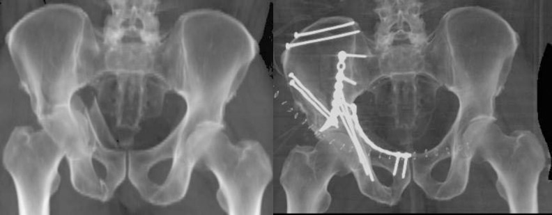
The fracture fixation constructs were predictably constant across the study time period. With the two primary fracture patterns noted, (AO/OTA 62B3 and 62C or ABC and AC/PHT), the constructs consisted predominantly of 3.5 mm malleable pelvic reconstruction plates (Zimmer, Warsaw, IN) located within the internal iliac fossa paralleling the pelvic brim, 3.5mm cortical lag screws directed into the posterior column stabilization both through the plate and independent from the plate [Figure 1a and 1b]. An intrapelvic 3.5 mm reconstruction plate was applied in those patients with intrusion of the quadrilateral surface (5). The average plate length was 7 holes (ranged from 6-9) for the pelvic brim plate and 10 holes (ranged from 9-14) for the intrapelvic plate. Standard 3.5 mm pelvic reconstruction plates and screws were used in all patients. The patients with transverse patterns (62B1 and B2) were treated with a Kocher-Langenbeck exposure and one or two 7-8 holed posterior column plates. The superior pubic ramus anterior column portion of the fracture was stabilized with 4.5 mm cortical screws (Synthes, Paoli, PA) located between the supra-acetabular region and the ipsilateral pubic symphysis. Occasionally, two medullary screws were placed depending on the individual osteology.
Figures 1.

(a) Significant intrusion of the quadrilateral surface giving a ‘protusio-type’ fracture pattern. (b) This is best addressed with the utilization of an intrapelvic plate buttressing the quadrilateral surface. This can assist with restoring the hip’s native offset.
Mortality
Patient mortality at one-year post-injury was 32.7% with 78.4% of these patients expiring within 75 days from injury. In patients treated non-operatively, one-year mortality was 44%. In the operative group, one-year mortality was 12%. One-year mortality and 90 day mortality were 79% and 47% respectively for patients treated with traction alone. The mortality data was stratified by age, and our data demonstrated a relatively even distribution among 5-year groups. Utilizing a chi-square analysis, there were no significant differences among the groups with 30-day, 90-day, or one-year mortality.
Discussion
As our aging population expands and the number of geriatric fractures increases, evidence-based treatment regimens will continue to be important in their management. There is a paucity of literature focused on defining acetabular fracture management in geriatric patients. In order to derive the best treatment strategies, we must first identify the population of interest and provide information relevant to the formulation of such appropriate strategies. The objective of this study was to determine the demographic and epidemiological data relating to this patient population, mortality rates at different time points in non-operatively and operatively treated patients, common fracture configurations, and if routine fixation strategies could be utilized in this patient population.
While this study reports on one of the largest numbers of senior patients with acetabular fractures, it has major limitations. First, this study has all the inherent weaknesses that accompany a retrospective study. Second, we did not collect clinical outcomes from this patient cohort. Our institution has a multi-state referral network, and locating and requesting patients to return for evaluation was not a reasonable option. For the same reason we could not perform critical long-term radiographic assessment. Additionally, our routine utilization of CT scans in the post-operative evaluation of joint reductions discriminates beyond what can be seen with plain radiographs thus negating commonly used reduction criteria (6). Therefore, one cannot conclude that the described fixation constructs may result in having positive outcomes. Based on our own clinical practice these constructs allowed maintenance of fracture reduction, but there is no long-term data to report utilizing these techniques. Despite these obvious limitations, our findings are very useful for orthopedic surgeons in counseling and treating senior patients with acetabular fractures.
Our study provides epidemiologic and demographic details on a five-year experience treating 156 senior patients with acetabular fractures. In our series, geriatric patients, aged 65 years or older, only accounted for 14% of our patients with acetabular fractures, while 72% of our patients were male. The average age of our patients was 77 years, which is comparable to a number of other studies reported on the treatment of acetabular fractures in elderly. Helfet et al. reviewed their experience for the treatment of 18 patients, aged 60-81 years, who had undergone ORIF for an acetabular (7). The injury mechanisms for those series were falls in 50% and MVCs for the rest. Anglen reported their experience with the operative treatment of acetabular fractures in 48 patients aged 60 and older (average age 72)(8). Eighteen patients (38%) had sustained their injuries in falls, and 24 (50%) had been involved in a motor-vehicle crash. Hessmann reviewed 27 patients aged 65 years or older who had sustained an acetabular fracture over a four-year period (9). The mean age was 72.5 years. 67% sustained acetabular fractures from low-energy mechanisms such as ground-level falls, and 19% of patients were injured in MVCs. In our study, low energy falls accounted for 70% of acetabular fractures which is similar to Hessman’s study, but more common than in Helfet and Anglen’s series of patients. The lower energy mechanism should theoretically lead to decrease amount of associated injuries.
Alost reviewed a geriatric population of patients with pelvic trauma and found that significantly more older patients than younger patients (86% vs. 25%) sustained injuries from falling alone, and this was the inverse with injuries sustained from MVCs (10). Furthermore, they found significantly fewer associated injuries in the elder population compared to the younger population (40% vs. 61%). We report similar findings with 30% of senior patients with acetabular fractures having associated injuries. Both of our findings highlight the important role that caregivers and nursing facilities have to minimize falls in the senior population. This entails scrutinizing living quarters of the senior population to ensure that tripping hazards are eliminated, or at the very least, minimized. In addition, the use of ladders in the senior population should be strongly discouraged. Of our patients with falls, 15% fell from a ladder or rooftops.
Letournel and Judet reported one of the largest series of patients with acetabular fractures, and similar to our series with incidence of 14%, they reported 103 of their 940 patients were over 60 years old (3). They found a wide variety of fracture patterns. Posterior wall, AC/PHT and ABC patterns were the most common fracture configurations noted [Tables 2 and 3]. They attributed this to the most common mechanism in their series in the senior population: pedestrians being struck by a vehicle and receiving a direct blow in the trochanteric region. Our data demonstrates that 70% of senior patients with acetabular fractures will sustain injury patterns involving the anterior and posterior acetabular columns together; either ABC or AC/PHT. This distribution was also identified in the study by Helfet et al., but interestingly, in the study by Anglen et al., transverse patterns and posterior wall patterns were the most common (7, 8). Their study had no AC/PHT patterns and only 10% ABC patterns. In our experience, most geriatric patients who sustain acetabular fractures have a relatively predictable radiographic pattern that includes a low exiting-anterior column component and a posterior column fragment with a large portion of the quadrilateral surface displaced medially and cranially into the true pelvis [Figure 1]. This unique “senior” fracture pattern has been recognized and reported in previous studies (3, 11, 12). Identification and characterization of senior patients with hip pain and suspicious radiographs warrant special consideration and evaluation to ensure that occult injuries are not missed (13, 14). We would like to stress that variant patterns are common, and pure definitive classification of these fracture patterns is difficult.
In our retrospective review, 36.5% of the 156 patients underwent ORIF. We utilized a routine fixation construct and relied either upon the ilioinguinal with intrapelvic interval or the utilization of only the lateral (iliac) and middle (vascular) windows of the ilioinguinal exposure for the two most common fracture configurations. Often, we made adjustments to our incisions based upon previous surgeries that this population had undergone (i.e. hysterectomy, caesarean section, appendectomy, herniorrhaphy, etc.). In our operative cohort, post-operative reductions were routinely assessed with computed tomography (CT) scans. This technique allows us to critically evaluate our reductions and ensure the safety of screw/implant placement.
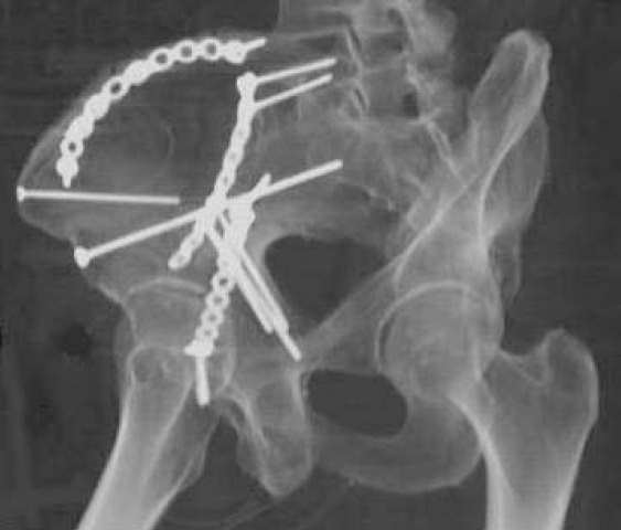
Our implant choices in the management of these patients were quite predictable and relied greatly on the involvement and position of the quadrilateral surface with anterior based fracture patterns. Typically, our goals led us to use a combination of plating techniques and positions, namely, the pelvic brim plate (to assist with stabilizing the anterior column and posterior column) and the intrapelvic plate (to assist with lateralization of the quadrilateral surface and further stabilization of the involved anterior column). A similar plating technique was recently described to effectively treat elderly patients with protrusio fractures of the acetabulum (15). It is not uncommon for us to supplement the fracture fixation with multiple screws placed independently from the plates and into osseous fixation pathways surrounding the fractures sites [Figure 2].
Figure 2.

Independent screws placed in pelvic osseus fixation pathways can assist the surgeon with maximizing fixation in osteopenic/osteoporotic bone.
The mortality rates for geriatric patients with acetabular fractures have been documented. Anglen reported an 85% mortality rate at one year after open reduction internal fixation in their senior patients with acetabular fractures (8). The average age of those who died was 77 years, and this was significantly older than those who survived (P=0.0098). Conversely, in our study, age stratification (65-69, 70-74, etc. to 90 and greater) did not identify any differences in mortality at any time period up to one year. Hessmann reported a lower mortality rate of 15% at 30 days and 33% during the study period for 27 geriatric patients with acetabular fractures (9). In our series, one-year mortality rate was 33% if all patients are included. Our patients who had non-operative management had a higher mortality rate of 44% compared to only 12% in those patients treated surgically. In contrast, Spencer reported an 8% (2/25) mortality rate in a series of geriatric patients with acetabular fractures treated in a non-operative manner at the 9 month time point (16). The discrepancy in the mortality rates is most likely due to a variety of factors, including concomitant injuries, medical comorbidities, and severity of injury. Additionally, one should not draw a conclusion from our study that operative intervention results in lower mortality. Many factors go into the decision making process for operative intervention, and a patient’s overall health can be a key component. As a result, one can assume that the morbidly sick patients were less likely to undergo operative management. However it is important to note that the patients who were placed on bed rest with traction for an extended period of time had significantly higher mortality rates. Our study provides epidemiologic and mortality details on a five-year experience treating the senior population. Multiple factors inherent to their population including bone quality, associated comorbidities, and often fragile physiologic reserves make their subsequent care challenging to the orthopaedic surgeon charged with their care.
In summary, acetabular fractures in the senior population present in a predictable pattern involving primarily the anterior column and quadrilateral surface with predominant fracture patterns being ABC and AC/PHT. Varying degrees of posterior column involvement are typically present and should be critically evaluated when determining treatment strategies. Implant choices and plate/screw positioning are important considerations given the often compromised bone quality in the senior population, and fixation constructs should reflect the surgeon’s understanding of this variable (5, 17). It is very common for this patient population to present with medical comorbidities, and the utilization of a multidisciplinary team may be beneficial in their care. Patients and their families need to be counseled in regards to high mortality rates. Ultimately, as with other fractures in this population, goals of treatment should be early mobilization to avoid complications of recumbency and the return to pre-injury function as quickly as possible.
Acknowledgments
The authors would like to thank Julie Agel for her assistance with statistical analysis.
The authors do not have any disclosures to report in relation to this manuscript.
References
- 1.Cole JD, Bolhofner BR. Acetabular fracture fixation via a modified stoppa limited intrapelvic approach. Description of operative technique and preliminary treatment results. Clin Orthop Relat Res. 1994;305(11):112–23. [PubMed] [Google Scholar]
- 2.Stoppa RE. The treatment of complicated groin and incisional hernias. World J Surg. 1989;13(5):545–54. doi: 10.1007/BF01658869. [DOI] [PubMed] [Google Scholar]
- 3.Letournel É, Judet R. Fractures of the acetabulum. 2nd ed. Heidelberg, Germany: Springer-Verlag; 1993. [Google Scholar]
- 4.Smith-Petersen MN. Approach to and exposure of the hip joint for mold arthroplasty. J Bone Joint Surg Am. 1949;31A(1):40–6. [PubMed] [Google Scholar]
- 5.Qureshi AA, Archdeacon MT, Jenkins MA, Infante A, DiPasquale T, Bolhofner BR. Infrapectineal plating for acetabular fractures: a technical adjunct to internal fixation. J Orthop Trauma. 2004;18(3):175–8. doi: 10.1097/00005131-200403000-00009. [DOI] [PubMed] [Google Scholar]
- 6.Matta JM. Fractures of the Acetabulum: accuracy of reduction and clinical results in patients managed operatively within three weeks after the injury. J Bone Joint Surg Am. 1996;78(11):1632–45. [PubMed] [Google Scholar]
- 7.Helfet DL, Borrelli J, Jr, DiPasquale T, Sanders R. Stabilization of acetabular fractures in elderly patients. J Bone Joint Surg Am. 1992;74(5):753–65. [PubMed] [Google Scholar]
- 8.Anglen JO, Burd TA, Hendricks KJ, Harrison P. The “Gull Sign”: a harbinger of failure for internal fixation of geriatric acetabular fractures. J Orthop Trauma. 2003;17(9):625–34. doi: 10.1097/00005131-200310000-00005. [DOI] [PubMed] [Google Scholar]
- 9.Hessmann MH, Nijs S, Rommens PM. Acetabular fractures in the elderly. Results of a sophisticated treatment concept. Unfallchirurg. 2002;105(10):893–900. doi: 10.1007/s00113-002-0437-0. [DOI] [PubMed] [Google Scholar]
- 10.Alost T, Waldrop RD. Profile of geriatric pelvic fractures presenting to the emergency department. Am J Emerg Med. 1997;15(6):576–8. doi: 10.1016/s0735-6757(97)90161-3. [DOI] [PubMed] [Google Scholar]
- 11.Mears DC. Surgical treatment of acetabular fractures in elderly patients with osteoporotic bone. J Am Acad Orthop Surg. 1999;7(2):128–41. doi: 10.5435/00124635-199903000-00006. [DOI] [PubMed] [Google Scholar]
- 12.Ferguson TA, Patel R, Matta JM. 24th Annual Meeting of the Orthopaedic Trauma Association. Denver, CO: 2008. Fractures of the acetabulum in patients over 60 years of age: epidemiology, fracture patterns and radiographic morphology. [Google Scholar]
- 13.Kakar R, Sharma H, Allcock P, Sharma P. Occult acetabular fractures in elderly patients: a report of three cases. J Orthop Surg (Hong Kong) 2007;15(2):242–4. doi: 10.1177/230949900701500225. [DOI] [PubMed] [Google Scholar]
- 14.Törnkvist H, Schatzker J. Acetabular fractures in the elderly: an easily missed diagnosis. J Orthop Trauma. 1993;7(3):233–5. doi: 10.1097/00005131-199306000-00006. [DOI] [PubMed] [Google Scholar]
- 15.Archdeacon MT, Kazemi N, Collinge C, Budde B, Schnell S. Treatment of protrusio fractures of the acetabulum in patients 70 years and older. J Orthop Trauma. 2013;27(5):256–61. doi: 10.1097/BOT.0b013e318269126f. [DOI] [PubMed] [Google Scholar]
- 16.Spencer RF. Acetabular fractures in older patients. J Bone Joint Surg Br. 1989;71(5):774–6. doi: 10.1302/0301-620X.71B5.2584245. [DOI] [PubMed] [Google Scholar]
- 17.Culemann U, Holstein JH, Kohler D, Tzioupis CC, Pizanis A, Tosounidis G, et al. Different stabilisation techniques for typical acetabular fractures in the elderly: a biomechanical assessment. Injury. 2010;41(4):405–10. doi: 10.1016/j.injury.2009.12.001. [DOI] [PubMed] [Google Scholar]


