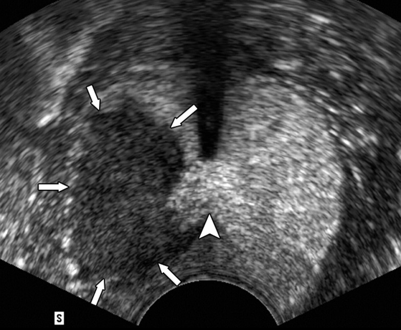
Figure 1c:
Transverse contrast-enhanced pulse-inversion harmonic US images. (a) Image of the prostate prior to contrast material injection. (b) Image shows uniform enhancement of the prostatic parenchyma with a spoke-like perfusion pattern after bolus injection (0.04 mL/kg) of contrast material. (c) Image after ablation of the right side of the prostate shows an avascular nonperfusion area (arrows), representing an RF-created coagulative tissue. Blood flow of the urethral wall exists and appears normal. Shadowing from urethra catheter (arrowhead) is noted.

