Abstract
Rubber tree (Hevea brasiliensis Muell. Arg) is an important economic crop in Thailand. Leaf fall and black stripe diseases caused by the aggressive oomycete pathogen Phytophthora palmivora, cause deleterious damage on rubber tree growth leading to decrease of latex production. To gain insights into the molecular function of H. brasiliensis subtilisin-like serine proteases, the HbSPA, HbSPB, and HbSPC genes were transiently expressed in Nicotiana benthamiana via agroinfiltration. A functional protease encoded by HbSPA was successfully expressed in the apoplast of N. benthamiana leaves. Transient expression of HbSPA in N. benthamiana leaves enhanced resistance to P. palmivora, suggesting that HbSPA plays an important role in plant defense. P. palmivora Kazal-like extracellular protease inhibitor 10 (PpEPI10), an apoplastic effector, has been implicated in pathogenicity through the suppression of H. brasiliensis protease. Semi-quantitative RT-PCR revealed that the PpEPI10 gene was significantly up-regulated during colonization of rubber tree by P. palmivora. Concurrently, the HbSPA gene was highly expressed during infection. To investigate a possible interaction between HbSPA and PpEPI10, the recombinant PpEPI10 protein (rPpEPI10) was expressed in Escherichia coli and purified using affinity chromatography. In-gel zymogram and co-immunoprecipitation (co-IP) assays demonstrated that rPpEPI10 specifically inhibited and interacted with HbSPA. The targeting of HbSPA by PpEPI10 revealed a defense-counterdefense mechanism, which is mediated by plant protease and pathogen protease inhibitor, in H. brasiliensis-P. palmivora interactions.
Introduction
Rubber tree (Hevea brasiliensis Muell. Arg) is the major source for natural latex production and one of economically important crops in Thailand. Phytophthora palmivora is known as an aggressive oomycete pathogen of many woody plants and food crops worldwide [1]. It attacks the petioles, leaf, root or tapping panel of the rubber tree, causing severe damages on plant growth and crop productivity, resulting in enormous economic loss [2–3]. Although this pathogen has been considered as a disastrous pathogen, molecular mechanisms involved in pathogenicity are largely unknown. An understanding of the molecular basis of host-pathogen interactions is expected to provide significant knowledge leading to the development of effective disease control measures.
To promote colonization, Phytophthora species interfere with host innate immune systems by secreting hundreds of effector proteins to manipulate host cell physiology, thereby facilitating infection and reproduction [4–5]. Some effector proteins possess biochemical activities that counteract their host targets to suppress primary defenses, such as P. infestans Kazal-like extracellular protease inhibitors EPI1 and EPI10 [6–7] and P. sojae glucanase inhibitor protein1 (GIP1) [8].
To successfully overcome pathogen invasion, plants develop effective defense strategies against challenging pathogens. One of the strongest plant defensive arsenals is the synthesis of pathogenesis-related (PR) proteins, which accumulate locally in the primarily infected and surrounding tissues, and also in systemic tissues [9]. So far at least 17 families of PR proteins are recognized according to their properties and functions [10]. Some of them, such as PR2 (β-1, 3-glucanases), PR3 (chitinases) and PR7 (endoproteases), might be directly antimicrobial, by catalyzing the degradation of structural components in pathogen cell walls. These hydrolytic enzymes are secreted to the plant extracellular matrix (intercellular space) where host-pathogen interaction, recognition, and signaling processes occur [10–11].
Subtilisin-like serine proteases or subtilases (EC 3.4.21.14) are serine endopeptidases containing a highly conserved catalytic triad motif that is comprised of aspartate, histidine, and serine [12]. They are ubiquitously found in plants and play essential roles in multiple physiological, morphological and biochemical processes, including responses to environmental stress [13]. The availability of the complete genomic sequence of some model species revealed that plant subtilases constitute a large gene family, comprising 56 genes in Arabidopsis (Arabidopsis thaliana), 63 genes in rice (Oryza sativa), 15 genes in tomato (Lycopersicon esculentum) and 80 genes in grape (Vitis vinifera) [14–17]. A more extensive expansion in the number of subtilase genes in plants compared to animals is accompanied by their diverse functions or various evolutionary mechanisms in plants [17].
The first evidence regarding the importance of plant subtilases in defense mechanisms was reported in tomato. P69B, an apoplastic subtilase known as a PR-protein (PR-7 class), is induced during infection by various plant pathogens, such as citrus exocortis viroid, Pseudomonas syringe, P. infestans, and salicylic acid treatment [6,18–21]. In addition, P69C, another isoform of the P69 subtilase family, is also induced by pathogen infection and is hypothesized to participate in pathogen perception and initiation of signaling cascades by specific processing of a LRP (leucine-rich repeat protein) in infected tomato plants [22]. Besides, the Arabidopsis SBT3.3 gene, encoding an extracellular subtilase homologous to the tomato P69C, is rapidly up-regulated in response to Pseudomonas syringae and implicated in the establishment of immune priming by induction of SA-responsive genes [23]. Moreover, the overexpression of an extracellular subtilase gene (GbSBT1) from Gossypium babardense enhances the transcripts of defense-related genes and the disease resistance of Arabidopsis to Fusarium oxysporum and Verticillium dahliae [24].
Interestingly, a critical role of subtilases in plant immunity is also implied by the observation that many pathogens produce various effectors that target these proteases. Examples are P. infestans EPI1 and EPI10, two Kazal-like extracellular serine protease inhibitors that are identified to specifically inhibit and bind to tomato P69B. Inhibition of P69B by two distinct and structurally divergent protease inhibitors is consistent with the hypothesis that P69B is potentially harmful to this pathogen and these pathogen-derived effectors function in counterdefense [6–7].
Furthermore, a Kazal-like extracellular serine protease inhibitor PpEPI10, a homolog of EPI10 from P. infestans, has been reported to be secreted by P. palmivora. Two Kazal domains (Kazal1 and Kazal2) of PpEPI10 inhibit and interact with a 95 kDa protease from rubber tree leaf extracts, suggesting a virulence role of PpEPI10 in pathogenicity [25]. However, the functional activity of PpEPI10 in suppressing immune responses in rubber tree remains largely unexplored. In addition, the 95kDa protease was not identified previously. The present study functionally characterized the biochemical activity of a novel subtilisin-like serine protease from H. brasiliensis (HbSPA) and its defensive role in plants against P. palmivora though Agrobacterium-mediated transient expression in N. benthamiana. P. palmivora PpEPI10 was found to inhibit and interact with HbSPA using in-gel zymogram and co-immunoprecipitation assays, suggesting that one of the antagonistic interactions between rubber tree and P. palmivora is mediated by a plant defensive protease and a pathogen protease inhibitor involved in counterdefense.
Materials and methods
Phytophthora palmivora culture
P. palmivora PP3-3 isolated from a diseased papaya plant [26] was cultured on 10% unclarified V8 agar (10% V8 juice, 0.1% CaCO3, 1.5% agar) at 25°C. For zoospore preparation, P. palmivora was cultured under fluorescent light for 5 days. Ten milliliters of cold (4°C) sterile distilled water was added to the growing P. palmivora mycelium. The plate was incubated at 4°C for 15 min and further shaken at 50 rpm at room temperature for 15 min to release the motile zoospores from sporangia. The concentration of zoospores was determined using a haemocytometer under a light microscope.
For RNA extraction, 1-cm agar plugs of P. palmivora mycelium from a 7-day-old culture were inoculated in 150 x 25 mm petri-dishes containing 20 ml of Plich liquid medium [26] and grown at 25°C in darkness for 7 days. The mycelium mats were harvested by vacuum filtration of the culture through a double-layer filter paper and washed twice with sterile water. The harvested mycelium mats were allowed to dry under sterile condition, fast-frozen in liquid nitrogen and stored at -80°C until use.
Plant growth and infection assay
Twenty-one-day-bud-grafted rubber seedlings (H. brasiliensis, RRIM600 cultivar) were propagated in a climate-controlled room at 25°C under 12h-light and 12h-dark cycle. For the time-course of P. palmivora colonization of rubber tree, the rubber leaves at the developmental B2C stage of plantlets with uniform growth were inoculated with 1 droplet (30-μL) containing ~3,000 zoospores of P. palmivora on the underside of the leaves. The treated rubber seedlings were further incubated in a climate-controlled room at 25°C, under 12h-light and 12h-dark cycle. The infected leaves were harvested at 0, 1, 2, 3, 4, and 5 days post-inoculation. Leaf discs of similar size (1.5 × 1.5 cm) were dissected around the inoculated regions with the inoculation spot at the center of the sampled area. The leaf samples were flash-frozen in liquid nitrogen and subsequently stored immediately to a -80°C freezer until RNA extraction.
N. benthamiana plants were grown in pots and maintained inside a growth chamber at 22°C, 60% humidity, under 16h-light and 8h-dark cycle. 6-week-old N. benthamiana plants were used for all agro-infiltration experiments.
Total RNA extraction and first-strand cDNA synthesis
P. palmivora mycelium mats and rubber tree leaf samples were ground in liquid nitrogen to a fine powder with a mortar and pestle. Total RNA was isolated using the RNeasy® Plant Mini Kit (Qiagen, CA, USA) according to the manufacturer’s instructions. The contaminating DNA was eliminated during total RNA extraction using an on-column RNase-free DNase I digestion set (Qiagen, CA, USA) according to the procedure of the RNeasy® Plant Mini Kit. First-strand cDNA synthesis was performed using the Superscript™ III (Invitrogen, CA, USA). The obtained first-strand cDNAs were kept at -20°C until use.
Expression pattern analysis of PpEPI10 and HbSPA
To determine the PpEPI10 gene expression during the infection of rubber tree leaves by P. palmivora using semi-quantitative reverse transcription polymerase chain reaction (semi-qRT-PCR), rubber tree leaves were inoculated with P. palmivora zoospores and total RNA was extracted from the infected leaves at 0, 1, 2, 3, 4, and 5 days post-inoculation as described previously. The specific primers, ExPpEPI10-F (5′CACCTAAATCGTCGGCAGACAACAGCTC3′), ExPpEPI10-R (5′ACCATCGTAATGTTTTTGCGCTCGTTGC3′), β-tubulin-F (5′GAAGTCATGTGCCTGGATAACGAG3′), and β-tubulin-R (5′CTCATGCGTCCGCGGAACATACAC3′) were designed based on the published P. palmivora PpEPI10 (PLTG_2.01611) [27] and β-tubulin (GenBank accession no. JN203065) gene sequences and used for determining the expression of PpEPI10 and β-tubulin transcripts, respectively. Amplification of β-tubulin gene was used as a constitutive control to determine the relative transcript level of PpEPI10 gene. Two μL of first-strand cDNAs were used as a template in a 30-μL PCR reaction containing Emerald Amp® GT PCR Master mix (Takara, Shiga, Japan), 0.2 μM of each gene-specific primer. The amplification was performed in a thermal cycler by preheating at 94°C for 5 min, followed by 30 cycles of denaturing at 94°C for 1 min, annealing at 60°C for 30 sec and extension at 72°C for 1 min, and a final extension step at 72°C for 10 min. The PCR products were analyzed by electrophoresis on a 1.2% (w/v) agarose gel, and visualized under the UV transilluminator and photographed by a Gel Document (UVP BioSpectrum® MultiSpectral Imaging System™, Cambridge, UK).
To determine the HbSPA gene expression during infection of rubber tree leaves by P. palmivora using real-time quantitative reverse transcription polymerase chain reaction (real-time qRT-PCR), The specific primers, ExHbSPA-F (5′GCCACTGATTTTCTTCAAGAGTCAAACC3′) and ExHbSPA-R (5′TTGGATGGACAGTTGTATGGCTTATCAG3′), ExHbYLS8-F (5′CAAGCAAGAGTTCATTGACATTATTGAGACTG3′) and ExHbYLS8-R (5′ATCACAGGTTCTTACATAGTCGGATAGTC3′) were designed for determining the expression of the H. brasiliensis subtilisin-like serine protease A (HbSPA, GenBank accession no. KU845302) and the H. brasiliensis mitosis protein YLS8 (HbYLS8, GenBank accession no. HQ323250), respectively. HbYLS8 is stably expressed in the rubber tree [28] and used as an endogenous control. The real-time RT-qPCR was performed with 2 μL of first-strand cDNAs, gene-specific primers (final concentration of 0.9 μM for HbSPA and 1.0 μM for HbYLS8), and (2x) Luminaris Color HiGreen Low ROX qPCR Master Mix (Thermo Fisher Scientific, MA, USA) according to the manufacturer’s instructions on the Stratagene™ Mx3005P qPCR System (Agilent Technologies, CA, USA). The amplification results were automatically analyzed using the MxPro™ QPCR Software. Transcript abundance was calculated using the linearized plasmid DNA standard method as previously described [29]. The experiment was performed with three independent biological replicates.
Bacterial strains and plasmids
E. coli DH5α (Invitrogen, CA, USA) or BL21 (DE3) (New England BioLabs, MA, USA) and Agrobacterium tumefaciens GV3101 [30] or C58-C1 [31] were used throughout this study and routinely maintained on Luria-Bertani (LB) agar or broth [32] at 37°C and 28°C, respectively. The pGD plasmid, a plant binary vector [33], was used to transiently express rubber tree serine proteases in N. benthamiana. The pJL3-P19 plasmid was used to express Tomato bushy stunt virus P19 protein (GeneBank accession number M21958), a suppressor of post-transcriptional gene silencing to enhance the protein expression level in N. benthamiana [34–35].
Plasmid construction and transient expression of Hevea serine proteases in N. benthamiana
The pGD-HbSPA, pGD-HbSPB, and pGD-HbSPC plasmids were constructed by cloning PCR-amplified DNA fragments corresponding to the open reading frames of HbSPA, HbSPB, and HbSPC fused with a hexahistidine(His)-tag encoding sequences at the C-terminus into the pGD vector [33]. The pJET-HbSPA, pJET-HbSPB, and pJET-HbSPC plasmids containing the open reading frames of H. brasiliensis subtilisin-like serine protease A (HbSPA), H. brasiliensis subtilisin-like serine protease B (HbSPB), and H. brasiliensis subtilisin-like serine protease C (HbSPC) (GenBank accession no. KU845302, KU845303, and KU845304, respectively) [29] were used as a template for PCR. Specific primers, HbSPA-F (5′GCGagatctATGAGGATCCGTAGCCATTCATG3′) and HbSPA-R (5′GCGgtcgacTTAgtggtgatggtgatggtgAGCTGAACCTGCATCTTTCTTCT3′), were used to amplify the open reading frame of HbSPA fragment fused with a hexahistidine(His)-tag encoding sequences at the C-terminus. The introduced BglII and SalI restriction sites were underlined. The italic letters represent the His-tag sequence. Primers HbSPBC-F (5′GCGgtcgacATGGAAAACAGAAGATGCAAAAT3′) and HbSPBC-R (5′GCGggatccCTAgtggtgatggtgatggtgTTTAAATAAGACAGCAATTGGGC3′) were used to amplify the open reading frame of either HbSPB or HbSPC fused with a hexahistidine(His)-tag encoding sequences at the C-terminus. The introduced SalI and BamHI restriction sites were underlined. The His-tag sequence is shown in italic. The PCR was performed using the Phusion®High-Fidelity DNA polymerase (New England BioLabs, MA, USA) by preheating at 98°C for 30 s, followed by 35 cycles of denaturing 98°C for 15 s, annealing 60°C for 15 s and extension at 72°C for 2.3 min, and a final extension step at 72°C for 10 min. The amplified fragments were digested with appropriated restriction enzymes and cloned into the corresponding restriction sites of pGD vector [33]. The pGD-HbSPA, pGD-HbSPB, and pGD-HbSPC plasmids containing HbSPA, HbSPB, and HbSPC gene insert with the correct nucleotide sequences were used to transform A. tumefaciens stain C58-C1 by electroporation. To inhibit the post-transcriptional gene silencing (PTGS) that suppresses foreign gene expression in N. benthamiana, A. tumefaciens stain GV3101 containing pJL3-P19 [34–35] was used in co-infiltration. Transient expression of Hevea serine protease genes in N. benthamiana using A. tumefaciens stain C58-C1 carrying pGD-HbSPA, pGD-HbSPB, or pGD-HbSPC, with or without A. tumefaciens stain GV3101 carrying pJL3-P19, was achieved by agroinfiltration following the methods previously described [36]. The expression of His-tagged HbSPA, HbSPB, and HbSPC proteins was analyzed by Western blot using HRP conjugated anti-His monoclonal antibody His-probe (H-3) (sc-8036HRP, Santa Cruz Biotechnology, INC., http://www.scbt.com) as described previously [36].
Isolation of intercellular fluids
Intercellular (apoplastic) fluids were extracted from N. benthamiana leaves either infiltrated with A. tumefaciens stain GV3101 carrying pJL3-P19 or a mixture of A. tumefaciens stain C58-C1 carrying pGD-HbSPA and A. tumefaciens stain GV3101 carrying pJL3-P19 using a vacuum infiltration method [37]. Briefly, detached leaves were submerged in a cold extraction buffer (300 mM NaCl, 50 mM NaPO4, pH 7.0) [38] in a plastic container with the help of a plastic lined wire mesh and vacuum was applied in a Nalgene vacuum desiccator. Vacuum-infiltrated leaves were blot dried with soft paper towels to remove the liquid on the leaf surface, followed by centrifugation at 2,000 g for 20 min at 4°C to collect the intercellular fluids. The recovered fluids were filter-sterilized and analyzed by 10% SDS-PAGE and Western blot, or stored at -80°C.
Plasmid construction, expression and purification of PpEPI10
Plasmid pET28a-PpEPI10 was constructed by cloning a PCR-amplified DNA fragment corresponding to the mature protein of PpEPI10 with the predicted signal peptide removed into BamHI and SalI sites of pET28a (Novogen). The specific primer pairs PpEPI10-F (5′GCGggatccATCGACGACGACAAGTGCTCA3′) and PpEPI10-R (5′GCGgtcgacTTACGACTGTTGCTCCTGCT3′) were designed based on the published P. palmivora PpEPI10 sequence (PLTG_2.01611) [27]. The introduced BamHI and SalI restriction sites are underlined. The PpEPI10 fragment was amplified by PCR using Phusion®High-Fidelity DNA polymerase (New England BioLabs, MA, USA) with P. palmivora mycelium cDNAs as the template. The resulting plasmid pET28a-PpEPI10 was transformed into E. coli BL21 (DE3) cells by electroporation. The recombinant PpEPI10 protein expressed from pET28a-PpEPI10 contains a His tag followed by a T7-tag at its N-terminus. Expression and purification of recombinant PpEPI10 proteins were performed as described previously [39]. The protein concentration was determined using Bradford protein assay kit (Bio-RAD, CA, USA).
Protease activity and inhibition assay
The protease activity of HbSPA and its inhibition by PpEPI10 were determined by an in-gel protease assay using the Bio-Rad zymogram buffer system. Twenty microliters of the intercellular fluids containing the His-tagged HbSPA were incubated with or without various final concentrations (0.1, 0.4, 1.0, 4.0 μM) of the purified rPpEPI10 in a total volume of 22 μL for 30 min at 25°C and then mixed with equal volume of zymogram sample buffer (62.5 mM Tris-HCl, pH 6.8, 25% glycerol, 4% SDS, and 0.01% bromophenol blue). Ten microliters of the mixtures were loaded onto a 7.5% SDS-PAGE containing 0.1% gelatin in the absence of reducing reagent and boiling. After electrophoresis, the gel was incubated in 1x zymogram renaturation buffer (2.5% Triton X-100) for 30 min at room temperature and then equilibrated in 1x zymogram development buffer (50 mM Tris-HCl, pH 7.5, 200 mM NaCl, 5 mM CaCl2, and 0.02% Brij 35) for 2 h at 37°C. The gel was fixed and stained with 0.5% Coomassie Brilliant Blue R-250 and destained with destaining solution (5% methanol and 1% acetic acid). Areas of protease activity were visualized as clear bands against the blue background on the gel, as a result of the proteolytic digestion of the gelatin in the gel.
Co-immunoprecipitation assay
Co-immunoprecipitation (co-IP) was performed on the intercellular fluid containing the His-tagged HbSPA and the purified His-T7-tagged PpEPI10 (rPpEPI10) using the T7.tag®Affinity Purification Kit (Novagen, Merck Millipore, MA, USA) following the manufacturer’s instructions, with minor modifications. A total of 10 μg of purified rPpEPI10 was pre-incubated with 40 μL of 50% slurry T7.Tag Antibody covalently linked agarose beads for 2 h at 4°C with gentle agitation. The protein/bead complex was pelleted at 2,000 rpm for 10 min followed by three washes with 1x Bind/Wash Buffer to remove unbound protein. Five hundred microliters of the intercellular fluids containing the His-tagged HbSPA were added to the protein/bead complex followed by overnight incubation at 4°C with gentle agitation. The agarose beads were washed three times with 1x Bind/Wash Buffer, followed by centrifugation at 2,000 rpm for 10 min. The precipitated protein complexes were eluted with 40 μL of 1x Elution Buffer and neutralized by adding 6 μL of 1x Neutralization Buffer. The immunocomplexes were subjected to 10% SDS-PAGE and followed by Western blot with HRP conjugated anti-His monoclonal antibody.
Pathogen infection assay to evaluate the role of HbSPA in plant resistance
The role of HbSPA protein in plant immune response against P. palmivora was evaluated using droplet inoculation of P. palmivora zoospore suspension on N. benthamiana leaves transiently expressing HbSPA generated by agroinfiltration. Leaves of 6-week-old N. benthamiana plants were infiltrated with a mixture of Agrobacterium stain C58-C1 carrying pGD-HbSPA and Agrobacterium stain GV3101 carrying pJL3-P19 on one half leaf (delineated by the midvein), and on another half Agrobacterium stain GV3101 carrying pJL3-P19 was infiltrated to serve as a control, following the methods described previously [36]. After that, plants were maintained in the laboratory under continuous fluorescent lighting for protein expression. At 36 h post-agroinfiltration, the infiltrated leaves were inoculated with droplets (10-μL) of P. palmivora zoospores suspension (1x10⁴ spores per mL) on the abaxial surface of the leaf. The plants were kept at high humidity. The diameters of the necrotic lesions were measured at 3 dpi. At least three independent experiments were performed for this assay.
Statistical analysis
The transcript abundance of HbSPA was analyzed using the one-way analysis of variance (ANOVA) according to Tukey’s HSD test using SPSS Statistics 17.0 software. The disease lesion size measurements on N. benthamiana plants after inoculation with P. palmivora zoospores were analyzed by comparing the lesion size in the HbSPA-expressed half with the lesion size in the control half of each inoculated leaf using the paired t-test with R.
Results
Expression of PpEPI10 and HbSPA genes during infection of rubber tree by P. palmivora
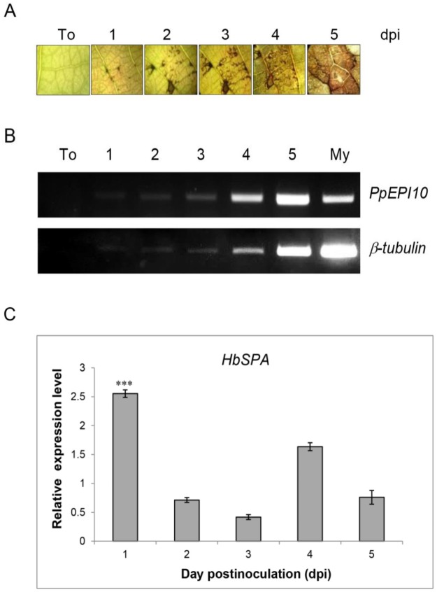
To investigate a possible role of HbSPA in defense against P. palmivora and a possible role of PpEPI10 in P. palmivora virulence, we determined the expression of PpEPI10 and HbSPA genes during infection of rubber tree by P. palmivora. We collected the infected leaf tissues across the infection time course, during which increased disease and increased growth of P. palmivora were observed (Fig 1A). Semi-quantitative RT-PCR analysis was performed to determine the expression of PpEPI10 gene during colonization of the rubber tree by P. palmivora and in mycelium cultured in vitro. The PpEPI10 gene was expressed in P. palmivora across the time course of colonization on rubber tree leaves. Notably, the expression of PpEPI10 was remarkably induced during infection of rubber tree in contrast to its expression in mycelium grown in vitro and relative to the constitutive P. palmivora β-tubulin gene (Fig 1B), suggesting that PpEPI10 plays a role in pathogenesis. The expression of HbSPA was examined using real-time qRT-PCR analysis. HbSPA was significantly up-regulated by 2.5 fold at 1 dpi, abruptly declined thereafter and slightly increased at 4 dpi (1.6 fold), when compared to the relative transcript level of the non-infected control (Fig 1C). The rapid increase in HbSPA expression during early infection of rubber tree by P. palmivora suggests that HbSPA plays a potential role in plant defense.
Fig 1. Expression of PpEPI10 and HbSPA genes in rubber tree infected by P. palmivora.
(A) Symptoms observed on noninfected rubber leaves (To), and infected rubber leaves after inoculation with zoospores of P. palmivora under a light microscope at 1, 2, 3, 4, and 5 days post-inoculation (dpi). (B) Semi-quantitative RT-PCR analysis of PpEPI10 and P. palmivora β-tubulin expression during colonization of rubber tree by P. palmivora. Total RNAs isolated from noninfected leaves (To), infected leaves of rubber tree at 1, 2, 3, 4, or 5 dpi, and P. palmivora mycelium grown in synthetic medium (My) were used for the analysis. The constitutive P. palmivora β-tubulin gene was used as a control to determine the relative expression of PpEPI10. (C) Real-time RT-PCR analysis of HbSPA expression during colonization of rubber tree by P. palmivora. Total RNA from a time course similar to the one described in (B) was used. The expression of HbSPA gene was expressed as a relative transcript fold change to their non-infected controls. The columns and vertical bars represent a mean ± standard errors (SE) of the relative expression levels calculated from the three independent experiments. Three asterisks (***) indicate statistically significant differences in the relative expression levels of HbSPA in the infected condition compared to its expression levels in the uninfected condition as a control (P-value < 0.01) determined by paired t-test.
Functional expression of Hevea serine proteases genes in N. benthamiana
In order to express Hevea subtilisin-like serine protease genes in N. benthamiana, the coding sequences of HbSPA, HbSPB, and HbSPC fused to the hexahistidine (His)-tag at the C-terminus were cloned into pGD and transiently expressed in N. benthamiana leaves by A. tumefaciens mediated transient expression (agroinfiltration). The expression of HbSPA, HbSPB, and HbSPC proteins was analyzed in total protein extracts of agroinfiltrated N. benthamiana leaves by Western blot with HRP conjugated anti-His monoclonal antibody. Only HbSPA gene was successfully expressed in N. benthamiana leaves as visualized by Western blot showing a corresponding protein band of approximately 75-kDa (Fig 2, lanes 3 and 4). Moreover, an increase in the expression level of HbSPA protein was observed when co-expressing HbSPA and P19 compared to expressing HbSPA alone, demonstrating that the employment of gene silencing suppressor P19 protein derived from Tomato bushy stunt virus (TBSV) to suppress the onset of post-transcriptional gene silencing was able to enhance the expression level of HbSPA in N. benthamiana (Fig 2, lane 4).
Fig 2. Agrobacterium-mediated transient expression of Hevea serine protease genes in N. benthamiana.
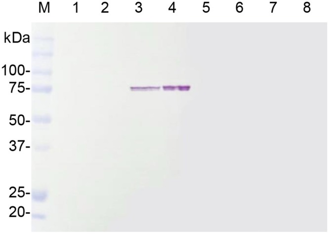
Total proteins were extracted from agroinfiltrated N. benthamiana leaves and subjected to SDS-PAGE followed by Western blot with HRP conjugated anti-His monoclonal antibody. Lane M represents the protein standard and the numbers on the left indicate the size of molecular weight markers. Lanes1-8 represent total proteins extracted from N. benthamiana leaves infiltrated with the infiltration media (Mock, lane 1), A. tumefaciens GV3101 carrying the pJL3-P19 (lane 2), A. tumefaciens C58-C1 carrying the pGD-HbSPA (lane 3), a mixture of A. tumefaciens strains carrying the pGD-HbSPA and the pJL3-P19 (lane 4), A. tumefaciens C58-C1 carrying the pGD-HbSPB (lane 5), a mixture of A. tumefaciens strains carrying the pGD-HbSPB and the pJL3-P19 (lane 6), A. tumefaciens C58-C1 carrying the pGD-HbSPC (lane 7), a mixture of A. tumefaciens strains carrying the pGD-HbSPC and the pJL3-P19 (lane 8), respectively.
To identify whether the recombinant HbSPA protein was expressed in the apoplast of N. benthamiana, the intercellular fluids were recovered from the whole leaves of N. benthamiana plants infiltrated with A. tumefaciens GV3101 carrying pJL3-P19 or a mixture of A. tumefaciens strains carrying pGD-HbSPA and pJL3-P19 using a vacuum infiltration method, and subjected to Western blot analysis. The protein band of HbSPA was detected in samples from leaves co-infiltrated with A. tumefaciens strains carrying pGD-HbSPA and pJL3-P19, but not from leaves infiltrated with A. tumefaciens GV3101 carrying only pJL3-P19, indicating that the recombinant HbSPA protein was an extracellular protein (Fig 3A).
Fig 3. Transient expression of HbSPA in N. benthamiana.
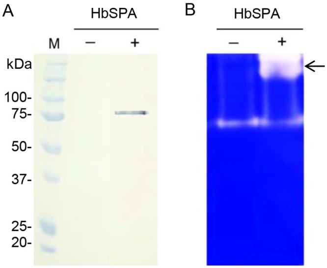
Intercellular fluids were isolated from N. benthamiana leaves infiltrated with A. tumefaciens GV3101 carrying the pJL3-P19 as a control (- HbSPA), a mixture of A. tumefaciens strains carrying pGD-HbSPA and pJL3-P19 (+ HbSPA). (A) Samples were analyzed by SDS-PAGE followed by Western blot with HRP conjugated anti-His monoclonal antibody. Lane M represents the protein standard and the numbers on the left indicate the size of molecular weight markers. (B) Samples were analyzed by zymogram in-gel protease assay. Samples were loaded on a SDS-PAGE containing 0.1% of gelatin. After electrophoresis, proteins were renaturated in the gel and the protease activity was detected after a 2-h incubation required for gelatin degradation. The clear and distinct bands against the blue background on the gel corresponding to the protease activity associated with the presence of the HbSPA protein was indicated by an arrow.
To further determine whether the recombinant HbSPA protein was a functional protease, the intercellular fluids isolated from N. benthamiana leaves infiltrated with A. tumefaciens GV3101 carrying pJL3-P19 or a mixture of A. tumefaciens strains carrying pGD-HbSPA and pJL3-P19 were subjected to zymogram in-gel protease assay. The zymogram gel revealed an additional protease band in the sample containing HbSPA protein, but not in the control, indicating that the recombinant HbSPA protein was a functional extracellular protease (Fig 3B).
Ectopic expression of HbSPA in N. benthamiana enhances resistance to P. palmivora
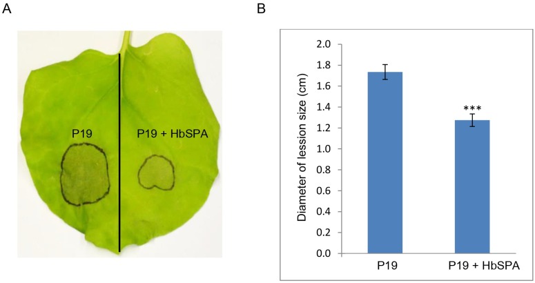
To evaluate the effect of HbSPA-expressing N. benthamiana plants against P. palmivora infection, we expressed HbSPA protein in N. benthamiana leaves via Agrabacterium-mediated transient transformation. N. benthamiana leaf was separated into two halves delineated by the midvein. The first half was infiltrated with A. tumefaciens GV3101 carrying pJL3-P19 and the other half was infiltrated with a mixture of A. tumefaciens strains carrying pGD-HbSPA and pJL3-P19. The agroinfiltrated leaves were then inoculated with droplets (10-μL per droplet) containing ~100 zoospores of P. palmivora. Obvious differences in disease symptoms were observed at 3 dpi. Ectopic expression of HbSPA protein significantly reduced the localized necrotic area (Fig 4). The average lesion size in the halves expressing HbSPA protein (1.27-cm-diameter) was smaller than in the control halves of the same leaves (1.73-cm-diameter) (Fig 4B). This result suggested that HbSPA gene plays a significant role in plant resistance against P. palmivora infection.
Fig 4. Increased resistance mediated by HbSPA expressed in N. benthamiana to P. palmivora.
(A) Infection symptoms of P. palmivora on a representative N. benthamiana leaf with one half transiently co-expressing HbSPA and P19 genes and another half expressing P19 gene only as a control. The photograph was taken at 3 days post inoculation (dpi). (B) Average lesion size at 3dpi on N. benthamiana leaf halves expressing P19 gene only or both P19 and HbSPA following drop inoculation with 100 zoospores of P. palmivora. The histograms correspond to the mean ± standard errors (SE) of lesion diameter calculated from the three independent experiments (n = 150). Three asterisks (***) indicate statistically significant differences in the lesion diameters derived from N. benthamiana leaves with one half co-expressing HbSPA and P19 genes in the infected condition compared to another half expressing P19 gene only in the same condition as a control (P-value < 0.01) determined by paired t-test.
Inhibition of protease activity of HbSPA by PpEPI10
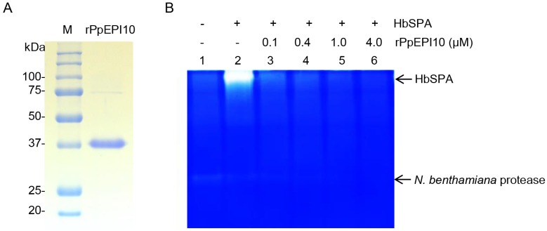
To determine whether P. palmivora PpEPI10 targets HbSPA, the recombinant PpEPI10 (rPpEPI10) protein, which is composed of mature PpEPI10 fused with a hexahistidine (His) tag followed by a T7 tag at the N-terminus, was expressed and purified from pET28a-PpEPI10 in E. coli BL21 (DE3). The purified rPpEPI10 appeared as a major band of 37 kDa and a minor, barely detectable band of 75 kDa on a SDS-PAGE gel, indicating high purity of rPpEPI10 (Fig 5A).
Fig 5. The protease activity of HbSPA is inhibited by rPpEPI10.
(A) Affinity-purified rPpEPI10 visualized on 12% SDS-PAGE followed by Coomassie Brilliant Blue staining. Lane M represents the protein standard and the numbers on the left indicate the size of molecular weight markers. Lane rPpEPI10 represents the purified rPpEPI10 eluted from the Ni-NTA His-Bind® affinity column. (B) Inhibition of the protease activity of HbSPA by rPpEPI10. The intercellular fluids isolated from N. benthamiana leaves infiltrated with A. tumefaciens GV3101 carrying the pJL3-P19 were incubated with buffer (25 mM HEPES, pH 7.5, 150 mM NaCl, 10% glycerol) (lane 1), and the intercellular fluids isolated from N. benthamiana leaves infiltrated with a mixture of A. tumefaciens strains carrying pGD-HbSPA and pJL3-P19 were incubated with buffer (lane 2) or the purified rPpEPI10 at concentrations indicated (lanes 3–6). The remaining protease activity of each sample was determined using zymogram in-gel protease assay. The upper and lower arrows indicate the band location corresponding to the protease activity of HbSPA and a N. benthamiana protease, respectively.
To investigate the antagonistic activity of rPpEPI10 against HbSPA, the intercellular fluids isolated from agroinfiltrated N. benthamiana leaves expressing HbSPA protein were incubated in the absence of protease inhibitor (buffer) or the presence of various concentrations of rPpEPI10. The remaining protease activity of HbSPA protein in each incubation sample was detected using the zymogram in-gel protease assay (Bio-RAD, CA, USA). Increasing concentrations of rPpEPI10 exhibited dose-dependent inhibition of the protease activities of HbSPA and a N. benthamiana protease as these protease bands diminished on the gel after incubation with rPpEPI10 (Fig 5B). This suggests that PpEPI10 encodes a functional protease inhibitor that inhibits the protease activity of HbSPA.
Interaction of HbSPA with PpEPI10
To determine whether HbSPA protein is targeted by PpEPI10, co-immunoprecipitation (co-IP) was performed on the intercellular fluids extracted from N. benthamiana leaves expressing HbSPA-His protein incubated with the purified His-T7-tagged rPpEPI10 or buffer control using the T7 antibody covalently linked agarose beads. The eluted solutions from co-IP were analyzed by Western blot with HRP conjugated anti-His monoclonal antibody. The HbSPA-His protein expressed in the intercellular fluids could only be pulled down in the presence of rPpEPI10 but not in the absence of rPpEPI10 (Fig 6), indicating rPpEPI10 interacted with HbSPA protein which represented a plant protease target of PpEPI10.
Fig 6. Co-immunoprecipitation of HbSPA and rPpEPI10.
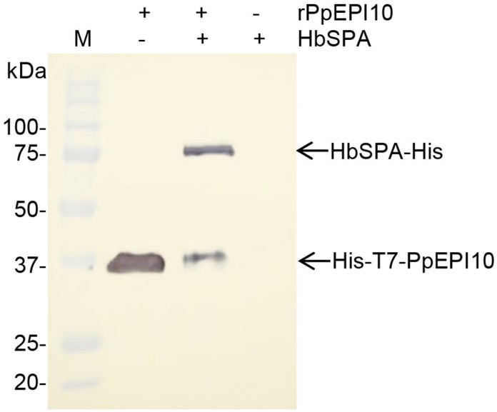
The T7 antibody covalently linked agarose beads were incubated with the purified His-T7-tagged PpEPI10 (+ rPpEPI10) or buffer (- rPpEPI10). The mixture was added to buffer (- HbSPA) or the intercellular fluids isolated from N. benthamiana leaves expressing HbSPA-His protein (+ HbSPA). After washing, the eluates were analyzed by Western blot with HRP conjugated anti-His monoclonal antibody. Lane M represents the protein standard. The numbers on the left indicate the size of molecular weight markers.
Discussion
Like other plant pathogens, oomycete Phytophthora species secrete various effector proteins to suppress host defense responses and promote colonization [40–42]. Elucidation of the functions of effectors and the underlying molecular mechanisms involved in pathogenicity remains challenging. Previously, the PpEPI10 gene, encoding a Kazal-like extracellular serine protease inhibitor from P. palmivora, has been implicated in pathogenicity through suppressing plant protease activity in rubber tree [25]. However, the inhibited rubber tree protease(s) was not identified. In this study, we expressed H. brasiliensis subtilisin-like serine protease genes (HbSPA, HbSPB, and HbSPC) in N. benthamiana plant using Agrobacterium-mediated transient expression to elucidate their possible biological role in plant defense responses. We also examined whether these Hevea serine proteases could be targets of PpEPI10. Our results indicated that HbSPA was involved in plant defense responses against P. palmivora, and targeted by PpEPI10 effector, which is induced and secreted into the plant apoplast from P. palmivora during infection.
A large number of subtilisin-like serine proteases have been linked to plant immune responses [7,13,23]. Tomato subtilases P69B and P69C are induced and accumulated in the plant extracellular matrix during pathogen attack of tomato [6,19–20]. It was proposed that P69C proteolytically processes an extracellular matrix associated leucine-rich repeat protein (LRP) in viroid-infected tomato to activate plant defense signal transduction processes [22]. In Arabidopsis, an extracellular subtilase (SBT3.3) homologous to tomato P69C was found to be a crucial component mediating the pathogen recognition and activating the expression of SA-responsive defensive genes for priming immune responses [23]. Transgenic Arabidopsis plants overexpressing SBT3.3 gene showed enhanced resistance to P. syringae and the oomycete Hyaloperonospora arabidopsidis [23]. In cotton (G. babardense), the expression of GbSBT1 gene is induced by V. dahliae infection, jasmonic acid, and ethylene treatments and the GbSBT1 protein interacts with the effector PHB protein secreted from V. dahliae [24]. Ectopic expression of GbSBT1 gene in Arabidopsis exhibited the enhanced resistance to F. oxysporum and V. dahliae [24]. Similarly, the expression of HbSPA was found to be increased during infection of rubber tree by P. palmivora. In addition, N. benthamiana leaves expressing HbSPA protein were more resistant to P. palmivora infections, substantiating its value in establishing effective defense mechanisms of the rubber tree. However, the exact biochemical mechanism by which HbSPA protein confers plant defense is unclear and needs to be further elucidated. It may participate in pathogen recognition, immune signaling activation, and/or the proteolytic degradation of pathogen proteins.
In this study, we found the elevated PpEPI10 gene expression during colonization of the rubber tree by P. palmivora, which is similar to the expression of epi1 and epi10 genes that were previously reported to be highly induced during infection of tomato by P. infestans [6–7]. We also found that PpEPI10 interacts and inhibits HbSPA via co-immunoprecipitation and zymogram in-gel protease assay. Altogether, these results suggest that PpEPI10 potentially functions as a disease effector that contributes to the pathogenicity of P. palmivora by targeting plant defensive protease HbSPA. As the expression of HbSPA is also induced during infection, the concurrent expression of PpEPI10 and HbSPA during infection and the facts that both encode secreted proteins support the possibility of the direct interaction between PpEPI10 and HbSPA at the infection interface. Moreover, it has been demonstrated that the two Kazal domains of PpEPI10 inhibit and interact with a 95 kDa rubber tree protease [25], implying that PpEPI10 may possibly target multiple rubber tree proteases in the pathogenic process.
The inhibition of plant defensive proteases is one of the multitudinous countermeasures that plant pathogens have developed to shut down host defense mechanisms and colonize plant tissue. For example, P. infestans secreted two distinct and structurally divergent Kazal-like extracellular protease inhibitors (EPI1 and EPI10) to recognize the same tomato defense-related protease P69B [6–7]. In addition, P. infestans secretes cystatin-like extracellular protease inhibitors, called EPICs. EPIC1 and EPIC2B were found to bind and inhibit PIP1 and RCR3, two tomato defense papain-like cysteine proteases in the apoplast during infection [43–44]. The inhibition of host proteases by P. infestans-derived inhibitors may represent a major virulence mechanism in this pathogen. Our finding that the rubber tree defensive protease HbSPA is targeted by P. palmivora PpEPI10 suggests that a similar virulence mechanism is employed by another Phytophthora spp. Plant defense and pathogen counterdefense mediated by interactions of plant proteases and pathogen protease inhibitors may represent a common mechanism of the antagonistic interactions between various Phytophthora spp. and their hosts.
Acknowledgments
The authors would like to express special thanks to Dr. Dongliang Wu for his assistance with the statistical analysis using paired t-test and valuable advice during our conducting the experiments at University of Hawaii at Manoa, USA. We also wish to thank Dr. Nion Chirapongsatonkul for providing valuable and helpful suggestions.
Data Availability
All relevant data are within the paper.
Funding Statement
This work was financially supported in part by the Thailand Research Fund (TRF) through the Royal Golden Jubilee Ph.D. Program (RGJ-PHD) to Miss Kitiya Ekchaweng (Grant No. PHD/0030/2555; www.trf.or.th); the University Academic Excellence Strengthening Program in Biochemistry, Faculty of Science, Prince of Songkla University, Thailand (www.sci.psu.ac.th); the Graduate School of Prince of Songkla University, Thailand (www.psu.ac.th). The funders had no role in study design, data collection and analysis, decision to publish, or preparation of the manuscript.
References
- 1.Widmer TL. Phytophthora palmivora. Forest Phytophthoras. 2014; 4 (1): 3557. [Google Scholar]
- 2.Erwin DC, Ribeiro OK. Phytophthora Diseases Worldwide. The American Phytopathological Society Press; 1996. ISBN: 0-89054-212-0 [Google Scholar]
- 3.Liyanage NIS, Wheeler BEJ. Comparative morphology of Phytophthora species on rubber. Plant Pathol. 1989; 38: 592–597. [Google Scholar]
- 4.Kamoun S. A catalogue of the effector secretome of plant pathogenic oomycetes. Annu. Rev. Phytopathol. 2006; 44: 41–60. 10.1146/annurev.phyto.44.070505.143436 [DOI] [PubMed] [Google Scholar]
- 5.Giraldo MC, Valent B. Filamentous plant pathogen effectors in action. Nat. Rev. Microbiol. 2013; 11: 800–814. 10.1038/nrmicro3119 [DOI] [PubMed] [Google Scholar]
- 6.Tian M, Huitema E, Da Cunha L, Torto-Alalibo T, Kamoun S. A Kazal-like extracellular serine protease inhibitor from Phytophthora infestans targets the tomato pathogenesis-related protease P69B. J. Biol. Chem. 2004; 279(25): 26370–26377. 10.1074/jbc.M400941200 [DOI] [PubMed] [Google Scholar]
- 7.Tian M, Benedetti B, Kamoun S. A second Kazal-like protease inhibitor from Phytophthora infestans inhibits and interacts with the apoplastic pathogenesis-related protease P69B of tomato. Plant Physiol. 2005; 138(3): 1785–1793. 10.1104/pp.105.061226 [DOI] [PMC free article] [PubMed] [Google Scholar]
- 8.Rose JK, Ham KS, Darvill AG, Albersheim P. Molecular cloning and characterization of glucanase inhibitor proteins: coevolution of a counterdefense mechanism by plant pathogens. Plant Cell. 2002; 14(6): 1329–1345. 10.1105/tpc.002253 [DOI] [PMC free article] [PubMed] [Google Scholar]
- 9.Van Loon LC, Pierpoint WS, Boller T, Conejero V. Recommendations for naming plant pathogenesis-related proteins. Plant Mol. Biol. Rep. 1994; 12: 245–264. [Google Scholar]
- 10.Van Loon LC, Rep M, Pieterse CMJ. Significance of inducible defense-related proteins in infected plants. Annu. Rev. Phytopathol. 2006; 44: 135–162. 10.1146/annurev.phyto.44.070505.143425 [DOI] [PubMed] [Google Scholar]
- 11.Dixon RA, Lamb CJ. Molecular communication in interaction between plants and microbial pathogens. Annu. Rev. Plant Physiol. Plant Mol. Biol. 1990; 41: 339–367. [Google Scholar]
- 12.Dodson G, Wlodawer A. Catalytic triads and their relatives. Trends Biochem. Sci. 1998; 23(9): 347–352. [DOI] [PubMed] [Google Scholar]
- 13.Figueiredo A, Monteiro F, Sebastiana M. Subtilisin-like proteases in plant-pathogen recognition and immune priming: a perspective. Front. Plant Sci. 2014; 5: 739 10.3389/fpls.2014.00739 [DOI] [PMC free article] [PubMed] [Google Scholar]
- 14.Meichtry J, Amrhein N, Schaller A. Characterization of the subtilase gene family in tomato (Lycopersicon esculentum Mill.). Plant Mol. Biol. 1999; 39(4): 749–760. [DOI] [PubMed] [Google Scholar]
- 15.Rautengarten C, Steinhauser D, Bussis D, Stintzi A, Schaller A, Kopka J, et al. Inferring hypotheses on functional relationships of genes: analysis of the Arabidopsis thaliana subtilase gene family. PLoS Comput. Biol. 2005; 1(4): e40 10.1371/journal.pcbi.0010040 [DOI] [PMC free article] [PubMed] [Google Scholar]
- 16.Tripathi LP, Sowdhamini R. Cross genome comparisons of serine proteases in Arabidopsis and rice. BMC Genomics. 2006; 7: 200 10.1186/1471-2164-7-200 [DOI] [PMC free article] [PubMed] [Google Scholar]
- 17.Cao J, Han X, Zhang T, Yang Y, Huang J, Hu X. Genome-wide and molecular evolution analysis of the subtilase gene family in Vitis vinifera. BMC Genomics. 2014; 15: 1116 10.1186/1471-2164-15-1116 [DOI] [PMC free article] [PubMed] [Google Scholar]
- 18.Tornero P, Conejero V, Vera P. Primary structure and expression of a pathogen-induced protease (PR-P69) in tomato plants: similarity of functional domains to subtilisin-like endoproteases. Proc. Natl. Acad. Sci. U.S.A. 1996; 93(13): 6332–6337. [DOI] [PMC free article] [PubMed] [Google Scholar]
- 19.Tornero P, Conejero V, Vera P. Identification of a new pathogen-induced member of the subtilisin-like processing protease family from plants. J. Biol. Chem. 1997; 272(22): 14412–14419. [DOI] [PubMed] [Google Scholar]
- 20.Jorda L, Coego A, Conejero V, Vera P. A genomic cluster containing four differentially regulated subtilisin-like processing protease genes is in tomato plants. J. Biol. Chem. 1999; 274(4): 2360–2365. [DOI] [PubMed] [Google Scholar]
- 21.Zhao Y, Thilmony R, Bender CL, Schaller A, He SY, Howe GA. Virulence systems of Pseudomonas syringae pv. tomato promote bacterial speck disease in tomato by targeting the jasmonate signaling pathway. Plant J. 2003; 36(4): 485–499. [DOI] [PubMed] [Google Scholar]
- 22.Tornero P, Mayda E, Gomez MD, Canas L, Conejero V, Vera P. Characterization of LRP, a leucine-rich repeat (LRR) protein from tomato plants that is processed during pathogenesis. Plant J. 1996; 10(2): 315–330. [DOI] [PubMed] [Google Scholar]
- 23.Ramirez V, Lopez A, Mauch-Mani B, Gil MJ, Vera P. An extracellular subtilase switch for immune priming in Arabidopsis. PLoS Pathog. 2013; 9(6): e1003445 10.1371/journal.ppat.1003445 [DOI] [PMC free article] [PubMed] [Google Scholar]
- 24.Duan X, Zhang Z, Wang J, Zuo K. Characterization of a novel cotton subtilase gene GbSBT1 in response to extracellular stimulations and its role in Verticillium resistance. PLoS ONE. 2016; 11(4): e0153988 10.1371/journal.pone.0153988 [DOI] [PMC free article] [PubMed] [Google Scholar]
- 25.Chinnapun D, Tian M, Day B, Churngchow N. Inhibition of a Hevea brasiliensis protease by a Kazal-like serine protease inhibitor from Phytophthora palmivora. Physiol. Mol. Plant Pathol. 2009; 74: 27–33. [Google Scholar]
- 26.Wu D, Navet N, Liu Y, Uchida J, Tian M. Establishment of a simple and efficient Agrobacterium-mediated transformation system for Phytophthora palmivora. BMC microbiology. 2016; 16(1): 204 10.1186/s12866-016-0825-1 [DOI] [PMC free article] [PubMed] [Google Scholar]
- 27.Evangelisti E, Gogleva A, Hainaux T, Doumane M, Tulin F, Quan C, et al. Time-resolved dual root-microbe transcriptomics reveals early induced Nicotiana benthamiana genes and conserved infection promoting Phytophthora palmivora effectors. bioRxiv. 2017 Jan 6. [DOI] [PMC free article] [PubMed]
- 28.Li H, Qin Y, Xiao X, Tang C. Screening of valid reference genes for real-time RT-PCR data normalization in Hevea brasiliensis and expression validation of a sucrose transporter gene HbSUT3. Plant Sci. 2011; 181(2): 132–139. 10.1016/j.plantsci.2011.04.014 [DOI] [PubMed] [Google Scholar]
- 29.Ekchaweng K, Khunjan U, Churngchow N. Molecular cloning and characterization of three novel subtilisin-like serine protease genes from Hevea brasiliensis. Physiol. Mol. Plant Pathol. 2017; 97: 79–95. [Google Scholar]
- 30.Koncz C, Schell J. The promoter of TL-DNA gene 5 controls the tissue-specific expression of chimaeric genes carried by a novel type of Agrobacterium binary vector. Mol. Gen. Genet. 1986; 204(3): 383–396. [Google Scholar]
- 31.Ashby AM, Watson MD, Loake GJ, Shaw CH. Ti plasmid-specified chemotaxis of Agrobacterium tumefaciens C58C1 toward vir-inducing phenolic compounds and soluble factors from monocotyledonous and dicotyledonous plants. J. Bacteriol. 1988; 170(9): 4181–4187. [DOI] [PMC free article] [PubMed] [Google Scholar]
- 32.Sambrook J, Fritsch EF, Maniatis T. Molecular cloning: a laboratory manual. 2nd ed Cold Spring Harbor: Cold Spring Harbor Laboratory; 1989. ISBN: 0879693096 [Google Scholar]
- 33.Goodin MM, Dietzgen RG, Schichnes D, Ruzin S, Jackson AO. pGD vectors: versatile tools for the expression of green and red fluorescent protein fusions in agroinfiltrated plant leaves. Plant J. 2002; 31(3): 375–383. [DOI] [PubMed] [Google Scholar]
- 34.Voinnet O, Pinto YM, Baulcombe DC. Suppression of gene silencing: a general strategy used by diverse DNA and RNA viruses of plants. Proc. Natl. Acad. Sci. U.S.A. 1999; 96: 14147–14152. 10.1073/pnas.96.24.14147 [DOI] [PMC free article] [PubMed] [Google Scholar]
- 35.Lindbo JA. High-efficiency protein expression in plants from agroinfection-compatible Tobacco mosaic virus expression vectors. BMC Biotechnol. 2007; 7: 52 10.1186/1472-6750-7-52 [DOI] [PMC free article] [PubMed] [Google Scholar]
- 36.Khunjan U, Ekchaweng K, Panrat T, Tian M, Churngchow N. Molecular cloning of HbPR-1 gene from rubber tree, expression of HbPR-1 gene in Nicotiana benthamiana and its inhibition of Phytophthora palmivora. PLoS ONE. 2016; 11(6): e0157591 10.1371/journal.pone.0157591 [DOI] [PMC free article] [PubMed] [Google Scholar]
- 37.De Wit PJGM, Spikman G. Evidence for the occurrence of race and cultivar-specific elicitors of necrosis in intercellular fluids of compatible interactions of Cladosporium fulvum and tomato. Physiol. Plant Pathol. 1982; 21(1): 1–8. [Google Scholar]
- 38.Kruger J, Thomas CM, Golstein C, Dixon MS, Smoker M, Tang S, et al. A tomato cysteine protease required for Cf-2-dependent disease resistance and suppression of autonecrosis. Science. 2002; 296(5568): 744–747. 10.1126/science.1069288 [DOI] [PubMed] [Google Scholar]
- 39.Vlot AC, Liu PP, Cameron RK, Park SW, Yang Y, Kumar D, et al. Identification of likely orthologs of tobacco salicylic acid-binding protein 2 and their role in systemic acquired resistance in Arabidopsis thaliana. Plant J. 2008; 56(3): 445–456. 10.1111/j.1365-313X.2008.03618.x [DOI] [PubMed] [Google Scholar]
- 40.Kamoun S. Groovy times: filamentous pathogen effectors revealed. Curr. Opin. Plant Biol. 2007; 10(4): 358–365. 10.1016/j.pbi.2007.04.017 [DOI] [PubMed] [Google Scholar]
- 41.Wawra S, Belmonte R, Lobach L, Saraiva M, Willems A, van West P. Secretion, delivery and function of oomycete effector proteins. Curr. Opin. Microbiol. 2012; 15(6): 685–691. 10.1016/j.mib.2012.10.008 [DOI] [PubMed] [Google Scholar]
- 42.Fawke S, Doumane M, Schornack S. Oomycete interactions with plants: infection strategies and resistance principles. Microbiol. Mol. Biol. Rev. 2015; 79(3): 263–280. 10.1128/MMBR.00010-15 [DOI] [PMC free article] [PubMed] [Google Scholar]
- 43.Tian M, Win J, Song J, van der Hoorn R, van der Knaap E, Kamoun S. A Phytophthora infestans cystatin-like protein targets a novel tomato papain-like apoplastic protease. Plant Physiol. 2007; 14: 364–377. [DOI] [PMC free article] [PubMed] [Google Scholar]
- 44.Song J, Win J, Tian M, Schornack S, Kaschani F, Ilyas M, et al. Apoplastic effectors secreted by two unrelated eukaryotic plant pathogens target the tomato defense protease Rcr3. Proc. Natl. Acad. Sci. U.S.A. 2009; 106: 1654–1659. 10.1073/pnas.0809201106 [DOI] [PMC free article] [PubMed] [Google Scholar]
Associated Data
This section collects any data citations, data availability statements, or supplementary materials included in this article.
Data Availability Statement
All relevant data are within the paper.


