Endoscopic submucosal dissection (ESD) has become a standard therapy for early gastric cancer (EGC); however, ESD for lesions located on the greater curvature of the gastric body remains technically challenging, because of the direction of gravity [1]. Pooled water or blood hinders the field of view and interrupts the electric current. Pooled blood also makes it difficult to identify the bleeding point of a hemorrhage during the procedure. Herein, we present a case of EGC resected using underwater ESD (UESD) [2] to overcome these technical difficulties (Video S1).
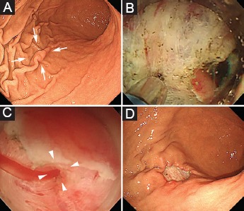
An 84-year-old man was diagnosed with an EGC on the greater curvature of the gastric body (Fig. 1A). After a circumferential marking, a mucosal incision was made as in conventional ESD. The gastric lumen was then filled with saline using a water jet and the lesion was resected underwater (Fig. 1B-D). We used a bipolar device (Jet B-knife, Zeon Medical Co. Tokyo, Japan), because electrical energy using monopolar devices is dispersed in saline, which has an electrical conductivity higher than that of body tissue. The UESD technique achieved complete en bloc resection without any adverse event (Fig. 2).
Figure 1.

Underwater endoscopic submucosal dissection (UESD) procedure. (A) Conventional endoscopic view of the lesion (arrow). (B) Underwater view during the submucosal dissection. (C) Underwater view at hemorrhage during procedure. The bleeding point was well visualized (arrow head). (D) The ulcer bed after performing UESD
Figure 2.

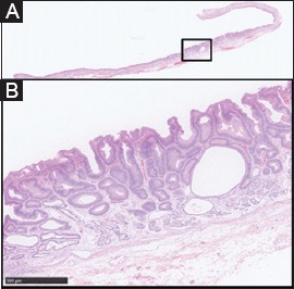
Histological examination of the resected specimen revealed an intramucosal well-differentiated adenocarcinoma with tumor-free resection margins. (A) Loupe view (hematoxylin and eosin [H&E] stain). (B) High-power microscopic view of the area outlined in black in loupe view (H&E stain)
This is the first report to describe UESD for an EGC. The floating effect of the mucosa and submucosa provides good traction against gravity. Water immersion improves visualization by reducing glare and dirt on the lens; in addition, in the case of hemorrhage the bleeding point is well visualized [3]. UESD can be a useful strategy for EGC located on the greater curvature of the gastric body.
Biography
Osaka University Graduate School of Medicine, Japan
Footnotes
Conflict of interest: None
References
- 1.Mori G, Nonaka S, Oda I, et al. Novel strategy of endoscopic submucosal dissection using an insulation-tipped knife for early gastric cancer: near-side approach method. Endosc Int Open. 2015;3:E425–E431. doi: 10.1055/s-0034-1392567. [DOI] [PMC free article] [PubMed] [Google Scholar]
- 2.Yoshii S, Hayashi Y, Matsui T, et al. “Underwater” endoscopic submucosal dissection: a novel technique for complete resection of a rectal neuroendocrine tumor. Endoscopy. 2016;48(Suppl 1 UCTN):E67–E68. doi: 10.1055/s-0042-101855. [DOI] [PubMed] [Google Scholar]
- 3.Frossard JL, Gervaz P, Huber O. Water-immersion sigmoidoscopy to treat acute GI bleeding in the perioperative period after surgical colorectal anastomosis. Gastrointest Endosc. 2010;71:167–170. doi: 10.1016/j.gie.2009.07.018. [DOI] [PubMed] [Google Scholar]


