

Abstract
Background:
Melaleuca alternifolia (Myrtaceae) is a well-known, commonly used, tall shrub plant in Ayurvedic medicine. Traditionally, it is used for its antimicrobial potential to treat cutaneous infections. No attempts have been made regarding pharmacognostic investigation of the plant till date. So, the present study was aimed to establish standards with the help of different pharmacognostic parameters.
Methods:
Various pharmacognostic parameters (morphological, microscopic, physicochemical evaluations and preliminary phytochemical screening) were studied along with fluorescent and thin layer chromatographic analysis of the extract.
Result:
Morphologically Melaleuca alternifolia is a shrub having height of 7 m with layered and papery bark. Leaves have an arranged pattern, petiole is 1 mm in length; linear-acute with dimensions of 10-35 mm x 1 mm. Organoleptic features shows that leaves have characteristic odour and astringent taste. The transverse section of the leaf reveals the existence of epidermal layers, mesophyll tissues, vascular bundles and secretory cavities. The stomata are anomocytic and leaf constants such as stomatal number is 180-200-225, stomatal index is 3.8-4.4-5.9, vein islet number is 18.68 (average), veinlet termination number 20.3 (average) and palisade ratio is 5.5-6.4-6.9. The results of phytochemical screening showed the occurrence of different phytoconstituents (flavonoids, phenolic tannins, phytosterol and terpenoids).
Conclusion:
The present study evaluated various pharmacognostic parameters which will help in quality control (standardization) of Melaleuca alternifolia leaves in crude form, in herbal formulation and also aid in the preparation of an herbal monograph for the species.
Keywords: Melaleuca alternifolia, pharmacognostic, organoleptic, microscopy, physicochemical
Introduction
The beneficial role of plant or plant-derived products in human therapy is a significant breakthrough in the history of humankind. However, it is not easy to know the accurate number of medicinal plants available on earth till date (Tiwary & Upadhyay, (2009). As per reports, it was seen that around 35,000-70,000 plants were used across the world to treat various ailments. In the last few decades, pharmaceutical manufacturer companies have focused on research and development of newly occurring plant-derived drugs. The estimated global market for medicinal plants is around the US $60 billion per year with a growth rate of 7% annually with developed and developing countries having varying shares (Farnsworth & Soejarto, (1991). Dissemination of knowledge about the importance and beneficial medicinal properties of such plants by the researcher is creating awareness among people. Accountability about the standardization and its biological activities are the few main factors to be dealt with so that the beneficial therapeutic benefits of the traditional medicine are scientifically validated (Rastogi & Rastogi, (2009).
The genus Melaleuca has more than 250 species of plants throughout the world but very few species are of Indian origin (Brophy et al., (2013) and have multiple applications in different traditional and folk medicines as well as economic importance.
Melaleuca alternifolia is also known as tea tree oil (TTO) and the volatile essential oil is derived largely from the plants which are native to Australia. The TTO possesses antimicrobial activity and employed as an active component in many of the formulations for treating cutaneous infections. Formulations containing Melaleuca alternifolia are OTC products in countries like Australia, Europe and North America where they are marketed for different remedies (Carson et al., (2006). This plant is very important for medicinal uses and herbal products. The leaves of “tea trees” were used for the treatment of cough or were spread on wounds, (Shemash& Mayo, (1991) the infusion of the leaves is utilised in treating sore throats and skin ailments (Low, (1990).
A thorough literature survey carried out on Melaleuca alternifolia revealed that it is an evergreen shrub to tree found as native to India. The entire plant is traditionally used in the management of many diseases. Despite many reports on the ethnomedicinal properties of the species, few investigations have been carried out on the selected plant in respect of the pharmacognostic standardization and pharmacological exploration. The work performed so far is insufficient to support the traditional claims of treating various ailments. In respect to the above, we have evaluated the pharmacognostic parameters for the first time which will help in the quality control of the herbal formulations and also in the preparation of the herbal monograph of the leaves of plant in near future.
Materials and Methods
Collection and authentication of plant material
The leaves of Melaleuca alternifolia (Myrtaceae) were collected from the hills of the Nilgiris district of Tamil Nadu, India in January 2016 from the healthy plants. The herbarium was prepared by using collected plant material and Dr. K. Madhava Chetty (Assistant Professor, Department of Botany, Sri Venkateswara University, Tirupati, Andhra Pradesh, India) authenticated the plant under voucher specimen no. 1241, dated May 28, 2016. A voucher specimen of plant material has been retained in the Department of Pharmacognosy and Phytochemistry, IKG Punjab Technical university, Kapurthala, Punjab, India
Macroscopical studies
Morphological observations of the Melaleuca alternifolia leaves at the macroscopic level were carried out in the laboratory.
Microscopic studies
The leaves of Melaleuca alternifolia were taken. Free hand section of the leaves was cut with the help of new sharp blade. The various tissues were distinguished using the staining technique (Figure 1).
Figure 1.
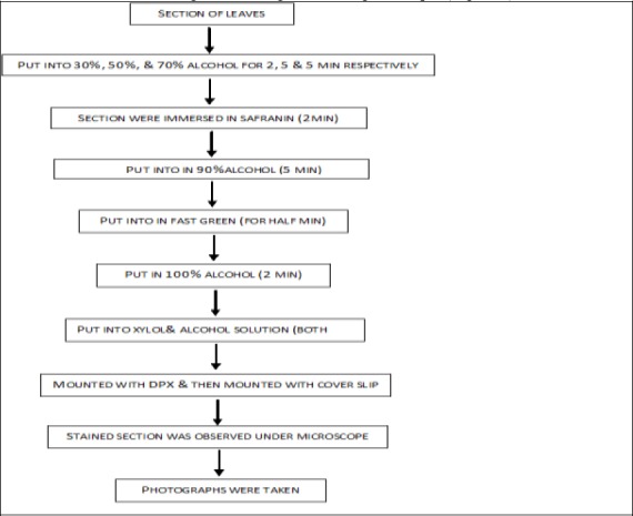
Scheme for preparation of transverse section of Melaleuca alternifolia leaves. Powder microscopy.
For the microscopic details of different tissues micrographs were prepared. Photographs at different magnification power were clicked using Nikon labphot-2 Microscopic unit. The bright field was used for normal observations. The study of crystals, starch grains and lignified cells was carried out using polarized light. Since these structures have birefringent property, observing them under polarized light they seems to be bright against dark background (Easu, (1964).
Determination of leaf constants
Following leaf constants were determined (Evans, (1997); Kokate, (2001); Kokate et al., (2003).
a) Stomatal number: The average amount of stomata per square millimeter of epidermis is termed the stomatal number.
b) Stomatal index: The ultimate divisions of the epidermis of a leaf percentage which have been changed into stomata is termed the stomatal index.
S = quantity of stomata per unit area and E = amount of ordinary epidermal cells in the same unit area.
c) Vein-islet number: Vein-islets per sq. mm calculated from four contiguous squares in the central portion of the lamina, midway in the middle of the midrib and the margin.
d) Veinlet termination number: The quantity of veinlet terminations was determined per sq. mm of the leaf surface.
e) Palisade ratio: The average number of palisade cells under each higher epidermal cell is called the palisade ratio.
Physicochemical parameters
Various parameters were evaluated for identity, purity and strength according to IP, 1996. All the readings were taken in duplicate (Gupta, (2003).
a) Loss on drying: About 3 g of sample was transferred to the separate bottle and distributed evenly by gently side wise shaking to a depth not exceeding 10 mm. Loaded bottle was placed in a drying chamber not exceeding 100-105°C (the stopper was removed and left in the chamber). The bottle along with the content was weighed (the process was repeated until the successive weights get constant (drying to constant weight). They were allowed to cool in desiccator and the percentage loss of weight was calculated with reference to the fresh drug.
b) Total ash: Accurately weigh plant material (3 g) was taken in silica crucible (previously ignited, cooled and weighed). Incinerate the plant material by gradually increasing the heat, not exceeding 450°C until free from carbon. Afterwards cooled in a desiccator and weighed to the constant weight and calculated the percentage (%) of total ash with reference to air dried drug as per Indian pharmacopoeia.
c) Acid insoluble ash: Total ash was boiled for 5 minutes with 25.0 mL of 2 N HCl (~70 g/L). Ignite the insoluble matter on an ash less filter paper at about 500°C to constant weight. The weight of insoluble matter was subtracted from the weight of total ash. The difference in weight represents the acid insoluble ash and the percentage of acid insoluble ash calculated with reference to the air dried drug.
d) Water soluble ash: The total ash obtained was boiled with 25 mL of water for 5 minutes. The insoluble matter was collected on an ash less filter paper. Washed with hot water and ignited the insoluble matter on an ash less filter paper for 15 minutes at a temperature not exceeding 450°C. The weight of insoluble matter was subtracted from the weight of total ash. The difference in weight represents the water soluble ash and the percentage of water soluble ash calculated with reference to the air dried drug.
e) Sulphated ash: Silica crucible was warmed to redness for 10 minutes and was permitted to cool in a desiccator and 3 g of sample was precisely weighed and transferred to the crucible. It was touched off delicately at to start with until the substance was altogether scorched. At that point residue was cooled and soaked with l mL concentrated sulphuric acid, warmed tenderly until white fumes did not advance anymore and touched off at 800°C±25°C until every single black particles have vanished. The ignition was led in a muffle furnace. The crucible was permitted to cool and a couple drops of concentrated sulphuric acid were included and warmed. Touched off as before and was permitted to cool and weighed. The operation was repeated until two successive readings with not more than 0.5 mg of variation were observed.
f) Ethanol soluble extractive: Powdered drug (5 g) was macerated with 100 ml of ethanol in a closed flask for 24 hrs with successive shaking for initial 6 hrs, the flask was permitted to remain for 18 hrs. The concentrate was separated and 25 ml of filtrate was vanished to dryness in a shallow dish. Then dried at 105°C and weighed. The rate of ethanol-soluble extractive with reference to the air dried drug was figured.
g) Water soluble extractive: Powdered drug (5 g) was macerated with 100 ml of chloroform water in a closed flask for 24 hours with successive shaking for the initial 6 hours, the flask was permitted to remain for 18 hours. The concentrate was separated and 25 ml of filtrate was dissipated to dryness in a shallow dish. The build-up was dried at 105°C and weighed. The rate of water solvent extractive with reference to the air dried drug was figured.
Thin Layer Chromatography
30 g of silica gel “G” was weighed and made to a homogenous suspension with 60 ml of refined water for two minutes. This suspension was filled in TLC implement which was changed in accordance with 0.25 mm thickness (20 x 20 cm). TLC plate was air dried until the straightforwardness of the layer vanishes. The plates were dried in hot air stove at 110°C for 30 minutes and afterward put away in dry air and utilized at whatever point required (Ravishankar, (1998).
The powder drug stayed regarded with acids, for example-1 N HCl and 50 % H2SO4, basic arrangements sodium hydroxide (aqueous), sodium hydroxide (alcoholic) and othe different solvents like nitric corrosive, picric corrosive, acidic corrosive, ferric chloride, and nitric corrosive with smelling salts and subjected to fluorescence investigation in daytime and in the bright UV light (254 nm and 365 nm) (Gupta et al., (2006).
Fluorescence analysis Preliminary phytochemical analysis
Phytochemial screening of methanolic extract of Melaleuca alternifolia leaves was carried out (Ronsenthaler, (1930); Middeltone, (1956).
Results
Organoleptic description
The characteristics observed are as follows (Figure 2)
Figure 2.
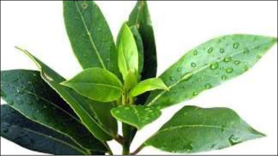
Melaleuca alternifolia leaves.
Type - simple leaf
Colour - upper surface: dark green and lower surface: light green
Odour - characteristic
Taste - astringent and bitter
Size - length is 5-8 cm and width is 3-6 cm
Shape - ovate
Petiole - narrowly winged
Margin - serrate
Apex - obtuse
Surface - glabrous
Microscopy
Anatomy of leaf
In cross section view, the leaf is more or less uniformly thick throughout its breadth, except slightly thick midrib Figure 3. The two margins are as thick as the lamina and are semicircular at the edges Figure 4. The leaf is 315 μm in thickness by the side of the midrib and 250 μm along the submarginal part. The leaf surface is smooth and even. The leaf consists of epidermal layers, mesophyll tissues, vascular bundles and secretory cavities.
Figure 3.
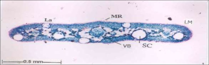
A. T.S. of leaf entire view. [La-Lamina, Lm-Leaf margin, MR-Mid ribs, Sc-Secretory Cavity, VB-Vascular bundle].
Figure 4.
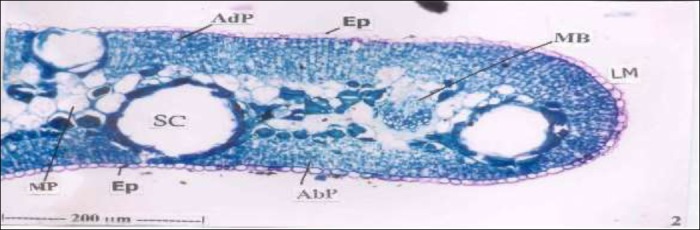
T.S of the margin enlarged.[Abp - Abaxial palisade cells, Adp - Adaxial palisade cells, Ep- Epidermis, Lm - Leaf margin, Mp - Middle parenchyma cells, Sc - Secretory Cavity]
(a). Epidermal layers:
Both abaxial and adaxial epidermal layers are thin; they consist of semicircular cells with hemispherical outer walls. The cuticle is thick Figure 5. The epidermal layer is nearly 10 μm thick.
Figure 5.
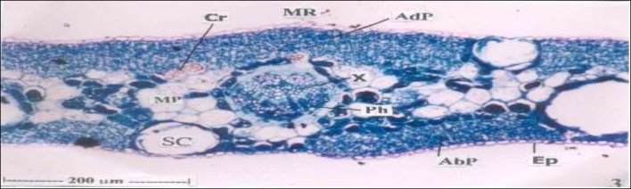
T.S. of leaf midrib enlarged. [Abp - Abaxial palisade cells, Adp - Adaxial palisade cells, Cr - Crystals, Ep-Epidermis, Mp - Middle parenchyma cells, MR - Mid ribs, Ph - Phloem, Sc - Secretory Cavity, X - Xylem]
b.Mesophyll tissue
The mesophyll tissues exhilit isobilateral symmetry; that is, it consists of palisade zones both on the adaxial and abaxial regions (figure 3 and 4). The adaxial palisade zone is 58 μm is height and consists of four or five layers short, norrowly rectangular chlorenchymatous cells. The aleaxial palisasade zone is 42 μm in height and has three or four layers of compact rectangular cells. In between the two palisade zones occens a median, nonchlorophyllous parenchyma zone; the cells of the median zone are angular, large and compact, Some of the cells have tannin contents which appear dark in the figure (figure 5).
(c). Vascular bundles
These are three prominent and large vascular bundles, one placed in the midrib other two placed along the margins of the lamina (figure 3, 5). The midrib bundle is larger than the marginal bundles and measures 166 x 125 μm in size. The marginal bundles are 85 μm in size. The bundles are circular and collateral. They have fan shaped, vertical parallel files of xylem elements and an arch of phloem elements. The bundle has an outer layer of tanniniferous, parenchymatous sheath and inner two or three layers of sclerenchymatous sheath. The inner sheath cells have thick lignified walls; this is indicated by bright walls of the cells when viewed under the polarized light microscope (figure 8, 9). The adaxial palisade zone continues above the midrib bundle as transcurrent - band, on the abaxial of the vascular bundle in seem a mass of parenchyma cells supporting the bundle.
Figure 8.
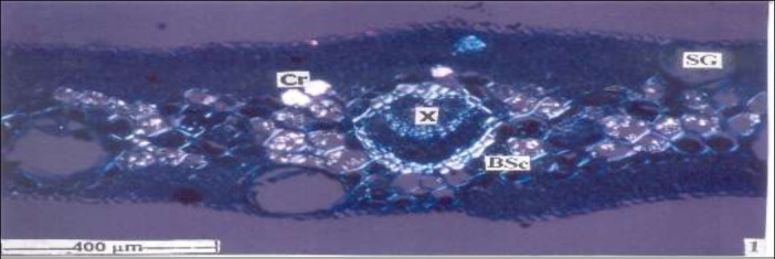
T.S. of showing sclerenchyma bundle sheath, crystals and starch grains. [BSc - Bundle Sheath cells, Cr -Crystal, SG - Starchgrains, X - Xylem]
Figure 9.
Druses in the adaxial palisade mesophyll with starch grains.
(d). Secretory cavities:
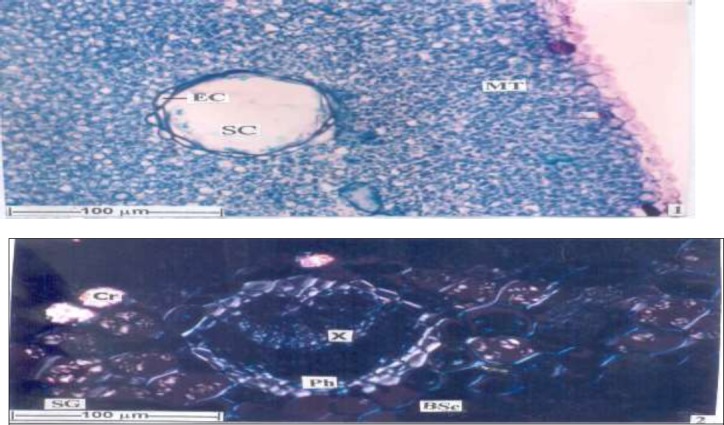
Much wide, circular secretary cavities are seen embedded in the palisade mesophyll zones. The cavities are schizogenous type; that is they arise by moving apart of a group of cells leaving a central space. The space widens gradually and forms the cavity surrounding the cavity is a circle of cells called epithelial cells. These cells are spindle shaped and have dense cytoplasm and secretary in functions (figure 6). The secretary cavities are 83
Figure 6.
Secretory cavity showing in Paradermal section.
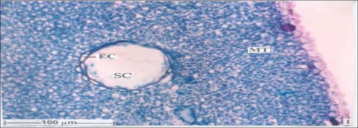
(e) Stomata
The leaf is amphistomatic with stomata seen on both upper and lower sides. The stomata are small and circular measuring 12 mm in vertical and horizontal planes. The stomata are anomocytic and no distinct subsidiary cells can be seen associated with the guard cells. The epidermal cells are small, polygonal in outline; their antielinal walls are thin and straight (figure 7)
Figure 7.
Abaxial epidermis with stomata [Ep- Epidermis, MT - Mesophyll tissue, SC- Secretory Cavity, St- Stomata] Cell inclusions (Figure 8 - 9).
There are two types of cell inclusions in the leaf-:
i) Calcium oxalate crystals:
Two types of calcium oxalate crystals are seen Druses or sphaerocrystals are seen in the median parenchyma cells of the mesophyll. Prismatic types of crystals are abundant along the sclerenchyma sheath of the vascular strands.
ii) Starch grains are abundant in the median parenchyma cells. They are circular, concentric or exocentric types. Many starch grains seen in a cell.
Powder microscopy
(a)Venation of fragmental leaf: Portions of leaf were seen with veins and veinlets. The lateral veins were thick, the veinlets were also prominent these were distinct polygonal vein islets. The vein islets end in vein termination, which were unbranched or branched once (Figure 10).
Figure 10.
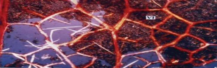
Vein and vein-islets [VI-Vein islets].
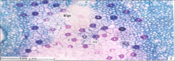
(b) Stomata: Some epidermal fragments with stomata were observed in the powder. Anomocytic type of stomata was found. Every stomata was enclosed by 2 or 3 whorls of subsidiary cells having each whorl of 4 to 6 rectangular cells. The guard cells were circular with or without stomatal aperture. The epidermal cells were rectangular small and have dense anticlinal walls (Figure 11).
Figure 11.
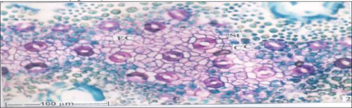
Abaxial epidermis with stomata [AdE-Adaxial epidermis, EC-Epidermal cell, SC-Secretory cavity, St-Stomata, Ve-Vein].
Leaf constants
The parameters evaluated are presented in Table 1.
Table 1.
Leaf constants of Melaleuca alternifolia.
| Leaf constant | Value per sq. mm |
|---|---|
| Stomatal Number | 125 |
| Stomatal Index | 20 |
| Palisade Ratio | 5 |
| Vein Islet Number | 4.2 |
| Vein Termination Number | 6.9 |
Fluorescence analysis
The result of fluorescence analysis is shown in Table 2. The leaf powder fluoresced green under daylight with short UV light (254 nm), dark green under long UV light (365 nm). The Melaleuca alternifolia leaf showed the characteristic fluorescent green treated with 50% H2SO4, concentrated HNO3, 50% HNO3, 40% NaOH+10% lead acetate, NH3, acetone, ethanol under short UV light (254 nm).
Table 2.
Fluorescence analysis of Melaleuca alternifolia leaf
| S. no. | Experiments | Visible/daylight | UV Low (254 nm), | UV High (365nm) |
|---|---|---|---|---|
| 1 | Powder | Green | Green | Dark green |
| 2 | Powder+1N Aqueous NaOH | Yellowish green | Greenish yellow | Dark blue |
| 3 | Powder+1 N Alcoholic NaOH | Yellowish green | Dark green | Dark brown |
| 4 | Powder+50% HNO3 | Greenish yellow | Fluorescent green | Violet |
| 5 | Powder+Conc. H2SO4 | Light brown | Light green | Brown |
| 6 | Powder+50% H2SO4 | Brown | Fluorescent green | Dark green |
| 7 | Powder+Conc. HNO3 | Light green | Fluorescent green | Dark green |
| 8 | Powder+Conc. HCl | Green | Light green | Dark green |
| 9 | Powder+50% HNO3 | Greenish yellow | Fluorescent green | Violet |
Physicochemical evaluation
The powdered leaves of Melaleuca alternifolia were evaluated for physicochemical parameters. Results are presented in Table 3.
Table 3.
Physical parameters of Melaleuca alternifolia powdered leaves
| S. No | Analytical parameters | Value (% w/w) |
|---|---|---|
| 1. | Total ash | 3.7838 |
| 2. | Acid insoluble ash | 0.982 |
| 3. | Water soluble ash | 1.642 |
| 4. | Sulphated ash | 2.142 |
| 5 | Loss on drying | 8.2 |
| 6 | Ethanol soluble extractive | 19.242 |
| 7 | Water soluble extractive | 12.140 |
Thin Layer Chromatography
The extract obtained from the leaves of Melaleuca alternifolia were subjected to TLC in toluene: ethyl acetate (93:7) as mobile phase. The colour of the spots and Rf values are given in the (Table 4).
Table 4.
Thin layer chromatographic result of the of Melaluca alternifolia.
| Absorbent | Mobile phase | Spraying reagent | No. of Spots | Rf Values | Blue |
|---|---|---|---|---|---|
| Silica gel | Toluene: Ethyl acetate (93:7) | Vanillin sulphuric acid | |||
| 1 | 0.9 | Light brown | |||
| 2 | 0.21 | Pale brown | |||
| 3 | 0.36 | Blue | |||
| 4 | 0.48 | Violet | |||
| 5 | 0.61 | Deep blue | |||
| 6 | 0.82 | Blue |
Phytochemical Screening
Phytochemical analysis of leaves extract of Melaluca alternifolia reveals the presence of different phytoconstituents (terpenoids, flavonoids, steroids, phenolic compounds and tannins) (Table 5).
Table 5.
Phytochemical screening of leaves extracts of Melaluca alternifolia
| S. No. | Test | Presence (+)/Absence (-) |
|---|---|---|
| 1 |
Carbohydrates
a. Molisch’s test |
- |
| 2 |
Glycosides a. Keller-Killiani test |
- |
| 3 |
Saponins a. Foam test |
+ |
| 4 |
Alkaloids a.Mayer’s test b.Dragendroff’s test |
- |
| 5 |
Flavonoids a. Alkaline reagent test |
+ |
| 6 |
Phenolics and Tannins a.Ferric chloride test b.Test for Tannins |
+ + |
| 7 |
Phytosterols and Triterpenoids a.Leiberman-Bucharat test b.Salkowaski test |
+ + |
| 8 |
Fixed oils and Fats a. Oily spot test |
- |
Discussion
Regardless of ethnomedicinal significance, still no attempt has been made for the pharmacognostical standardization of leaves of Melaleuca alternifolia. The standardization is a very important step in establishing the correct identity, purity, safety and quality of crude drug and it should be established before the inclusion of crude drug in herbal pharmacopoeia (Gupta et al., 2012). Recently, there has been an emphasis in standardization of medicinal plants of therapeutic potential. In spite of modern techniques, pharmacognostical evaluation is still more reliable for identification and evaluation of plants. World Health Organisation (WHO) recommends that the macroscopic and microscopic evaluation is most important in establishing the identity and purity of the plant (Kumar et al., 2012). In the present study, pharmacognostical standardization of leaves was carried out for the first time. Morphological, microscopic and physicochemical evaluation provides the simplest, quickest and cheapest means to establish the identity and purity of drug and also acts as a reliable tool for detecting adulteration. As the adulteration of the genuine raw material is the main cause of degradation of desired therapeutic effect of plant species used in traditional systems of medicine (Chinmay et al., 2011; Kumar et al., 2012).
In physicochemical parameters, ash values and extractive values serve as a reliable aid for detecting adulteration and in identification of plant. Ash values give an idea about inorganic composition or earthy matter or other impurities present along with drug while extractive value are useful for the determination of exhausted and adulterated drug. Extractive value also helps in indication of chemical constituents and in estimation of specific constituents soluble in particular solvents in a crude drug (Kumar et al., 2011; Thomas et al., 2008). Moisture content indicates about the storage of crude drug (Khandelwal, 2010). In the present study, the various pharmacognostical parameters such as morphological, microscopic, physicochemical evaluations and preliminary phytochemical screening were studied along with fluorescent and thin layer chromatographic analysis of the extract.
Melaleuca alternifolia has simple ovate glabrous leaves of 5-8 x 3-6 cm. The microscopic evaluation provides a clear picture of existence of epidermal layers, mesophyll tissues, vascular bundles and secretory cavities. The leaves are amphistomatic with anomocytic type of stomata. The preliminary phytochemical screening shows the presence of terpenoids, steroids, flavonoids, phenolic compounds and tannins. Fluorescent analysis, thin layer chromatography and the leaf constants were also reported which can be considered as characteristic parameters to authenticate the crude drug.
Conclusion
In conclusion, the parameters which are evaluated here can be considered as unique enough to identify and decide the authenticity of leaves of Melaleuca alternifolia which will serve in the development of pharmacopoeial standards for the future studies.
Referengces
- 1.Brophy J.J, Craven L.A, Doran J.C. Melaleucas: their botany, essential oils and uses. Australian Centre for International Agricultural Research (ACIAR) 2013 [Google Scholar]
- 2.Carson C.F, Hammer KA, Riley T.V. Melaleuca alternifolia (Tea Tree) Oil: a Review of Antimicrobial and Other Medicinal Properties. Clinical Microbiology Reviews. 2006;19(1):50–62. doi: 10.1128/CMR.19.1.50-62.2006. [DOI] [PMC free article] [PubMed] [Google Scholar]
- 3.Easu K. Plant Anatomy. New York: John Wiley and Sons; 1964. p. 767. [Google Scholar]
- 4.Evans W. C. In: In Trease and Evan ’s Pharmacognosy. 14th edition. Evans W. C, Evans D, editors. London, New York, UK: W.B. Saunders; 1997. pp. 576–578. [Google Scholar]
- 5.Farnsworth N.R, Soejarto D.D. Global importance of medicinal plants. The conservation of medicinal plants. 1991;26:25–51. [Google Scholar]
- 6.Gupta AK. Quality standards of Indian Medicinal Plants. Vol. 1. New Delhi: Indian Council of Medical Research 2003; 2003. pp. 236–7. [Google Scholar]
- 7.Gupta M.K, Sharma P. K, Ansari S. H, Lagarkha R. Pharmacognostical evaluation of Grewiaasiatica fruits. International Journal of Plant Sciences 2006. 2006;1(2):249–251. [Google Scholar]
- 8.Kokate C.K. In: In Practical Pharmacognosy. 4th edition. Kokate C. K, editor. Vallabh Prakashan; 2001. pp. 107–111. 115-121. [Google Scholar]
- 9.Kokate C.K, Purohit A.P, Gokhale S.B. In: Pharmacognosy. 24th edition. Prakashan: Nirali; 2003. pp. 100–336. [Google Scholar]
- 10.Low T. Bush medicine. North Ryde, NSW, Australia: Harper Collins Publishers; 1990. [Google Scholar]
- 11.Middeltone H. Systematic Qualitative Analysis. London: Edward Arnnold Publishers Ltd; 1956. pp. 91–94. [Google Scholar]
- 12.Rastogi S, Rastogi R, Rastogi R. Standardization and simplification of physico-therapeutic procedures in Ayurveda and naturopathy—A preliminary study with JalaNeti. Indian Journal Traditional Knowledge. 2009;8(3):464–70. [Google Scholar]
- 13.Ravishankar S. Textbook of Pharmaceutical Analysis. Second ed. Ootacamund Rx Publication; 1998. pp. 2–12. 5-2 to 5-7, 14-2 to 14-9. [Google Scholar]
- 14.Rosenthaler L. Chemical investigations of Plants. London: G. Bell and Sons; 1930. pp. 23–29. 119-132. [Google Scholar]
- 15.Shemesh A, Mayo W.L. Australian tea tree oil: a natural antiseptic and fungicidal agent. Australian Journal of Pharmacy. 1991;72:802–803. [Google Scholar]
- 16.Tiwari B.G, Upadhyay B.N. Concept of aging in Ayurveda. Indian Journal of Traditional Knowledge. 2009;8(3):396–9. [Google Scholar]
- 17.Khandelwal K.R. Practical Pharmacognosy Techniques and Experiments. Prakashan, Pune, India: Nirali; 2010. [Google Scholar]
- 18.Chinmay R, Kumari S, Dhar B, Mohanty R.C, Dixit R, Padhi M.M, Babu R. Phyto-Pharmacognostical studies of two endangered species of Malaxis (Jeevak and Rishibhak) Pharmacognosy Journal. 2011;3:77–85. [Google Scholar]
- 19.Gupta C.P, Sharma N, Rao C.V. Pharmacognostic studies of the leaves and stem of Careya arborea Roxb. Asian Pacific Journal of Tropical Biomedicine. 2012;2:404–8. doi: 10.1016/S2221-1691(12)60065-3. [DOI] [PMC free article] [PubMed] [Google Scholar]
- 20.Kumar D, Kumar A, Prakash O. Pharmacognostic evaluation of stem bark of Pongamia pinnata (L.) Pierre. Asian Pacific Journal of Tropical Biomedicine. 2012;2:543–6. [Google Scholar]
- 21.Kumar S, Kumar V, Prakash O.M. Pharmacognostic study and anti-inflammatory activity of Allistemon lanceolatus leaf. Asian Pacific Journal of Tropical Biomedicine. 2011;1:177–81. doi: 10.1016/S2221-1691(11)60022-1. [DOI] [PMC free article] [PubMed] [Google Scholar]
- 22.Thomas S, Patil D.A, Patil A.G, Chandra N. Pharmacognostic evaluation and physicochemical analysis of Averrhoa carambola L. fruit. Journal of Herb Toxicology. 2008;2:51–4. [Google Scholar]