Abstract
We report 2 families with undiagnosed recessive presynaptic congenital myasthenic syndrome (CMS). Whole exome or genome sequencing identified segregating homozygous variants in VAMP1: c.51_64delAGGTGGGGGTCCCC in a Kuwaiti family and c.146G>C in an Israeli family. VAMP1 is crucial for vesicle fusion at presynaptic neuromuscular junction (NMJ). Electrodiagnostic examination showed severely low compound muscle action potentials and presynaptic impairment. We assessed the effect of the nonsense mutation on mRNA levels and evaluated the NMJ transmission in VAMP1 lew/lew mice, observing neurophysiological features of presynaptic impairment, similar to the patients. Taken together, our findings highlight VAMP1 homozygous mutations as a cause of presynaptic CMS. Ann Neurol 2017;81:597–603
The congenital myasthenic syndromes (CMSs) are a heterogeneous group of inherited diseases of the neuromuscular junction (NMJ), with fatigable muscle weakness as the clinical hallmark.1 Several molecular causes can be implicated in CMS pathophysiology, including mutations in genes encoding proteins associated with the muscle nicotinic acetylcholine receptor and the synaptic basal lamina, or (more rarely) involved in the NMJ presynaptic transmission.2, 3, 4, 5, 6
We describe 2 families from Kuwait and Israel where 2 of the siblings in each family presented clinical and neurophysiological features typical of a presynaptic CMS. Whole exome sequencing (WES) or whole genome sequencing (WGS) followed by Sanger sequencing unraveled either a homozygous frameshift or missense variants in VAMP1 segregating with the phenotype in the 2 families. Screening a cohort of 63 undiagnosed CMS individuals failed to show any further causative variant in VAMP1.
Materials and Methods
Subjects
This study was approved by the institutional review boards of the participating centers. Informed consent was obtained from the families. Clinical details were obtained from medical records. Neurophysiological studies were performed according to standard procedures.7, 8
Genetic Studies
Before WES, the Kuwaiti probands (Family 1) underwent extensive molecular investigations that included sequencing of AGRN, mitochondrial DNA (mtDNA) sequencing, and deletion/duplication analysis and array comparative genome hybridization, which were all negative. Clinical trio‐based WES of Family 1 and WGS of the Israeli probands and their parents (Family 2) were performed as previously described.9, 10 Immortalized lymphoblastoid cell lines were used for RNA extraction, reverse transcription polymerase chain reaction (RT‐PCR) analysis, and semiquantitative RT‐PCR assay. Sanger sequencing was performed to analyze segregation of the variants identified by WES/WGS.
Vamp1 lew/lew Mice
Breeder pairs of Vamp1 +/lew mice (C3H/HeDiSnJ‐Vamp1 lew/GrsrJ, stock # 004626) were obtained from the Jackson Laboratory (Bar Harbor, ME) and mated to generate homozygous mutant (Vamp1 lew/lew) mice. Electrophysiological and morphological analyses of the NMJ in the Vamp1 lew/lew mice were performed as previously reported.11, 12 All experimental protocols were approved by the University of Texas Southwestern Medical Center institutional animal care and use committee.
Results
Clinical and Neurophysiological Characteristics
Family 1
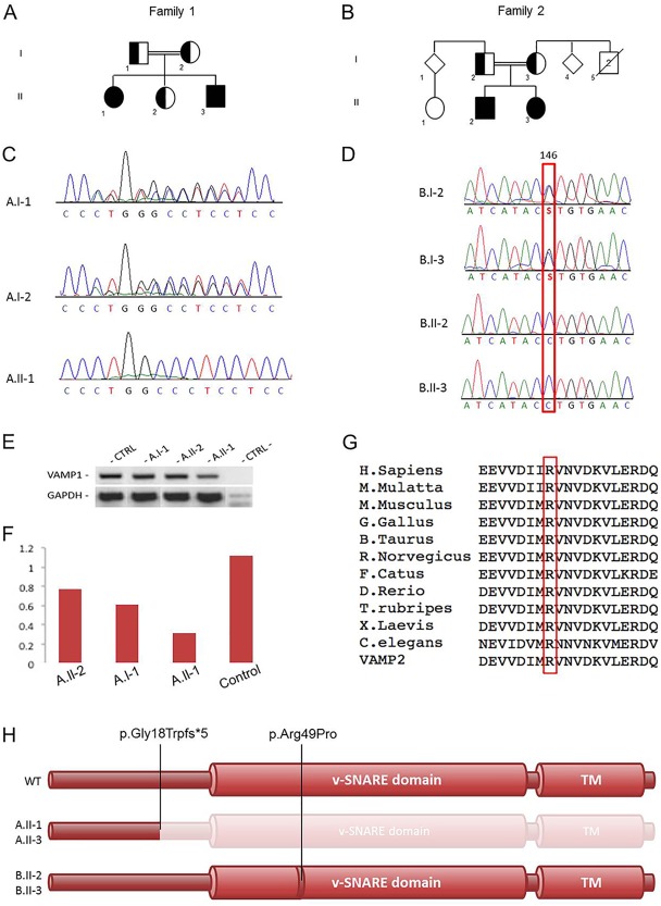
Both affected individuals A.II‐1 and A.II‐3 (Fig 1A) presented shortly after birth with hypotonia and muscular weakness. Feeding difficulties requiring gavage feeding, delayed motor development, and ophthalmoparesis characterized the disease course. A.II‐3 also presented joint contractures. Creatine kinase and plasma lactate were normal in the 2 children. On initial evaluation of Patient A.II‐3, muscle biopsy showed myopathic features and borderline low complex IV activity (0.011; normal range = 0.014–0.034), but congenital myopathy gene panel and mtDNA analysis were negative. Although hypotonia slightly improved in Patient A.II‐1, at the age of 3 years she still had difficulties standing upright and was unable to walk without support. Electrodiagnostic examination (EDX) in the 2 individuals showed similar findings (Table), with marked reduction in the amplitude of the compound muscle action potentials (CMAPs) and an increase in the amplitude to >200% of baseline on repetitive nerve stimulation (RNS) to 20Hz, indicating presynaptic impairment of NMJ transmission. The children's weakness slightly ameliorated under pyridostigmine treatment.
Figure 1.

Family trees, Sanger sequencing, and VAMP1 mutation analysis. (A) Pedigree from Family 1. (B) Pedigree from Family 2. (C) Electropherograms of carrier parents and index case with the c.51_64delAGGTGGGGGTCCCC variant. (D) Electropherograms of carrier parents and the 2 patients with the c.146G>C variant. (E) Reverse transcription polymerase chain reaction (PCR) amplifying the mutant cDNA transcript from mRNA extracted from the immortalized lymphoblastoid cell lines of the index case, her father, and her healthy sister (both carriers of the heterozygous deletion), and a wild‐type control (CTRL). (F) Analysis of the semiquantitative PCR using the densitometry software ImageJ after normalization relative to a housekeeping gene (GAPDH) and calculation using a relative relationship method. (G) Multiple‐sequence alignment showing complete conservation of protein sequence across species and SNARE homolog VAMP2 in the v‐SNARE coiled coil homology, in which the disease‐segregating mutation p.Arg49Pro was found. (H) VAMP1 protein representative. The c.51_64delAGGTGGGGGTCCCC deletion causes a nonsense mutation, putatively producing a truncated protein lacking the v‐SNARE and the transmembrane (TM) domains, whereas the p.Arg49Pro mutation affects an active site of the conserved v‐SNARE domain.
Family 2
The affected individuals of this family (B.II‐2 and B.II‐3; see Fig 1B) showed severe hypotonia and muscle weakness since birth. Both siblings had feeding difficulties and required percutaneous endoscopic gastrostomy. They presented severe impairment of developmental milestones. B.II‐1 also showed joint laxity and kyphoscoliosis. B.II‐3 presented knee contractures and breathing difficulties. During disease course, both children showed markedly reduced ability to generate antigravity posture and movements. B.II‐2 never reached autonomous walk; his EDX showed severely low CMAPs and increased neuromuscular jitter, indicating NMJ transmission abnormalities (see Table 1). In both siblings, pyridostigmine treatment improved symptoms.
Table 1.
Clinical and Neurophysiological Features of VAMP1‐Associated Congenital Myasthenic Syndrome in Our Families
| Feature | A.II‐1 | A.II‐3 | B.II‐1 | B.II‐2 |
|---|---|---|---|---|
| Parental consanguinity | + | + | + | + |
| Onset | Birth | Birth | Antenatal, DFM | Birth |
| Muscle weakness | ++ | ++ | ++ | ++ |
| Developmental delay | ++ | ++ | ++ | ++ |
| Feeding difficulties | ++ | ++ | ++ | ++ |
| Ophthalmological abnormalities | Strabismus, mild ophthalmoplegia | Mild ophthalmoplegia | Strabismus | Strabismus |
| GI abnormalities | − | GERD | Dysphagia | Dysphagia |
| Skeletal joint abnormalities | − | Joint contractures | Joint laxity, kyphoscoliosis | Joint contractures |
| Chest infections, aspiration | + | + | + | + |
| Response to pyridostigmine | + | + | + | + |
| Sensory studies | Normal | Normal | Normal | NT |
| Motor studies | AH CMAP ↓↓ | AH CMAP ↓↓ | ACL CMAP ↓↓ | NT |
| EMG | Myopathic | Myopathic | Myopathic | NT |
| Repetitive stimulation | AH: 3Hz, + 32.8%; 20Hz, + 640% | AH: 3Hz, + 60%; 20Hz, + 207% | NA | NT |
| Jitter | EDC, no twitch | Orb oculi, no twitch | ↑↑ mean MCD = 74.3 µs | NT |
ACL = accessorius motor left; AH = abductor pollicis; CMAP = compound muscle action potential; DFM = decreased fetal movements; EDC = extensor digitorum communis; EMG = electromyogram; GERD = gastroesophageal reflux disease; GI = gastrointestinal; MCD = mean consecutive difference; NA = not available; NT = not tested; Orb oculi = orbicularis oculi.
Identification of the VAMP1 Mutation
Trio‐based WES of Family 1 (A.I‐1, A.I‐2, A.II‐1; see Fig 1A) indicated in the index case 3 genes (Supplementary Table 1) carrying homozygous exonic variants predicted to have a possible pathogenic effect on protein function, based on the guidelines for variant classification.13 Full Sanger‐based segregation analysis of the candidate variants reduced the gene list to only 1 mutation in VAMP1 (NM_014231: c.51_64delAGGTGGGGGTCCCC; p.Gly18TrpfsTer5*), which was found to be homozygous in the affected individuals and heterozygous in their healthy sister and in the unaffected parents (see Fig 1C; data shown for the index case and her parents).
WGS of the 4 members of Family 2 (B.I‐2, B.I‐3, B.II‐2, B.II‐3; see Fig 1B) identified 6 genes carrying rare (likely) damaging variants (Supplementary Table 2), which were homozygous in the affected individuals and heterozygous in the parents.12 Among these 6 variants, a homozygous missense mutation in VAMP1 (NM_014231: c.146G>C; p.Arg49Pro; see Fig 1D) emerged as the most likely explanation for the disease pathogenesis, as supported by protein function (the mutation affects a conserved amino acid within the active domain of the protein),14, 15 expression and role of this gene in the NMJ,12, 16 and the homozygous mutation identified in the patients from Family 1 presenting the same phenotype (see Fig 1C–G).
RT‐PCR assay (performed to analyze possible nonsense‐mediated decay associated with the VAMP1 truncating variant in Family 1) found a mild reduction of mutant cDNA expression in the index case compared to the heterozygous carriers and the wild‐type control (see Fig 1E, F).
Impairments of the Neuromuscular Junction in Vamp1 lew/lew Mice
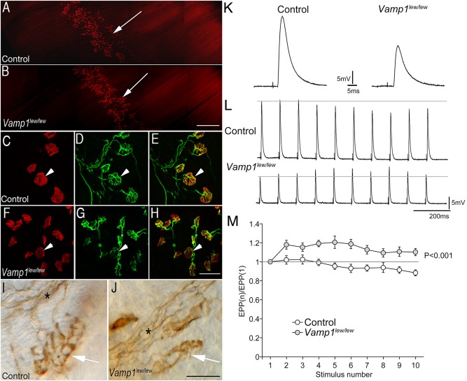
To further investigate whether a biallelic null mutation in VAMP1 in animal models may cause presynaptic NMJ abnormalities similarly to affected individuals, we re‐examined Vamp1 lew/lew mutant mice that were previously described.11, 12 The endplates were localized along the central regions of the muscle in both control and Vamp1 lew/lew mice (Fig 2). Individual neuromuscular synapses were found markedly smaller in Vamp1 lew/lew mice compared with control, and a severe reduction in endplate potentials (EPPs) was also observed in the mutant mice. Importantly, a low‐frequency, repetitive stimulation (10Hz) led to a run‐down of EPPs in control mice, but synaptic facilitation in Vamp1 lew/lew mice, indicating presynaptic defects.
Figure 2.

Synaptic defects at the neuromuscular junctions in Vamp1 lew/lew mice. (A, B) Low‐power images of the whole‐mount diaphragm muscles (P14) labeled by Texas Red—conjugated α‐bungarotoxin. The endplate band (arrow) is similarly localized along the central regions of the muscle in both control (A) and Vamp1 lew/lew mice (B). (C–H) High‐power confocal images of individual neuromuscular synapses in triangularis sterni muscles, labeled by Texas Red–conjugated α‐bungarotoxin (arrowheads in C and F) and antineurofilament NF150 and antisynaptotagmin2 antibodies (arrowheads in D and G point to the nerve terminals). Merged images are shown in E and H, for control and Vamp1 lew/lew mice, respectively. (I, J) Individual neuromuscular synapses (arrows) in triangularis sterni muscles labeled by antisyntaxin1 antibodies. The synapses are markedly smaller in Vamp1 lew/lew mice compared with the control. Asterisks indicate nerve bundles. (K) An example of endplate potentials (EPPs) recorded in the diaphragm muscle in control and Vamp1 lew/lew mice. (L) EPP traces responding to a low‐frequency, repetitive nerve stimulation (10Hz). (M) Quantitative measurement of the ratios of EPP amplitudes: EPP(n) to the first EPP amplitude, (EPP1). A low‐frequency, repetitive stimulation (10Hz) led to a run‐down of EPPs in control, but synaptic facilitation in (Vamp1 lew/lew) mice.
Discussion
Here, we report 4 children from 2 consanguineous families who presented with typical clinical and neurophysiological features of presynaptic CMS associated with homozygous mutations in VAMP1.
The protein encoded by this gene is a member of the synaptobrevin family.14 Synaptobrevins (eg, Vamp1, Vamp2), syntaxins, and the synaptosomal‐associated protein Snap25 represent the main components of the SNARE (soluble N ‐ethylmaleimide‐sensitive factor attachment protein receptors) complex, which is involved in docking and fusion of synaptic vesicles with the presynaptic membrane at the central and the neuromuscular synapses.15, 16 Proteins belonging to this complex are involved in vesicle docking through the evolutionarily conserved active v‐SNARE coiled coil homology domain and present high sequence similarity across the different SNAREs.17, 18, 19
Notably, the c.51_64delAGGTGGGGGTCCCC frameshift deletion identified in Family 1 leads to a change in the gene reading frame with the generation of a premature stop codon 5 amino acids downstream (see Fig 1H). The result is a putative VAMP1 product of only 21 amino acids, with a resulting function that is highly likely to be disrupted due to the absence of the downstream v‐SNARE domain (amino acids 33–93). The homozygous mutation identified in Family 2 consists of a substitution of a highly conserved arginine (Gerp++ score = 5.77) by a proline within the v‐SNARE domain. The mutated arginine residue in position 49 corresponds to the arginine in position 47 of the better‐studied SNARE homolog VAMP2, encoding another synaptobrevin with similar functions to VAMP1.20
Interestingly, it has been shown that disruption of this specific site in VAMP2 interferes with SNARE complex assembly, impairing neurotransmission, likely due to lack of association with other proteins involved in vesicle fusion.21, 22, 23 The Arg49Pro mutation is predicted as deleterious by SIFT, PolyPhen, and Mutation Taster and is carried in the heterozygous state by only 1 individual in the ExAC database (http://exac.broadinstitute.org, last accessed January 2017). Of note, in the ExAC database of 60,706 individuals there are only 17 individuals with heterozygous nonsynonymous single nucleotide substitutions within the v‐SNARE domain and 4 individuals in total carrying heterozygous truncating variants in VAMP1. None of these variants is present as homozygous, providing supportive evidence of pathogenicity for biallelic VAMP1 variants, either resulting in changes of the gene reading frame or affecting conserved active sites crucial to v‐SNARE domain function.
Interestingly, we also showed that the electrodiagnostic anomalies recorded in VAMP1‐associated CMS are consistent with the abnormal features of presynaptic transmission we recorded in the VAMP1 null mutant mice (including the incremental response to RNS; see Fig 2). These animals, of a model called lethal wasting (carrying a homozygous mutation that causes the truncation of half of the protein), lack movement because of an impaired NMJ transmission and die within 3 weeks of birth.11, 12
To date, biallelic variants in VAMP1 have never been reported, but heterozygous mutations in this gene have been described by a single study in association with a phenotype of autosomal dominant spastic ataxia.23 However, we have not observed any neurological phenotype in the heterozygous carriers from the 2 families.
In conclusion, the identification of biallelic variants in VAMP1 as a novel cause of CMS, in addition to other genes (eg, SNAP25B, SYT2) previously associated with similar presynaptic abnormalities of neuromuscular transmission,5, 6 highlights the crucial role of different SNAREs in NMJ physiology. Intriguingly, the relatively mild phenotype showed by our patients compared to the mouse model, which dies prematurely, suggests the possible existence of species‐specific compensation of vesicle fusion and release at the nerve terminal, perhaps through genetic modifiers in humans but not in mice or fruit flies. This highlights a promising area of future research aimed at the pathways involved in physiological presynaptic vesicle exocytosis at the motor endplate.
Author Contributions
Study concept and design: W.L., J.E.R., B.W., H.H. Data acquisition and analysis: A.M., A.S., K.M., L.B.‐V., M.W.H., S.A., M.P., S.E., S.W., A.Y.M., M.S., O.B., S.S.K. Drafting the manuscript and figures: V.S., A.D.V., Y.L., Q.Y.
Potential Conflicts of Interest
Nothing to report.
Supporting information
Additional supporting information can be found in the online version of this article.
Supporting Information 1
Acknowledgment
This study was supported by the Wellcome Trust (WT093205MA, WT104033AIA), Medical Research Council (H.H.), European Community's Seventh Framework Programme (FP7/2007‐2013, under grant agreement No. 2012‐305121; B.W., H.H.), National Institute for Health Research University College London Hospitals Biomedical Research Centre (H.H. and National Institutes of Health (NIH, NS‐055028; W.L.).
We thank the families for enthusiastic collaboration in this study.
Contributor Information
Brunhilde Wirth, Email: brunhilde.wirth@uk-koeln.de.
Henry Houlden, Email: h.houlden@ucl.ac.uk.
References
- 1. Engel AG, Shen XM, Selcen D, Sine SM. Congenital myasthenic syndromes: pathogenesis, diagnosis, and treatment. Lancet Neurol 2015;14:420–434. [DOI] [PMC free article] [PubMed] [Google Scholar]
- 2. Beeson D. Congenital myasthenic syndromes: recent advances. Curr Opin Neurol 2016;29:565–571. [DOI] [PubMed] [Google Scholar]
- 3. Schaaf CP. Nicotinic acetylcholine receptors in human genetic disease. Genet Med 2014;16:649–656. [DOI] [PubMed] [Google Scholar]
- 4. O'Grady GL, Verschuuren C, Yuen M, et al. Variants in SLC18A3, vesicular acetylcholine transporter, cause congenital myasthenic syndrome. Neurology 2016;87:1442–1448. [DOI] [PMC free article] [PubMed] [Google Scholar]
- 5. Herrmann DN, Horvath R, Sowden JE, et al. Synaptotagmin 2 mutations cause an autosomal‐dominant form of Lambert‐Eaton myasthenic syndrome and nonprogressive motor neuropathy. Am J Hum Genet 2014;95:332–339. [DOI] [PMC free article] [PubMed] [Google Scholar]
- 6. Shen XM, Selcen D, Brengman J, Engel AG. Mutant SNAP25B causes myasthenia, cortical hyperexcitability, ataxia, and intellectual disability. Neurology 2014;83:2247–2255. [DOI] [PMC free article] [PubMed] [Google Scholar]
- 7. Pitt M. Neurophysiological strategies for the diagnosis of disorders of the neuromuscular junction in children. Dev Med Child Neurol 2008;50:328–333. [DOI] [PubMed] [Google Scholar]
- 8. Aharoni S, Sadeh M, Shapira Y, et al. Congenital myasthenic syndrome in Israel: genetic and clinical characterization. Neuromuscul Disord 2017;27:136–140. [DOI] [PMC free article] [PubMed] [Google Scholar]
- 9. Mencacci NE, Kamsteeg EJ, Nakashima K, et al. De novo mutations in PDE10A cause childhood‐onset chorea with bilateral striatal lesions. Am J Hum Genet 2016;98:763–771. [DOI] [PMC free article] [PubMed] [Google Scholar]
- 10. Delle Vedove A, Storbeck M, Heller R, et al. Biallelic loss of proprioception‐related PIEZO2 causes muscular atrophy with perinatal respiratory distress, arthrogryposis, and scoliosis. Am J Hum Genet 2016;99:1406–1408. [DOI] [PMC free article] [PubMed] [Google Scholar]
- 11. Nystuen AM, Schwendinger JK, Sachs AJ, et al. A null mutation in VAMP1/synaptobrevin is associated with neurological defects and prewean mortality in the lethal‐wasting mouse mutant. Neurogenetics 2007;8:1–10. [DOI] [PubMed] [Google Scholar]
- 12. Liu Y, Sugiura Y, Lin W. The role of synaptobrevin1/VAMP1 in Ca2+‐triggered neurotransmitter release at the mouse neuromuscular junction. J Physiol 2011;589(pt 7):1603–1618. [DOI] [PMC free article] [PubMed] [Google Scholar]
- 13. Richards S, Aziz N, Bale S, et al. Standards and guidelines for the interpretation of sequence variants: a joint consensus recommendation of the American College of Medical Genetics and Genomics and the Association for Molecular Pathology. Genet Med 2015;17:405–424. [DOI] [PMC free article] [PubMed] [Google Scholar]
- 14. Sutton RB, Fasshauer D, Jahn R, Brunger AT. Crystal structure of a SNARE complex involved in synaptic exocytosis at 2.4 A resolution. Nature 1998;395:347–353. [DOI] [PubMed] [Google Scholar]
- 15. Cupertino RB, Kappel DB, Bandeira CE, et al. SNARE complex in developmental psychiatry: neurotransmitter exocytosis and beyond. J Neural Transm (Vienna) 2016;123:867–883. [DOI] [PubMed] [Google Scholar]
- 16. Kittel RJ, Heckmann M. Synaptic vesicle proteins and active zone plasticity. Front Synaptic Neurosci 2016;8:8. [DOI] [PMC free article] [PubMed] [Google Scholar]
- 17. Weimbs T, Low SH, Chapin SJ, et al. A conserved domain is present in different families of vesicular fusion proteins: a new superfamily. Proc Natl Acad Sci U S A 1997;94:3046–3051. [DOI] [PMC free article] [PubMed] [Google Scholar]
- 18. Hong W. SNAREs and traffic. Biochim Biophys Acta 2005;1744:120–144. [DOI] [PubMed] [Google Scholar]
- 19. Rothman JE. The principle of membrane fusion in the cell (Nobel lecture). Angew Chem Int Ed Engl 2014;53:12676–12694. [DOI] [PubMed] [Google Scholar]
- 20. Zimmermann J, Trimbuch T, Rosenmund C. Synaptobrevin 1 mediates vesicle priming and evoked release in a subpopulation of hippocampal neurons. J Neurophysiol 2014;112:1559–1565. [DOI] [PubMed] [Google Scholar]
- 21. Hao JC, Salem N, Peng XR, et al. Effect of mutations in vesicle‐associated membrane protein (VAMP) on the assembly of multimeric protein complexes. J Neurosci 1997;17:1596–1603. [DOI] [PMC free article] [PubMed] [Google Scholar]
- 22. Trimbuch T, Xu J, Flaherty D, et al. Re‐examining how complexin inhibits neurotransmitter release. Elife 2014;3:e02391. [DOI] [PMC free article] [PubMed] [Google Scholar]
- 23. Trimbuch T, Rosenmund C. Should I stop or should I go? The role of complexin in neurotransmitter release. Nat Rev Neurosci 2016;17:118–125. [DOI] [PubMed] [Google Scholar]
Associated Data
This section collects any data citations, data availability statements, or supplementary materials included in this article.
Supplementary Materials
Additional supporting information can be found in the online version of this article.
Supporting Information 1


