Abstract
Psychiatric genetic studies have identified genome-wide significant loci for schizophrenia. The AKT3/1q44 locus is a principal risk region and gene-network analyses identify AKT3 polymorphisms as a constituent of several neurobiological pathways relevant to psychiatric risk; the neurobiological mechanisms remain unknown. AKT3 shows prenatal enrichment during human neocortical development and recurrent copy number variations involving the 1q43-44 locus are associated with cortical malformations and intellectual disability, implicating an essential role in early brain development. Here, we investigated the role of AKT3 as it relates to aspects of learning and memory and behavioral function, relevant to schizophrenia and cognitive disability, utilizing a novel murine model of Akt3 genetic deficiency. Akt3 heterozygous (Akt3-/+) or null mice (Akt3-/-) were assessed in a comprehensive test battery. Brain biochemical studies were conducted to assess the impact of Akt3 deficiency on cortical Akt/mTOR signaling. Akt3-/+ and Akt3-/- mice exhibited selective deficits of temporal order discrimination and spatial memory, tasks critically dependent on intact prefrontal-hippocampal circuitry, but showed normal prepulse inhibition, fear conditioned learning, memory for novel objects and social function. Akt3 loss-of-function, reduced brain size and dramatically impaired cortical Akt Ser473 activation in an allele-dose dependent manner. Such changes were observed in the absence of altered Akt1 or Akt2 protein expression. Concomitant reduction of the mTORC2 complex proteins, Rictor and Sin1 identifies a potential mechanism. Our findings provide novel insight into the neurodevelopmental role of Akt3, identify a non-redundant role for Akt3 in the development of prefrontal cortical-mediated cognitive function and show that Akt3 is potentially the dominant regulator of AKT/mTOR signaling in brain.
Introduction
Schizophrenia is a common neuropsychiatric disorder, characterized by positive (i.e. hallucinations, delusions) and negative symptomatology (i.e. flat affect, social withdrawal, lack of motivation) and cognitive disability. Deficits ascribed to abnormal development of the prefrontal cortex (PFC) and hippocampal formation, including working memory, executive function and cognitive flexibility, represent core features of the illness with an unknown etiology [1–4].
Large-scale genetic studies of schizophrenia have identified several genomic loci and gene pathways that increase risk. A recent genome-wide association study (GWAS) of 36,989 cases and 113,075 controls identified 108 independent associations [5], and network analyses of these data identified risk in several gene pathways involved in neuronal, immune and histone biology [6]. Interestingly, overlap of risk loci and antipsychotic drug gene targets has recently been reported [7], suggesting a link between disease etiology and antipsychotic mechanisms of action. Given the increasing understanding of the genetic basis of schizophrenia, a critical next step in mental illness research is identification of mechanisms and characterization of the in vivo function of risk genes. Such validation is necessary to identify novel therapeutic targets of pathophysiological relevance.
Genetic variation in the AKT3 locus (chr1:243503719–244002945) is a top GWAS signal in schizophrenia [5,8] and pathway analysis identified 50 single nucleotide polymorphisms (SNPs) within the AKT3 gene that contribute to four of the top pathways associated with risk for schizophrenia and bipolar disorder [6]. Moreover, recent investigations testing for enrichment of the 108 schizophrenia-risk loci, and overlapping rare singleton disruptive mutations in gene sets coding for proteins targeted by antipsychotic drugs, identified AKT3 as a potential target gene of relevance to antipsychotic treatment and response [7]. These studies identify AKT3 as a promising risk gene for schizophrenia and further highlight the AKT signaling pathway as a potential target for improved treatment development [9–11].
AKT (also known as protein kinase B (PKB)) is a critical intracellular serine/threonine kinase that translates signals from extracellular stimuli including growth factors, cytokines and neurotransmitters in response to activation of the intracellular second messenger, phosphatidylinositol 3-kinase (PI3K). AKT signaling plays critical roles in organogenesis, influencing cell growth, proliferation, survival, differentiation and metabolism [12–14] and in mammals, consists of a tripartite pathway which includes members AKT1, AKT2, and AKT3, each with a high level of protein homology [15] and encoded by independent genes (14q32.32; 19q13.2 and 1q44, respectively). The key regulatory phosphorylation sites on AKT (Ser473and Thr308) are conserved across isoforms and recent evidence suggests that phosphorylation of Ser473, which is governed by mTORC2, determines signaling to specific substrates [16,17]. Although the cellular function of each isotype is poorly understood, emerging in-vitro data suggests that AKT isoforms are present at distinct subcellular locations [18,19], exhibit different substrate specificity and importantly enzyme activity, with AKT3 being 15–40 fold more active than AKT1 or AKT2 [20,21].
Murine studies of single and double isoform Akt knockout (KO) mice have also provided critical information on the developmental role of individual Akt isoforms, with Akt1 being a key regulator of whole body organismal growth [22]; Akt2 being critical for normal glucose homeostasis [23], and Akt3 playing a specific role in attainment of normal brain size, specifically through regulation of cell size and number [24,25]. Consistently, recent data in rat stroke models demonstrate a critical neuroprotective role for Akt3 [26].
In addition to the recent AKT3 association, genetic variation in AKT1 has previously been implicated in risk for neuropsychiatric phenotypes. Specifically, AKT1 is associated in humans with schizophrenia [27–33] and abnormal PFC and hippocampal- structure and function [34–37]. Furthermore, evidence from postmortem brain and peripheral cells of patients with schizophrenia has identified reduced AKT activity, either through reduced expression of AKT1 [27,30], AKT3 [38] or phosphorylated AKT Ser473 [39,40]. It is important to note that it is currently unknown whether the decrease in AKT Ser473 phosphorylation observed in patients with schizophrenia [39,40] is accounted for by alterations in the activity of AKT1, AKT2 or AKT3.
In this context, mice with genetic deletion of Akt1 or Akt2 have previously been examined for neurobehavioral deficits relevant to psychiatric illness. Akt1 deficiency results in deficits of sensorimotor gating, hippocampal/PFC-dependent learning (affecting contextual fear and spatial and working memory), synaptic plasticity and electrophysiological measures [27,40–42]. In contrast, preliminary analyses show that Akt2 deletion produces selective anxiety and depression-like phenotypes in mice [43], thus suggesting that the Akt genes play differential roles in brain development and function. It is currently unknown whether Akt3 deficiency affects neurobehavioral and cognitive function and what signaling pathways may be involved.
AKT3 is highly expressed in both human and mouse brain [24,25,44], represents up to 50–60% of total AKT in the murine cortex and hippocampus [24] and shows differential prenatal expression patterns, with expression being higher during human fetal brain development [44]. Together these observations suggest a pivotal role for AKT3 in neocortical development and cognitive function. Consistently, numerous genomic studies have suggested a critical role for AKT3 in the developing human central nervous system, specifically in the attainment of normal brain size and intellectual ability. Notably, human germline and somatic mutations and copy-number variations (CNVs) impacting the 1q44 locus and AKT3 have been identified in a range of developmental brain malformation syndromes, including microcephaly [45,46], hemimegalencephaly [47,48] and megalencephalic syndromes [49], all of which are characterized by intellectual disability. Furthermore, studies have consistently shown that Akt3 knockout mice exhibit organ-specific reductions in brain size, with ~20–25% reductions compared to wildtype (WT) mice [24,25].
Here, we examined the role of Akt3 in cognitive function, learning and memory utilizing a novel murine model of Akt3 genetic deficiency and studied the biochemical signaling correlates. Akt3 mice with one (Akt3-/+) or zero alleles (Akt3-/-) were generated on the C57BL/6 background and compared to WT littermates (Akt3+/+). Examination of heterozygote Akt3 mice is an important advance of the current study and provides biologically relevant data on the function of Akt3 in the context of human genetic conditions of Akt3 dysfunction. A comprehensive battery of behavioral and physiological tests relevant to schizophrenia and cognitive dysfunction, chosen to allow dissection of underlying neuroanatomical system deficits were performed along with comprehensive brain biochemical studies of the Akt/mTOR signaling pathway. Our results identify a novel and non-redundant role for Akt3 in prefrontal cortical and hippocampal-mediated cognitive function, identify potential neurobiological mechanisms underlying association of the AKT3 gene with schizophrenia relevant to cortical development, and identify Akt3 as a critical determinant of AKT signaling in the brain.
Materials and methods
Subjects and ethics statement
All animal breeding, maintenance and experimental procedures were in accordance with and approved by the National Institute Health Animal Care and Use Committee and followed the National Institutes of Health Using Animals in Intramural Research and the University of Colorado Denver Institutional Animal Care and Use Committee (IACUC). Akt3-/- mice were generated via germ line excision of murine exon 3 (corresponding to nucleotides 209 to 320 of the mouse cDNA), which introduces a frame shift in the open reading frame (first described [24]). Akt3-/- mice were originally generated by Dr. Thomas Ludwig (Columbia University) and Dr. Morris Birnbaum (University of Pennsylvania) and obtained via Material Transfer Agreement with Columbia University. We successfully established Akt3 mutants on the C57BL/6 background (>25 generation of backcrosses) in our laboratory which are maintained as stable colonies. Wild-type (WT, Akt3+/+), heterozygous (Het, Akt3-/+) and knockout (KO, Akt3-/-) mice were generated and included in all assessments and represent littermates derived from multiple independent litters. Mice were group housed (up to 5 animals per cage) and maintained on a 14 hour light/10 hour dark cycle in a climate-controlled animal facility (22±2°C) with food and water ad libitum. Animals were sacrificed one week after all behavioral testing by guillotine without anesthesia, according to our approved IACUC method of sacrifice for tissue collection.
Genotyping
Genotyping was performed by PCR analysis of DNA extracted from tail clips. In brief, DNA extracted by the HOT SHOT method was used as a template for PCR amplification of Akt3 using the following primers: Akt3 forward, Primer 1, (5’-GAT AGG TGG CTT GTG AGT TC-3’); Akt3 wild-type reverse, Primer 2, (5’-CCA GGG TAA GGC CTA AAG CT-3’), which produced a 192kb band from DNAs of wild-type mice. Primers 1 and Akt3 knockout reverse Primer 3, (5’-GCT CAT TCC TCC CAC TCA TGA-3’) produced a 340kb band from DNA isolated from Akt3-/- mice (S1A Fig). Amplification was carried out using Invitrogen Platinum Taq (Thermo Fisher Scientific, PN#10966–018; Grand Island, NY, USA) per the manufacturers protocol, under the following thermal cycling conditions: Initial denaturation: 94°C for 3 min, 40 PCR cycles of: 94°C for 30 sec, 60°C for 30 sec, 72°C for 30 sec, final extension: 72°C for 7 min.
General physical health
Measures of general health and neurological reflexes were assessed in adult mice, as previously described [50,51]. Mice were observed for measures including posture, fur condition and empty cage behavior to detect irregular behaviors such as excessive grooming, digging, rearing, or stereotypy. Neurological reflexes included trunk curl, limb strength (wire hang test), forepaw reaching, corneal reflex, ear twitch, whisker twitch, and the righting reflex were also examined.
Behavioral testing
All behavioral tasks were performed on adult male mice, 3 to 5 months old during the light phase. Mice were handled by the experimenter the week preceding testing. To prevent influences of task-related anxiety, animals were split into independent cohorts with the most aversive task performed last. Animals were tested as follows: Cohort 1 (general health, open field, temporal order object recognition, and startle prepulse inhibition); Cohort 2 (social cognition tasks and fear conditioning); Cohort 3 (open field, novel object and fear extinction) and Cohort 4 (open field, object location). All mice were habituated one hour prior to testing. Locomotor activity was performed the day before temporal order object recognition, novel object recognition and object location tasks and served as habituation to the open field. All sessions for open field locomotor activity, sociability, temporal order object recognition, novel object recognition and object location tasks were video recorded with a top mounted CCTV camera (Security Cameras Direct, Luling, TX, USA) using Ethovision XT video tracking system (Noldus; Leesburg, VA, USA).
Locomotor activity (open field)
The open field test systematically assesses novel environment exploration, general locomotor activity, and anxiety-related emotional behaviors. Baseline locomotor activity was assessed as total distance traveled in an open field experimental arena made of gray polyvinyl (42×42×30cm) under red light luminescence for 60 mins, as previously described [50]. Time spent and distance traveled in the center portion of the arena was assessed as a measure of anxiety.
Temporal order object recognition memory
The temporal order object recognition task was assessed as previously described [50–53] and as described in detail in S1 Methods. Temporal order measures recency discrimination, defined as a rodent’s ability to differentiate between two familiar objects presented at different intervals and is dependent on information flow between the medial PFC (mPFC), perirhinal (PRH) cortex and hippocampus [52]. Briefly, the task was performed in three, 5-minute sessions, in the same experimental arena used for locomotor activity, under red light luminescence (5±2 lux). If temporal order discrimination memory is intact subjects would exhibit a discrimination ratio > 0. Individual discrimination ratios were calculated as time spent by the animal exploring the object from sample phase 1 minus the time spent with the object from sample phase 2, divided by the total time spent exploring both objects during the test period.
Novel object recognition
The novel object recognition test was as previously described with small changes [51,53]. Novel object preference requires intact function of the PRH cortex (but not the mPFC or hippocampus [52]). Briefly, the subject was introduced for 10 minutes to two identical objects for the acquisition trial. Two hours later, the mouse was placed back into the same arena with a duplicate of the old object and a new object of the opposite color and shape and allowed to explore for 5 mins. Individual discrimination ratios were calculated as time spent by the animal exploring the novel object minus the time spent with the object from phase 1, divided by the total time spent exploring both objects during the test period. Discrimination ratio > 0 show preference for the novel object.
Object location memory
Object location was performed to specifically assess hippocampal function and spatial memory [52]. The subject was introduced for 10 minutes to two identical objects for the acquisition trial. One hour later, the mouse was allowed to explore the same two objects for 5 minutes, with one object in its original location and the other counter-opposite to its original position. The object locations were counterbalanced across trials to eliminate the chance of side preference. Individual discrimination ratios were calculated as time spent by the animal exploring the relocated object minus the time spent with the unmoved object from phase 1, divided by the total time spent exploring both objects during the test period. Discrimination ratio > 0 show preference for the moved object and intact spatial memory.
Acoustic startle and Prepulse Inhibition (PPI)
Startle responses and PPI were measured using SR-Lab Systems (San Diego Instruments; San Diego, CA, USA) as previously described [50,53]. The background level was maintained at 70 dB. The trials consisted of measurements of the startle response to no stimulus (Nulstim), to an auditory startle stimulus (Startle; 40 ms, 120 dB), and of the PPI in response to an acoustic prepulse of 20 ms at 74, 78, 82, 86, and 90 dB.
Fear conditioning, cued and context
Fear conditioning, dependent on hippocampal and/or amygdala function, was assessed using Pavlovian methodology as previously described [53]. The task was performed using a San Diego Instruments Freeze Monitor System (San Diego Instruments; San Diego, CA, USA) conditioning chamber and is described in detail in S1 Methods.
Fear extinction
Fear extinction testing was used to assess amygdala depotentiation [54]. Mice were assessed over a total of 3 days. Day 1 (Conditioning Phase), consisted of five trials of five auditory tones (80db) lasting 30 seconds with variable intervals (ranging from 80 to 220 sec) followed by five additional trials of 30 sec tones preceding a 0.5 second foot shock. Day 2 and 3 (Extinction Phases) used identical protocols. On ‘Extinction Phase’ days, mice were exposed to thirty tones lasting 30 seconds each, at variable intervals without any foot shock. Freezing behavior was calculated by the average freezing time (time frozen/allotted time x 100). Normal freezing behavior in mice should progressively decrease over trial days (i.e. % Freezing on Day 1> Day 2> Day 3).
Social function
Social behavior was tested using methods previously described for assessment of sociability and preference for social novelty [53,55,56]. Social approach was tested in an automated three-chambered apparatus. Mice used as the novel stimulus target were C57BL/6 matched to the subject mice by sex and age.
Biochemical assessment of AKT signaling in Akt3-deficient mice
Fresh, frozen mPFC and hippocampus from behaviorally naïve mice was homogenized with a sonicator (Ultrasonic Processor model GE50 Sonics, Newtown, CT, USA) in a lysis buffer containing DTT 1 M (Dithiothreitol, Sigma-Aldrich, St. Louis, MO, USA), protease inhibitor cocktail and T-PER protein Extraction (PN 78510, Thermo Scientific, Grand Island, NY, USA). Thirty or fifty micrograms of protein was resolved per standard Western blotting procedure, as previously described [11,50]. A FluorChemQ image analyzer from Protein Simple (San Jose, CA, USA) was used for Chemiluminescence detection method. The optical density of the protein bands were calculated using Image J software. β-actin was used as a standard loading control for normalization. A full list of primary and secondary antibodies are described in Supplementary material.
Brain to body weight measurements
Male Akt3 mice were weighed at postnatal day 105 using an Ohaus CS 200 balance (Parsippany, NJ, USA) immediately before sacrifice for brain collection and weighing.
Quantitative real-time PCR
To quantitatively assess SDCCAG8 expression, total RNA was extracted from PFC and of male Akt3 mice 100 using the TRIzol Extraction method (Qiagen). Quantitative Real-Time RT-PCR was carried out as previously described using the standard curve method [53,57] and TaqMan assay (Hs01104613_m1). For each sample the expression levels of the gene of interest were normalized to the housekeeping gene GAPDH (Mm99999915_g1, Life Technologies).
Statistical analyses
Data was analyzed using SPSS statistical software (IBM Corp, Armonk, NY, USA v.22). Statistical significance levels were indicated by p<0.05. Fisher LSD posthocs were used to investigate significant findings. General health measures and discrimination ratios for recognition tasks were analyzed with Univariate ANOVA. Repeated Measures ANOVA was used for all other behavioral tasks unless indicated. Linear Regression Analysis was used to examine effects of genotype on quantitative protein traits.
Results
Akt3 deficiency results in reduced brain size, with no overall impact on body size or general health
Akt3 mice with one (Akt3-/+) or zero (Akt3-/-) alleles did not differ from WT (Akt3+/+) mice in a comprehensive general health screen (S1 Table), demonstrating physical capability for performing the subsequent battery of behavioral tasks. Consistent with previous reports in Akt3 null mice [24,25], Akt3 deficiency impacted total brain weight. Our data identify that Akt3 deficiency is consistently associated with reduced brain weight, expressed in absolute terms (Main effect of genotype: F (2, 21) = 51.337, p<0.001; posthoc LSD, Akt3+/+ vs. Akt3-/+ and Akt3+/+ vs. Akt3-/-, p<0.001; S1b Fig) and as a ratio to body weight (main effect of genotype: F (2, 21) = 49.497, p<0.001; posthoc LSD, Akt3+/+ vs. Akt3-/+ and Akt3+/+ vs. Akt3-/-, p<0.001; S1c Fig). In addition, we report a novel allele dose-dependent effect on brain weight, with Akt3 heterozygosity being associated with a 7% reduction in brain weight and null- with a 24% reduction (S1 Fig).
Baseline locomotor activity is increased in Akt3-/- mice
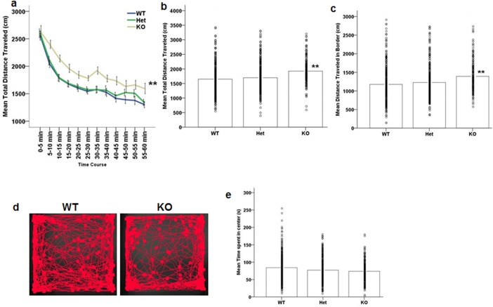
Repeated measures ANOVA revealed a significant decrease of distance traveled over time during the open field test (main effect of time: F (11, 759) = 136.238, p<0.001; Fig 1a), demonstrating that all mice show similar locomotive habituation. No time x genotype interaction was observed. A significant main effect of genotype was however observed (F (2, 69) = 5.954, p = 0.004) whereby Akt3-/- mice were hyperactive compared to their Akt3+/+ and Akt3-/+ littermates (posthoc LSD Akt3+/+ vs. Akt3-/-, p = 0.001; Fig 1b). Further investigation showed that Akt3-/- mice traveled significantly more distance in the border (F (2, 69) = 5.389, p = 0.007; posthoc LSD Akt3+/+ vs. Akt3-/-, p = 0.002; Fig 1c and 1d). However, there were no differences in distance traveled in the center among genotypes (p>0.1). We also measured time spent in the center of the apparatus to assess if Akt3 deficiency is associated with anxiety-like phenotypes. Our data indicate no effect of genotype on time spent in center (p>0.1, Fig 1e). These data reveal that genetic loss of Akt3 in the mouse results in hyperlocomotive behaviors similar to those observed in models of psychostimulant-mediated effects on increased dopaminergic function and suggestive of a novel role of Akt3 in dopaminergic signaling.
Fig 1. Increased locomotor activity but no anxiety-like phenotype in Akt3-/- mice.
(a) Ambulatory distance in 5 min bins displayed by WT and Akt3-deficient mice over the 60 minutes in the open field arena. (b) Akt3-/- (KO) mice display a hyperlocomotive phenotype, as defined by a significant increase in total distance traveled per segment. (c) Hyperlocomotion was accounted for by significant increased distance traveled in the border of the open field apparatus. (d) Representative screen shot of a WT and KO mouse performing the open field task. (e) No effect of genotype on time spent in the center of the apparatus was observed. (a) Data represents mean ± SEM. (b, c, e) Data bars represent the mean, with individual data points representing individual subject measures, n = 29 WT, 28 Het, 15 KO. **p≤0.002 compared to WT.
Deficits of temporal order object discrimination memory, object location memory, but not novel object preference in Akt3-deficient mice
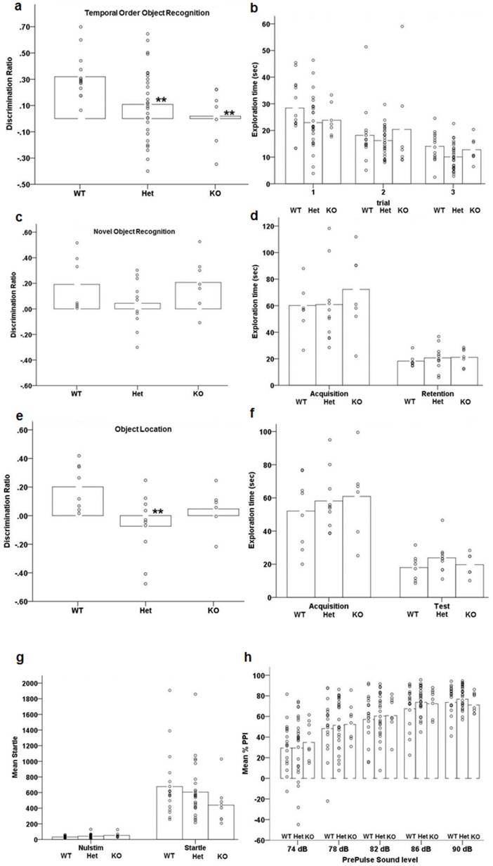
To determine whether Akt3 is involved in cognitive performance we tested Akt3 mice in a series of recognition memory tasks, including temporal order discrimination memory, novel object preference and the object location test. The combination of these tests can be used to examine the neuroanatomical components of dysfunction, with each task involving specific regions involved in components of the tests in question. Temporal order object recognition memory is critically dependent on the mPFC and integrated information flow with the perirhinal cortex and hippocampus [52]. Novel object preference requires intact function of the PRH cortex (but not the mPFC and the hippocampus), while object location is critically dependent on hippocampal function for spatial memory [52]. ANOVA revealed a significant effect of genotype on discrimination performance during the temporal order object recognition task (F (2, 46) = 4.805, p = 0.013; Fig 2a), driven by significant deficits in the Akt3-deficient mice (posthoc LSD, Akt3+/+ vs. Akt3-/+, p = 0.011; Akt3+/+ vs. Akt3-/-, p = 0.010). Akt3-deficient mice did not differ in the amount of exploration in the different phases of the task (Fig 2b; p>0.1).
Fig 2. Impaired temporal order object discrimination memory and spatial location memory in Akt3-deficient mice.
(a) Discrimination ratio displayed by Akt3-deficient mice during the 5 min test phase (sample 3) of the temporal order object recognition task. (** p≤0.01 by posthoc LSD; n = 14 WT, 28 Het, 7 KO). (b) Total time spent exploring the objects presented during the 5-min sample phases 1, 2 and 3. (c) Discrimination ratio for the novel object by Akt3-deficient mice during the novel object test (n = 9 WT, 11 Het, 7 KO). (d) Time spent exploring the two objects during the 10 min acquisition session and the 5 min retention session of the novel object recognition test. (e) Discrimination ratio for the moved object over the same-placed object (**p≤0.01; n = 9 WT, 10 Het, 6 KO). (f) Time spent exploring the two objects during the 10 min acquisition and the 5 min test session of the same object location test. (g) Absence of differences in baseline movement (Nulstim) or Startle responses in the pre-pulse inhibition test in Akt3-deficient mice. (n = 18 WT, 26 Het, 8 KO). (h) Percentage prepulse inhibition of the acoustic startle response displayed by the same mice after the presentation of prepulse (n = 18 WT, 26 Het, 8 KO). Data bars represent the mean, with individual data points representing individual subject measures.
Next, we tested a separate naïve cohort of mice in the novel object preference task. ANOVA revealed no effect of genotype on novel object discrimination (p>0.18; Fig 2c), with all mice significantly exploring the novel object more than the familiar during the test phase (main effect of trial; F (1, 24) = 102.298, p<0.001; Fig 2d). Exploration time within the different phases of the task (acquisition and retention) did not differ between genotype groups (p>0.6, Fig 2d). These findings suggest intact function of the perirhinal cortex in the context of Akt3 deficiency [52].
Finally, a third cohort of naïve mice were assessed in the object location task to examine spatial memory, dependent on intact hippocampal function [52]. We observed a significant effect of genotype on spatial memory (F (2, 21) = 3.755, p = 0.02). WT mice showed a positive discrimination ratio for the relocated object over the originally placed object (Fig 2e), which was significantly impaired in Akt3-deficient mice (posthoc LSD, Akt3+/+ vs. Akt3-/+, p = 0.006; Akt3+/+ vs. Akt3-/-, p = 0.14). Akt3-deficient mice did not differ in their extent of exploration of the objects during the acquisition and test phases of the task (p>0.2; Fig 2f). Together, these observations demonstrate deficits of prefrontal cortical and/or hippocampal-mediated cognitive function in the context of Akt3-deficiency.
Sensorimotor gating is normal in Akt3-deficient mice
Prepulse Inhibition (PPI) evaluates sensorimotor gating in the pre-pulse inhibition of the acoustic startle response and is a deficit observed commonly in neurodevelopmental disorders, including schizophrenia [58]. One-way ANOVA indicates no significant differences in nulstim (p>0.06) or startle (p>0.3) between genotypes (Fig 2g). Significant increases of inhibition across prepulse were observed in all groups (F (4, 196) = 78.345, p<0.001; Fig 2h), with no genotype effect or genotype x prepulse interaction (p>0.8).
Learned fear conditioning and fear extinction are intact in Akt3-deficient mice
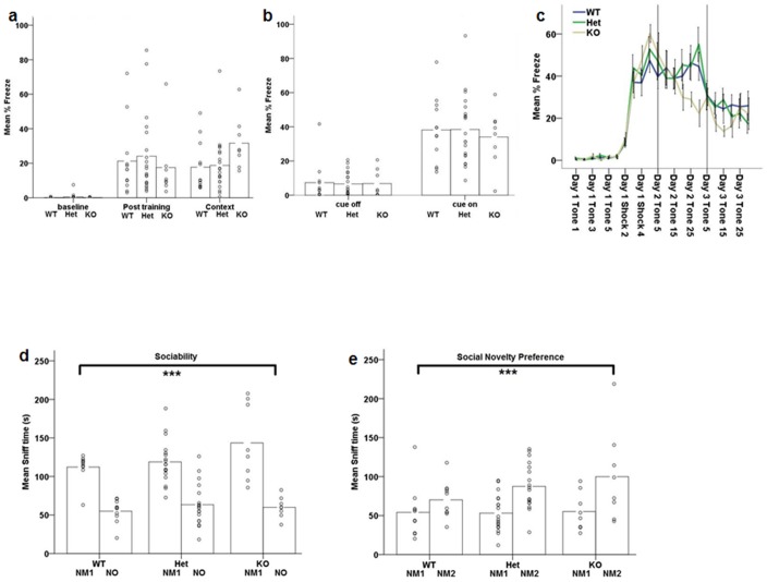
Using Pavlovian fear conditioning, we evaluated Akt3’s role in learning and memory mediated via hippocampal and amygdala neural circuitry. Notably, the hippocampus is a key mediator of the acquisition and expression of conditioned fear to a particular context, while the acquisition and extinction of auditory cued fear memories relies on the amygdala [59]. During the context fear test, we observed significant freezing behavior during the post training (acquisition) and contextual fear phases (main effect of phase: F (2, 74) = 35.972, p≤0.001; Fig 3a), with no significant genotype effects (p>0.8). At baseline, freezing was equal to zero in all groups. Re-exposure to the same context 24hrs, without cue, elicited a similar pattern of % freezing to that observed post training in all genotypes (Fig 3a). These data indicate that Akt3 does not impact fear learning and memory. Similarly, during the cued fear test all mice displayed normal cued conditioned freezing, revealed by a significant increase in freezing to the auditory cue (F(1, 37) = 118.634, p<0.001; Fig 3b) with no significant effect of genotype or interactions (p>0.2). To further assess synaptic efficacy within the amygdala, we used auditory fear extinction to assess depotentiation, the reversal of the learned condition-induced potentiation of the fear response [54]. All mice displayed a significant decrease of freezing over trials (F (21, 672) = 41.917, p<0.001; Fig 3c) with no effect of genotype (p>0.1).
Fig 3. Akt3-deficiency does not impact fear learning and extinction, or social behavior.
(a) Freezing behavior prior (baseline), subsequent (post training), and 24 hours later (context 24hr) after the cue-shock pairings. All genotypes increased freezing subsequent to the CS-US pairings and genotypes did not differ during the testing of contextual conditioning (n = 12 WT, 20 Het, 8 KO). (b) Freezing behavior during testing for cued conditioning in the altered context without (CS off) and plus the auditory cue (CS on; n = 12 WT, 20 Het, 8 KO). (c) Fear extinction after repeated exposure to the auditory cue (CS alone) shows no effect of genotype of genotype x day interaction (n = 13 WT, 13 Het, 9 KO) (d) Time spent sniffing the novel mouse during the 10-min test for sociability was greater than time spent sniffing the novel object for all mice (n = 10 WT, 18 Het, 8 KO). (e) Time spent sniffing the novel mouse 2 was greater than time spent sniffing novel mouse 1 (familiar) during the 10-min test for social novelty was greater for all mice (n = 10 WT, 18 Het, 8 KO). ***p≤0.001 main effect of chamber. (a,b,d,e) Data bars represent the mean, with individual data points representing individual subject measures. (c) Data represents mean ± SEM.
Akt3 deficiency does not impact social functioning
Abnormalities of social cognition are present in various neurodevelopmental disorders, including schizophrenia and autism [60–62]. To assess if Akt3 impacts social interaction behaviors, we examined sociability and social novelty utilizing the three-chamber sociability test as previously described in detail [50,53]. Analysis of sociability revealed that all mice exhibited a main chamber preference as assessed by exploration measured as sniff time (main effect of chamber: F (1, 33) = 121.336, p<0.001; Fig 3d) and time spent in the chamber containing the novel mouse vs. the novel object (F(1, 33) = 30.920, p<0.001), with no effect of genotype or interactions observed (p>0.2). For social novelty, all mice displayed significant preference for interaction with the novel mouse (2) vs. the familiar mouse (mouse 1) (main effect of chamber measured as exploration via sniff time: (F (1, 33) = 15.724, p<0.001; Fig 3e)), independent of genotype (p>0.3). These observations suggest that social functioning is preserved in the context of Akt3 deficiency.
Biochemical signaling in Akt3-deficient mice
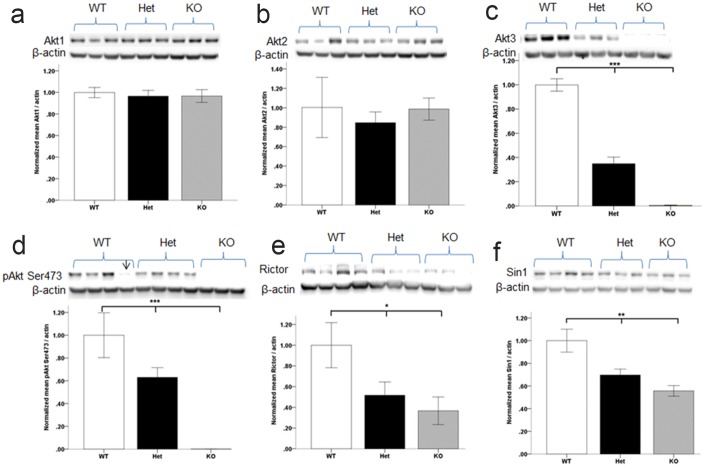
We next examined expression and activity of select proteins in the AKT/mTOR signaling network in the mPFC of Akt3-deficient mice. To address the issue of isoform redundancy and biological compensation, we first measured total Akt1 and Akt2. Linear regression analyses revealed no differences in levels of Akt1 (p = 0.8; Fig 4a) or Akt2 (p = 0.8; Fig 4b) protein, consistent with previous findings in Akt3 null mice [24, 25]. Our findings also demonstrate that hemizygous deletion of Akt3 does not result in changes in the level of Akt1 (Fig 4a) or Akt2 (Fig 4b). Linear regression revealed that 91% of the variance in Akt3 protein was explained by genotype (Model: F (1,16) = 160.30, p<0.0001; Genotype; β = −.0.95; t = −12.66; p<0.0001), with decreased levels in Akt3-/+ mice and absence of detection in Akt3-/- mice (Fig 4c). The activation status of Akt, as measured by phosphorylation of the pAkt Ser473 site, a conserved site across all three isotypes, was examined in Akt3-deficient mice. Linear regression revealed that 78% of the variance in pAKT Ser473 was explained by genotype (Model: F (1,9) = 33.33, p<0.001; Genotype; β = −0.89; t = −5.77; p<0.001), with decreased levels in Akt3-/+ mice and absence of detection in Akt3-/- mice (Fig 4d). Remarkably similar findings were observed in the hippocampus (Model: F (1,9) = 71.01, p<0.001; Genotype; β = −0.94; t = −8.4; p<0.001; S2 Fig). Phosphorylation of Ser473 is mediated by the PDK2/Rictor/Sin1-mTOR complex, mTORC2. Significant effects of genotype were observed on levels of Rictor and Sin1, critical components of mTORC2 (Rictor; Model: F(1, 9) = 6.96, p = 0.03; Genotype; β = −0.68; t = −2.63; p = 0.03; Sin1; Model: F(1, 9) = 16.88, p = 0.003; Genotype; β = −0.82; t = −4.1; p = 0.003), with reduced levels observed in the context of Akt3 deficiency (Fig 4e and 4f). Given the evidence for overall reduced activity of Akt in the context of Akt3 deletion, not compensated for by changes in total Akt1 or Akt2, we predicted altered expression/activation of downstream targets of Akt. The phosphorylation levels and total levels of glycogen synthase kinase 3 (GSK3β), pGSK3β/GSK3β were unaltered and proteins indicative of mTORC1 signaling were not altered in Akt3 mice (total mTOR; phosphorylation of mTOR at (Ser2448) and p70S6K). No changes were observed in the total levels of PSD95, PI3K, p110δ (PI3Kδ), AMPA receptor (GluR 2/3/4), NMDA receptor (NMDAR1) or PDK1. No effects of genotype were observed for expression levels of the SDCCAG8 gene (S3 Fig). Expression of the housekeeping gene and loading control, β-actin did not significantly differ across genotypes (Fig 4; p>0.9)
Fig 4. Biochemical changes in the mPFC of Akt3-deficient mice as determined by Western blot.
Absence of compensatory changes in Akt1 (a) or Akt2 (b) protein levels in Akt3-deficient mice. (c) Akt3 protein is significantly reduced in Akt3 (-/+) Het mice and absent in (-/-) KO mice. (d) Reduced pAKT Ser473 in Akt-deficient mice. (e) Total Rictor and Sin 1 (f) protein was decreased in Akt3 mutant mice. * p≤0.05, **p≤0.01, ***p≤0.001. Quantitative data derived from n = 3–6 per genotype. Data represents mean ± SEM. Representative Western blots are shown. Arrow denotes excluded sample due to gel bubble.
Discussion
Akt3 is the predominant Akt isotype in the adult brain [24,25], transcription is enriched in human fetal neocortex [44] and haploinsuffiency in humans is linked to cortical malformations, including microcephaly and cognitive dysfunction [45,46,63]. Despite this evidence, little is known about the neurobiological role and signaling specificity of AKT3. In the present study, we utilized Akt3 hemizygous and nullizygous mice in a pure C57BL/6J background and provide evidence for a causal relationship between Akt3 levels and the attainment of normal brain size, extending prior findings [24,25] to show that heterozygous reduction of Akt3 is associated with selective, subtle and dose-dependent decreases in brain size (7%). Moreover, we demonstrate that Akt3 is critical for neurocognitive performance linked to prefrontal cortical-hippocampal circuitry and that these changes are associated with a dramatic reduction of activated AKT (as measured by AKT Ser473 phosphorylation), deficits not accounted for by compensation of Akt1 or Akt2 levels. Together, these data implicate an indispensable and non-redundant role for Akt3 in brain development and cognitive function and identify neurobiological mechanisms of relevance to schizophrenia.
Contemporary psychiatric genetic studies have identified polymorphic variation in the AKT3 gene, including a putative functional SNP (rs14403) in the 3 untranslated region (UTR) of transcript NM_005465, as genome-wide associated to schizophrenia [5,8]. While the molecular mechanisms of risk remain to be determined, it is noteworthy in the context of our findings, that subtle whole brain- and cortical grey matter volume reductions have been observed in patients with schizophrenia [64–66], with meta-analyses of postmortem brain and structural imaging studies showing 2–3% global brain volume reductions, with larger reductions in the PFC and temporal lobe [64,66,67]. Although there is considerable debate about the origins of structural brain changes in schizophrenia, including secondary effects of antipsychotic drugs, drugs of abuse and illness [68], many changes are present to a lesser degree in first-episode patients [68–70]; and show evidence of heritability in patients and siblings [66,71]. Recent large-scale imaging genetic studies also provide evidence of a shared genetic basis of several neuroanatomical phenotypes and schizophrenia risk [72]. Evidence of association to genes which control key biological processes related to structural brain development, therefore implicate a potential neurobiological mechanism of risk. Consistently, a new genome-wide association study of 32,438 adults shows that variants determining human intracranial volume and related cognitive function are enriched in genes for PI3K-AKT signaling, including AKT3 [73]. Interestingly, rs7538011, an intronic variant associated with human intracranial volume [73], is in linkage-disequilibrium (LD) with the schizophrenia 3 UTR risk variant, rs14403 (D’ 0.92; R2 0.52) and has membership in one of the top pathways showing cross-disorder association to psychiatric risk [6].
Previous examination of Akt3 null mice shows that reduced brain size ([24] and confirmed in this study) is apparent at birth and histologically defined by a reduction in cortical cell size and density [24], potentially consistent with Akt3’s reported role in axonal and dendritic development [74,75]. Notably, similar cytoarchitectural changes have been reported in postmortem brain of patients with schizophrenia [76–80] and in conjunction with evidence for genetic association to biological pathways that regulate axonal, dendritic, and postsynaptic development [6], suggest that at least some neuropathological changes may be genetically determined, neurodevelopmental in origin, and potentially related to alterations in AKT signaling.
Mammalian brain size, in particular the magnitude of the cerebral cortex, is a significant determinant of cognitive ability [81,82] and several disorders of neurodevelopment, including schizophrenia are characterized by cognitive dysfunction [83]. Behavioral analysis revealed that genetic reduction of Akt3 produces selective deficits in mPFC and/or hippocampal mediated neurocognitive function, as evidenced by discrimination impairments of temporal order object recognition memory and spatial memory. Such findings are similar to temporal and spatial memory impairments observed in patients with schizophrenia [84–86] and consistent with abnormalities of prefrontal cortical function in the disorder [1,3, 87–89]. These novel results identify Akt3 as a critical determinant of prefrontal cortical and hippocampal development, not previously appreciated. Indeed, Akt3 is more transcriptionally active in cortex and hippocampus compared to other brain regions, [24,45] further supporting regional biological importance. Study of the effects of schizophrenia-risk associated AKT3 polymorphisms (as previously done for AKT1 [34,35,37]) on neuroanatomical structure and neurocognitive function in normal human subjects, is warranted.
The results presented here also highlight a key role for Akt3 in the expression of dopamine-mediated behaviors, with Akt3 null mice exhibiting a generalized increase in baseline locomotor activity in the open field. These findings are in contrast to observations in Akt2 null mice, whereby decreased ambulatory behavior was observed in the context of an anxiety like phenotype [43], and Akt1 null mice where no differences in locomotion were observed [90]. D2, dopamine receptors (the established target of antipsychotic drugs) are critical regulators of AKT signaling in brain [91,92], mediated via the β-arrestin 2/PP2A complex [93]; whereby dopamine-mediated signaling, and D2 receptor antagonism, result in inactivation and activation of AKT, respectively. [27, 91,92]. Which AKT (1, 2 or 3) mediates these effects is still unclear, although some evidence suggests a potential role for Akt1 [27]. Our data in this context implicate a novel role for Akt3 in dopamine neurotransmission and highlight more targeted approaches towards Akt3 for targeted neuroleptic drug development.
Biochemically, phosphorylation of Ser473 was dramatically diminished in the brain of Akt3 mutant mice, with Akt3-/+ mice showing 40% reduction and Akt3-/- showing complete abolishment, compared to WT. These effects were not accompanied by changes in total Akt1 or Akt2, supporting previous observations in Akt3 nullizygous mice [24,25], and suggest that Akt3 is a primary determinant of Ser473 activity in brain and a critical Akt isotype that does not exhibit redundancy. Phosphorylation status of Ser473 is used as an endogenous functional readout of mTORC2 kinase activity and in agreement, while the loss of Akt3 did not affect levels of mTOR, it did significantly abrogate levels of Rictor and Sin1, critical components of the PDK2/mTORC2 complex [16,94]. Importantly, all sites indicative of mTORC1 activation [94,95], including phospho-mTOR (Ser2448) and p70S6K were unaltered in Akt3 mice. These results suggest an mTORC2-dependant mechanism is responsible for the microcephaly and behavioral impairments in Akt3 mutant mice. Remarkably, ablation of the mTORC2 component, Rictor [94], produces dramatically reduced Akt Ser473 activation, microcephaly and alterations in neuronal morphology and function, without impact on mTORC1 signaling. Consistent results are also observed for Sin1 ablation [16]. These findings are strikingly reminiscent of Akt3 deletion and suggest a previously unappreciated feedback loop between Akt3 and mTORC2 and show that in brain, Akt3 may be the primary target for mTORC2 activity. The related mechanisms, however, remain to be determined. It is noteworthy that attenuated AKT activity and phosphorylated AKT Ser473 [39,40,96] is observed in patient brain and peripheral cells, suggesting that impaired PI3K-AKT signaling is relevant to etiopathogenesis of schizophrenia. Our data in mouse provide support for this hypothesis and demonstrate that reductions in Akt3 can result in attenuated brain AKT activity and phenotypic outcomes similar to observations in schizophrenia. Since cognitive deficits remain a significant therapeutic problem in schizophrenia, these findings highlight the AKT signaling pathway and Akt3 specifically, as a biologically relevant therapeutic target.
Finally, one potential shortcoming of in-vivo gene-targeting technologies is the issue of disrupting flanking genes [97]. This is particularly important for study of Akt3 function, given that SDCCAG8, a gene also implicated in schizophrenia [8, 98], flanks Akt3 both in humans and mouse and shares regulatory regions. We measured brain SDCCAG8 expression in Akt3 mice and show no differential expression based on genotype. This finding is important as it demonstrates that phenotypes observed in Akt3 mutant mice are the direct result of Akt3 disruption.
Conclusion
In summary, our study validates the essential role of Akt3 in the attainment of normal brain size and reveals a critical role for Akt3 in prefrontal cortical-hippocampal mediated cognitive function relevant to genetic risk for schizophrenia. Furthermore, we identify a central, non-redundant role for Akt3 in governing brain Akt signalling and identify Akt3 as potential novel pharmacological target for neuroleptic treatment development.
Supporting information
No general health abnormalities were observed. Values represent percentage or mean ± SEM. n = 14 WT, 21 Het, 4 KO.
(DOCX)
(a) PCR genotyping from wild-type (Akt3+/+), Akt3 knockout (Akt3-/-) mice, and Akt3 heterozygous (Akt3-/+) genomic DNA. Primers 1 and 2 generate a 192 kb band from wild-type DNA, while primers 1 and 3 generate a 340 kb band when Akt3 is the targeted allele. (b) Representative average brain weights for Akt3 male mice. Akt3 Het and KO mice show significant decreases in brain weight compared to littermate controls, indicating an allele dose impact on brain weight with a 7% and 24% reduction respectively. (c) There was a significant decrease in both Het and KO mice brain to body ratios when compared to WT littermate male Akt3 mice. n = 10 WT, 7 Het, 7 KO. ***p≤0.001 compared to WT; ### p≤0.001 compared to Het.
(TIF)
***p≤0.001. Quantitative data derived from n = 3 per genotype. Data represents mean ± SEM.
(JPG)
n = 6 per genotype.
(TIF)
(PDF)
Acknowledgments
We would like to thank Dr. Sara Palumbo, previously of the NIMH Intramural Program for the original colony back crossing, establishment and maintenance of the Akt3 mutant mice.
Data Availability
All relevant data are within the paper and its Supporting Information files.
Funding Statement
Research reported in this publication was supported by the National Institute of Mental Health of the National Institutes of Health under Award Number R01MH103716 (AJL) and via T32MH015442, an institutional postdoctoral research training program at University of Colorado, Denver, to KRH. The funders had no role in study design, data collection and analysis, decision to publish, or preparation of the manuscript.
References
- 1.Callicott JH, Mattay VS, Verchinski BA, Marenco S, Egan MF, Weinberger DR. Complexity of prefrontal cortical dysfunction in schizophrenia: more than up or down. Am J Psychiatry 2003; 160(12):2209–2215. 10.1176/appi.ajp.160.12.2209 [DOI] [PubMed] [Google Scholar]
- 2.Toulopoulou T, Goldberg TE, Mesa IR, Picchioni M, Rijsdijk F, Stahl D et al. Impaired intellect and memory: a missing link between genetic risk and schizophrenia? Arch Gen Psychiatry 2010; 67(9):905–913. 10.1001/archgenpsychiatry.2010.99 [DOI] [PubMed] [Google Scholar]
- 3.Rasetti R, Sambataro F, Chen Q, Callicott JH, Mattay VS, Weinberger DR. Altered cortical network dynamics: a potential intermediate phenotype for schizophrenia and association with ZNF804A. Arch Gen Psychiatry 2011; 68(12):1207–1217. 10.1001/archgenpsychiatry.2011.103 [DOI] [PubMed] [Google Scholar]
- 4.Schaefer J, Giangrande E, Weinberger DR, Dickinson D. The global cognitive impairment in schizophrenia: consistent over decades and around the world. Schizophr Res 2013; 150(1):42–50. 10.1016/j.schres.2013.07.009 [DOI] [PMC free article] [PubMed] [Google Scholar]
- 5.Schizophrenia Working Group of the Psychiatric Genomics Consortium. Biological insights from 108 schizophrenia-associated genetic loci. Nature 2014; 511(7510):421–427. 10.1038/nature13595 [DOI] [PMC free article] [PubMed] [Google Scholar]
- 6.Network and Pathway Analysis Subgroup of Psychiatric Genomics Consortium. Psychiatric genome-wide association study analyses implicate neuronal, immune and histone pathways. Nat Neurosci 2015; 18(2):199–209. 10.1038/nn.3922 [DOI] [PMC free article] [PubMed] [Google Scholar]
- 7.Ruderfer DM, Charney AW, Readhead B, Kidd BA, Kähler AK, Kenny PJ, et al. Polygenic overlap between schizophrenia risk and antipsychotic response: a genomic medicine approach. Lancet Psychiatry 2016; 3(4):350–357. 10.1016/S2215-0366(15)00553-2 [DOI] [PMC free article] [PubMed] [Google Scholar]
- 8.Ripke S, O'Dushlaine C, Chambert K, Moran JL, Kähler AK, Akterin S, et al. Genome-wide association analysis identifies 13 new risk loci for schizophrenia. Nat Genet. 2013; 45(10):1150–9. 10.1038/ng.2742 [DOI] [PMC free article] [PubMed] [Google Scholar]
- 9.Arguello PA and Gogos JA. A signaling pathway AKTing up in schizophrenia. J Clin Invest 2008; 118(6):2018–2021. 10.1172/JCI35931 [DOI] [PMC free article] [PubMed] [Google Scholar]
- 10.Beaulieu JM, Gainetdinov RR, Caron MG. Akt/GSK3 signaling in the action of psychotropic drugs. Annu Rev Pharmacol Toxicol 2009; 49:327–347. 10.1146/annurev.pharmtox.011008.145634 [DOI] [PubMed] [Google Scholar]
- 11.Law AJ, Wang Y, Sei Y, O'Donnell P, Piantadosi P, Papaleo F, et al. Neuregulin 1-ErbB4-PI3K signaling in schizophrenia and phosphoinositide 3-kinase-p110δ inhibition as a potential therapeutic strategy. Proc Natl Acad Sci U S A 2012; 109(30):12165–12170. 10.1073/pnas.1206118109 [DOI] [PMC free article] [PubMed] [Google Scholar]
- 12.Cohen MM Jr. The AKT genes and their roles in various disorders. Am J Med Genet A 2013; 161A(12):2931–2937. 10.1002/ajmg.a.36101 [DOI] [PubMed] [Google Scholar]
- 13.Song G, Ouyang G, Bao S. The activation of Akt/PKB signaling pathway and cell survival. J Cell Mol Med. 2005; 9(1):59–71. [DOI] [PMC free article] [PubMed] [Google Scholar]
- 14.Franke TF. Intracellular signaling by Akt: bound to be specific. Sci Signal. 2008; 1(24):pe29 10.1126/scisignal.124pe29 [DOI] [PubMed] [Google Scholar]
- 15.Masure S, Haefner B, Wesselink JJ, Hoefnagel E, Mortier E, Verhasselt P, Tuytelaars A, et al. Molecular cloning, expression and characterization of the human serine/threonine kinase Akt-3. Eur J Biochem 1999; 265(1):353–360. [DOI] [PubMed] [Google Scholar]
- 16.Jacinto E, Facchinetti V, Liu D, Soto N, Wei S, Jung SY, et al. SIN1/MIP1 maintains rictor-mTOR complex integrity and regulates Akt phosphorylation and substrate specificity. Cell. 2006; 127(1):125–37. 10.1016/j.cell.2006.08.033 [DOI] [PubMed] [Google Scholar]
- 17.Guertin DA, Stevens DM, Thoreen CC, Burds AA, Kalaany NY, Moffat J, et al. Ablation in mice of the mTORC components raptor, rictor, or mLST8 reveals that mTORC2 is required for signaling to Akt-FOXO and PKCalpha, but not S6K1. Dev Cell. 2006; 11(6):859–71 10.1016/j.devcel.2006.10.007 [DOI] [PubMed] [Google Scholar]
- 18.Santi SA and Lee H. The Akt isoforms are present at distinct subcellular locations. Am J Physiol Cell Physiol. 2010; 298(3):C580–91. 10.1152/ajpcell.00375.2009 [DOI] [PubMed] [Google Scholar]
- 19.Clark AR and Toker A. Signalling specificity in the Akt pathway in breast cancer. Biochem Soc Trans. 2014; 42(5):1349–55. 10.1042/BST20140160 [DOI] [PubMed] [Google Scholar]
- 20.Lee RS, House CM, Cristiano BE, Hannan RD, Pearson RB, Hannan KM. Relative Expression Levels Rather Than Specific Activity Plays the Major Role in Determining In Vivo AKT Isoform Substrate Specificity. Enzyme Res. 2011; 2011:720985 10.4061/2011/720985 [DOI] [PMC free article] [PubMed] [Google Scholar]
- 21.Diez H, Garrido JJ, Wandosell F. Specific roles of Akt iso forms in apoptosis and axon growth regulation in neurons. PLoS One. 2012; 7(4):e32715 10.1371/journal.pone.0032715 [DOI] [PMC free article] [PubMed] [Google Scholar]
- 22.Cho H, Thorvaldsen JL, Chu Q, Feng F, Birnbaum MJ. Akt1/PKBalpha is required for normal growth but dispensable for maintenance of glucose homeostasis in mice. J Biol Chem. 2001; 276(42):38349–52. 10.1074/jbc.C100462200 [DOI] [PubMed] [Google Scholar]
- 23.Cho H, Mu J, Kim JK, Thorvaldsen JL, Chu Q, Crenshaw EB 3rd, et al. Insulin resistance and a diabetes mellitus-like syndrome in mice lacking the protein kinase Akt2 (PKB beta). Science. 2001; 292(5522):1728–31. 10.1126/science.292.5522.1728 [DOI] [PubMed] [Google Scholar]
- 24.Easton RM, Cho H, Roovers K, Shineman DW, Mizrahi M, Forman MS, et al. Role for Akt3/protein kinase Bgamma in attainment of normal brain size. Mol Cell Biol 2005; 25(5):1869–1878. 10.1128/MCB.25.5.1869-1878.2005 [DOI] [PMC free article] [PubMed] [Google Scholar]
- 25.Tschopp O, Yang ZZ, Brodbeck D, Dummler BA, Hemmings-Mieszczak M, Watanabe T et al. Essential role of protein kinase B gamma (PKB gamma/Akt3) in postnatal brain development but not in glucose homeostasis. Development 2005; 132(13):2943–2954. 10.1242/dev.01864 [DOI] [PubMed] [Google Scholar]
- 26.Xie R, Cheng M, Li M, Xiong X, Daadi M, Sapolsky RM, et al. Akt isoforms differentially protect against stroke-induced neuronal injury by regulating mTOR activities. J Cereb Blood Flow Metab. 2013; 33(12):1875–85. 10.1038/jcbfm.2013.132 [DOI] [PMC free article] [PubMed] [Google Scholar]
- 27.Emamian ES, Hall D, Birnbaum MJ, Karayiorgou M, Gogos JA. Convergent evidence for impaired AKT1-GSK3beta signaling in schizophrenia. Nat Genet 2004; 36(2):131–137. 10.1038/ng1296 [DOI] [PubMed] [Google Scholar]
- 28.Schwab SG, Hoefgen B, Hanses C, Hassenbach MB, Albus M, Lerer B, et al. Further evidence for association of variants in the AKT1 gene with schizophrenia in a sample of European sib-pair families. Biol Psychiatry 2005; 58(6):446–450. 10.1016/j.biopsych.2005.05.005 [DOI] [PubMed] [Google Scholar]
- 29.Bajestan SN, Sabouri AH, Nakamura M, Takashima H, Keikhaee MR, Behdani F et al. Association of AKT1 haplotype with the risk of schizophrenia in Iranian population. Am J Med Genet B Neuropsychiatr Genet 2006; 141B(4):383–386. 10.1002/ajmg.b.30291 [DOI] [PubMed] [Google Scholar]
- 30.Thiselton DL, Vladimirov VI, Kuo PH, McClay J, Wormley B, Fanous A, et al. AKT1 is associated with schizophrenia across multiple symptom dimensions in the Irish study of high density schizophrenia families. Biol Psychiatry 2008; 63(5):449–457. 10.1016/j.biopsych.2007.06.005 [DOI] [PMC free article] [PubMed] [Google Scholar]
- 31.Norton N, Williams HJ, Dwyer S, Carroll L, Peirce T, Moskvina V, et al. Association analysis of AKT1 and schizophrenia in a UK case control sample. Schizophr Res 2007; 93(1–3):58–65. 10.1016/j.schres.2007.02.006 [DOI] [PubMed] [Google Scholar]
- 32.Mathur A, Law MH, Megson IL, Shaw DJ, Wei J. Genetic association of the AKT1 gene with schizophrenia in a British population. Psychiatr Genet 2010; 20(3):118–122. 10.1097/YPG.0b013e32833a2234 [DOI] [PubMed] [Google Scholar]
- 33.Zheng W, Wang H, Zeng Z, Lin J, Little PJ, Srivastava LK, et al. The possible role of the Akt signaling pathway in schizophrenia. Brain Res 2012; 1470:145–158. 10.1016/j.brainres.2012.06.032 [DOI] [PubMed] [Google Scholar]
- 34.Tan HY, Nicodemus KK, Chen Q, Li Z, Brooke JK, Honea R, et al. Genetic variation in AKT1 is linked to dopamine-associated prefrontal cortical structure and function in humans. J Clin Invest 2008; 118(6):2200–2208. 10.1172/JCI34725 [DOI] [PMC free article] [PubMed] [Google Scholar]
- 35.Tan HY, Chen AG, Kolachana B, Apud JA, Mattay VS, Callicott JH, et al. Effective connectivity of AKT1-mediated dopaminergic working memory networks and pharmacogenetics of anti-dopaminergic treatment. Brain 2012; 135 (Pt 5):1436–1445. 10.1093/brain/aws068 [DOI] [PMC free article] [PubMed] [Google Scholar]
- 36.Pietiläinen OP, Paunio T, Loukola A, Tuulio-Henriksson A, Kieseppä T, Thompson P, et al. Association of AKT1 with verbal learning, verbal memory, and regional cortical gray matter density in twins. Am J Med Genet B Neuropsychiatr Genet. 2009; 150B(5):683–692.. 10.1002/ajmg.b.30890 [DOI] [PMC free article] [PubMed] [Google Scholar]
- 37.Blasi G, Napolitano F, Ursini G, Taurisano P, Romano R, Caforio G, et al. DRD2/AKT1 interaction on D2 c-AMP independent signaling, attentional processing, and response to olanzapine treatment in schizophrenia. Proc Natl Acad Sci U S A. 2011; 108(3):1158–1163. 10.1073/pnas.1013535108 [DOI] [PMC free article] [PubMed] [Google Scholar]
- 38.van Beveren NJ, Buitendijk GH, Swagemakers S, Krab LC, Röder C, de Haan L, et al. Marked reduction of AKT1 expression and deregulation of AKT1-associated pathways in peripheral blood mononuclear cells of schizophrenia patients. PLoS One. 2012; 7(2):e32618 10.1371/journal.pone.0032618 [DOI] [PMC free article] [PubMed] [Google Scholar]
- 39.Sei Y, Ren-Patterson R, Li Z, Tunbridge EM, Egan MF, Kolachana BS, et al. Neuregulin1-induced cell migration is impaired in schizophrenia: association with neuregulin1 and catechol-o-methyltransferase gene polymorphisms. Mol Psychiatry. 2007; 12(10):946–957. 10.1038/sj.mp.4001994 [DOI] [PubMed] [Google Scholar]
- 40.Balu DT, Carlson GC, Talbot K, Kazi H, Hill-Smith TE, Easton RM et al. Akt1 deficiency in schizophrenia and impairment of hippocampal plasticity and function. Hippocampus 2012; 22(2):230–240. 10.1002/hipo.20887 [DOI] [PMC free article] [PubMed] [Google Scholar]
- 41.Lai WS, Xu B, Westphal KG, Paterlini M, Olivier B, Pavlidis P, et al. Akt1 deficiency affects neuronal morphology and predisposes to abnormalities in prefrontal cortex functioning. Proc Natl Acad Sci U S A 2006; 103(45):16906–16911. 10.1073/pnas.0604994103 [DOI] [PMC free article] [PubMed] [Google Scholar]
- 42.Featherstone RE, M Tatard-Leitman V, Suh JD, Lin R, Lucki I, Siegel SJ. Electrophysiological and behavioral responses to ketamine in mice with reduced Akt1 expression. Psychopharmacology (Berl). 2013; 227(4):639–649. [DOI] [PMC free article] [PubMed] [Google Scholar]
- 43.Leibrock C, Ackermann TF, Hierlmeier M, Lang F, Borgwardt S, Lang UE. Akt2 deficiency is associated with anxiety and depressive behavior in mice. Cell Physiol Biochem 2013; 32(3):766–777. 10.1159/000354478 [DOI] [PubMed] [Google Scholar]
- 44.Birnbaum R, Birnbaum R, Jaffe AE, Chen Q, Hyde TM, Kleinman JE, Weinberger DR. Investigation of the prenatal expression patterns of 108 schizophrenia-associated genetic loci. Biol Psychiatry 2015; 77(11):e43–51. 10.1016/j.biopsych.2014.10.008 [DOI] [PubMed] [Google Scholar]
- 45.Boland E, Clayton-Smith J, Woo VG, McKee S, Manson FD, Medne L et al. Mapping of deletion and translocation breakpoints in 1q44 implicates the serine/threonine kinase AKT3 in postnatal microcephaly and agenesis of the corpus callosum. Am J Hum Genet. 2007; 81(2):292–303. 10.1086/519999 [DOI] [PMC free article] [PubMed] [Google Scholar]
- 46.Ballif BC, Rosenfeld JA, Traylor R, Theisen A, Bader PI, Ladda RL, et al. High–Resolution array, CGH defines critical regions and candidate genes formicrocephaly, abnormalities of the corpus callosum, and seizure phe-notypes in patients with microdeletions of 1q43q44. Hum Genet 2012; 131:145–156. 10.1007/s00439-011-1073-y [DOI] [PubMed] [Google Scholar]
- 47.Poduri A, Evrony GD, Cai X, Elhosary PC, Beroukhim R, Lehtinen MK, et al. Somatic activation of AKT3 cause hemispheric developmental brain malformations. Neuron 2012; 74:41–48. 10.1016/j.neuron.2012.03.010 [DOI] [PMC free article] [PubMed] [Google Scholar]
- 48.Lee JH, Huynh M, Silhavy JL, Kim S, Dixon-Salazar T, Heiberg A et al. De novo somatic mutations in components of thePI3K-AKT3-mTOR pathway causes hemimegalencephaly. Nat Genet 2012; 44:941–945 10.1038/ng.2329 [DOI] [PMC free article] [PubMed] [Google Scholar]
- 49.Rivière JB, Mirzaa GM, O'Roak BJ, Beddaoui M, Alcantara D, Conway RL, et al. De novo germline and postzygotic mutations in AKT3, PIK3R2 and PIK3CA cause a spectrum of related megalencephaly syndromes. Nature Genet 2012; 44:934–940. 10.1038/ng.2331 [DOI] [PMC free article] [PubMed] [Google Scholar]
- 50.Paterson C, Law AJ. Transient Overexposure of Neuregulin 3 during Early Postnatal Development Impacts Selective Behaviors in Adulthood. PLoS ONE 2014; 9(8): e104172 10.1371/journal.pone.0104172 [DOI] [PMC free article] [PubMed] [Google Scholar]
- 51.Papaleo F, Crawley JN, Song J, Lipska BK, Pickel J, Weinberger DR, et al. Genetic dissection of the role of catechol-O-methyltransferase in cognition and stress reactivity in mice. J Neurosci 2008; 28: 8709–8723. 10.1523/JNEUROSCI.2077-08.2008 [DOI] [PMC free article] [PubMed] [Google Scholar]
- 52.Barker GR, Warburton EC. When is the hippocampus involved in recognition memory? J Neurosci 2011; 31: 10721–10731. 10.1523/JNEUROSCI.6413-10.2011 [DOI] [PMC free article] [PubMed] [Google Scholar]
- 53.Papaleo F, Yang F, Paterson C, Palumbo S, Carr GV, Wang Y, et al. Behavioral, Neurophysiological, and Synaptic Impairment in a Transgenic Neuregulin1 (NRG1-IV) Murine Schizophrenia Model. J Neurosci. 2016; 36(17):4859–75. 10.1523/JNEUROSCI.4632-15.2016 [DOI] [PMC free article] [PubMed] [Google Scholar]
- 54.Kim J, Lee S, Park K, Hong I, Song B, Son G, et al. Amygdala depotentiation and fear extinction. Proc Natl Acad Sci U S A 2007; 104(52):20955–20960. 10.1073/pnas.0710548105 [DOI] [PMC free article] [PubMed] [Google Scholar]
- 55.Moy SS, Nadler JJ, Perez A, Barbaro RP, Johns JM, Magnuson TR, et al. Sociability and preference for social novelty in five inbred strains: an approach to assess autistic-like behavior in mice. Genes Brain Behav 2004; 3(5):287–302. 10.1111/j.1601-1848.2004.00076.x [DOI] [PubMed] [Google Scholar]
- 56.Crawley JN. Mouse behavioral assays relevant to the symptoms of autism. Brain Pathol 2007; 17(4):448–459. 10.1111/j.1750-3639.2007.00096.x [DOI] [PMC free article] [PubMed] [Google Scholar]
- 57.Law AJ, Lipska BK, Weickert CS, Hyde TM, Straub RE, Hashimoto R, et al. Neuregulin 1 transcripts are differentially expressed in schizophrenia and regulated by 5' SNPs associated with the disease. Proc Natl Acad Sci U S A. 2006; 103(17):6747–52. 10.1073/pnas.0602002103 [DOI] [PMC free article] [PubMed] [Google Scholar]
- 58.Braff DL, Grillon C, Geyer MA. Gating and habituation of the startle reflex in schizophrenic patients. Arch Gen Psychiatry 1992; 49(3):206–215. [DOI] [PubMed] [Google Scholar]
- 59.Fendt M, Fanselow MS. The neuroanatomical and neurochemical basis of conditioned fear. Neurosci Biobehav Rev 1999; 23(5):743–760. [DOI] [PubMed] [Google Scholar]
- 60.Lee KH, Farrow TF, Spence SA, Woodruff PW. Social cognition, brain networks and schizophrenia. Psychol Med 2004; 34(3):391–400. [DOI] [PubMed] [Google Scholar]
- 61.Amodio DM and Frith CD. Meeting of minds: the medial frontal cortex and social cognition. Nat Rev Neurosci 2006; 7(4):268–277. 10.1038/nrn1884 [DOI] [PubMed] [Google Scholar]
- 62.Howes OD and Murray RM. Schizophrenia: an integrated sociodevelopmental-cognitive model. Lancet 2014; 383(9929):1677–16787. 10.1016/S0140-6736(13)62036-X [DOI] [PMC free article] [PubMed] [Google Scholar]
- 63.Gai D, Haan E, Scholar M, Nicholl J, Yu S. Phenotypes of AKT3 deletion: a case report and literature review. Am J Med Genet A. 2015; 167A(1):174–9. 10.1002/ajmg.a.36710 [DOI] [PubMed] [Google Scholar]
- 64.Harrison PJ, Freemantle N, Geddes JR. Meta-analysis of brain weight in schizophrenia. Schizophr Res. 2003; 64(1):25–34. [DOI] [PubMed] [Google Scholar]
- 65.Steen RG, Mull C, McClure R, Hamer RM, Lieberman JA. Brain volume in first-episode schizophrenia: systematic review and meta-analysis of magnetic resonance imaging studies. Br J Psychiatry. 2006; 188:510–8. 10.1192/bjp.188.6.510 [DOI] [PubMed] [Google Scholar]
- 66.Goldman AL, Pezawas L, Mattay VS, Fischl B, Verchinski BA, Chen Q, et al. Widespread reductions of cortical thickness in schizophrenia and spectrum disorders and evidence of heritability. Arch Gen Psychiatry. 2009; 66(5):467–77. 10.1001/archgenpsychiatry.2009.24 [DOI] [PMC free article] [PubMed] [Google Scholar]
- 67.Haijma SV, Van Haren N, Cahn W, Koolschijn PC, Hulshoff Pol HE, Kahn RS. Brain volumes in schizophrenia: a meta-analysis in over 18 000 subjects. Schizophr Bull. 2013; 39(5):1129–38. 10.1093/schbul/sbs118 [DOI] [PMC free article] [PubMed] [Google Scholar]
- 68.Zipursky RB, Reilly TJ, Murray RM. The myth of schizophrenia as a progressive brain disease. Schizophr Bull. 2013; 39(6):1363–72. 10.1093/schbul/sbs135 [DOI] [PMC free article] [PubMed] [Google Scholar]
- 69.Zipursky RB, Zhang-Wong J, Lambe EK, Bean G, Beiser M. MRI correlates of treatment response in first episode psychosis. Schizophr Res. 1998; 30(1):81–90. [DOI] [PubMed] [Google Scholar]
- 70.Bartholomeusz CF, Cropley VL, Wannan C, Di Biase M, McGorry PD, Pantelis C. Structural neuroimaging across early-stage psychosis: Aberrations in neurobiological trajectories and implications for the staging model. Aust N Z J Psychiatry. 2016; pii: 0004867416670522. [DOI] [PubMed] [Google Scholar]
- 71.Goldman AL, Pezawas L, Mattay VS, Fischl B, Verchinski BA, Zoltick B et al. Heritability of brain morphology related to schizophrenia: a large-scale automated magnetic resonance imaging segmentation study. Biol Psychiatry. 2008; 63(5):475–83. 10.1016/j.biopsych.2007.06.006 [DOI] [PubMed] [Google Scholar]
- 72.Lee PH, Baker JT, Holmes AJ, Jahanshad N, Ge T, Jung JY, et al. Partitioning heritability analysis reveals a shared genetic basis of brain anatomy and schizophrenia. Mol Psychiatry. 2016; [DOI] [PMC free article] [PubMed] [Google Scholar]
- 73.Adams HH, Hibar DP, Chouraki V, Stein JL, Nyquist PA, Rentería ME, et al. Novel genetic loci underlying human intracranial volume identified through genome-wide association. Nat Neurosci. 2016. October 3. [DOI] [PMC free article] [PubMed] [Google Scholar]
- 74.Markus A, Zhong J, Snider WD. Raf and akt mediate distinct aspects of sensory axon growth. Neuron. 2002; 35(1):65–76. [DOI] [PubMed] [Google Scholar]
- 75.Miao L, Yang L, Huang H, Liang F, Ling C, Hu Y. mTORC1 is necessary but mTORC2 and GSK3β are inhibitory for AKT3-induced axon regeneration in the central nervous system. Elife. 2016; 5. pii: e14908. [DOI] [PMC free article] [PubMed] [Google Scholar]
- 76.Benes FM. Altered glutamatergic and GABAergic mechanisms in the cingulate cortex of the schizophrenic brain. Arch Gen Psychiatry. 1995; 52(12):1015–8; discussion 1019–24. [DOI] [PubMed] [Google Scholar]
- 77.Selemon LD. Regionally diverse cortical pathology in schizophrenia: clues to the etiology of the disease. Schizophr Bull. 2001;27(3):349–77. [DOI] [PubMed] [Google Scholar]
- 78.Harrison PJ. The neuropathology of schizophrenia. A critical review of the data and their interpretation. Brain. 1999; April;122 (Pt 4):593–624 [DOI] [PubMed] [Google Scholar]
- 79.Harrison PJ, Lewis DA. GABAergic local circuit neurons and prefrontal cortical dysfunction in schizophrenia. Brain Res Brain Res Rev. 2000; 31(2–3):270–6. [DOI] [PubMed] [Google Scholar]
- 80.Falkai P, Malchow B, Wetzestein K, Nowastowski V, Bernstein HG, Steiner J et al. Decreased Oligodendrocyte and Neuron Number in Anterior Hippocampal Areas and the Entire Hippocampus in Schizophrenia: A Stereological Postmortem Study. Schizophr Bull. 2016; 42 Suppl 1:S4–S12. [DOI] [PMC free article] [PubMed] [Google Scholar]
- 81.Sun T, Hevner RF. Growth and folding of the mammalian cerebral cortex: from molecules to malformations. Nat Rev Neurosci. 2014; 15(4):217–32. 10.1038/nrn3707 [DOI] [PMC free article] [PubMed] [Google Scholar]
- 82.Gautam P, Anstey KJ, Wen W, Sachdev PS, Cherbuin N. Cortical gyrification and its relationships with cortical volume, cortical thickness, and cognitive performance in healthy mid-life adults. Behav Brain Res. 2015; 287:331–9. 10.1016/j.bbr.2015.03.018 [DOI] [PubMed] [Google Scholar]
- 83.Dickinson D, Goldberg TE, Gold JM, Elvevåg B, Weinberger DR. Cognitive factor structure and invariance in people with schizophrenia, their unaffected siblings, and controls. Schizophr Bull. 2011; 37(6):1157–67. 10.1093/schbul/sbq018 [DOI] [PMC free article] [PubMed] [Google Scholar]
- 84.Schwartz BL, Deutsch LH, Cohen C, Warden D, Deutsch SI. Memory for temporal order in schizophrenia. Biol Psychiatry. 1991; 29(4):329–39. [DOI] [PubMed] [Google Scholar]
- 85.Rizzo L, Danion JM, van der Linden M, Grangé D. Patients with schizophrenia remember that an event has occurred, but not when. Br J Psychiatry. 1996; 168(4):427–31. [DOI] [PubMed] [Google Scholar]
- 86.Folley BS, Astur R, Jagannathan K, Calhoun VD, Pearlson GD. Anomalous neural circuit function in schizophrenia during a virtual Morris water task. Neuroimage 2010. 49(4):3373–84. 10.1016/j.neuroimage.2009.11.034 [DOI] [PMC free article] [PubMed] [Google Scholar]
- 87.Meyer-Lindenberg AS, Olsen RK, Kohn PD, Brown T, Egan MF, Weinberger DR, et al. Regionally specific disturbance of dorsolateral prefrontal-hippocampal functional connectivity in schizophrenia. Arch Gen Psychiatry. 2005; 62(4):379–86. 10.1001/archpsyc.62.4.379 [DOI] [PubMed] [Google Scholar]
- 88.Toulopoulou T, Goldberg TE, Mesa IR, Picchioni M, Rijsdijk F, Stahl D, et al. Impaired intellect and memory: a missing link between genetic risk and schizophrenia? Arch Gen Psychiatry. 2010; 67(9):905–13. 10.1001/archgenpsychiatry.2010.99 [DOI] [PubMed] [Google Scholar]
- 89.Senkowski D, Gallinat J. Dysfunctional prefrontal gamma-band oscillations reflect working memory and other cognitive deficits in schizophrenia. Biol Psychiatry. 2015; 77(12):1010–9. 10.1016/j.biopsych.2015.02.034 [DOI] [PubMed] [Google Scholar]
- 90.Chen YW, Kao HY, Min MY, Lai WS. A sex- and region-specific role of Akt1 in the modulation of methamphetamine-induced hyperlocomotion and striatal neuronal activity: implications in schizophrenia and methamphetamine-induced psychosis. Schizophr Bull. 2014; 40(2):388–98. 10.1093/schbul/sbt031 [DOI] [PMC free article] [PubMed] [Google Scholar]
- 91.Beaulieu JM, Sotnikova TD, Yao WD, Kockeritz L, Woodgett JR, Gainetdinov RR, et al. Lithium antagonizes dopamine-dependent behaviors mediated by an AKT/glycogen synthase kinase 3 signaling cascade. Proc Natl Acad Sci U S A. 2004; 101(14):5099–104. 10.1073/pnas.0307921101 [DOI] [PMC free article] [PubMed] [Google Scholar]
- 92.Beaulieu JM, Del'guidice T, Sotnikova TD, Lemasson M, Gainetdinov RR. Beyond cAMP: The Regulation of Akt and GSK3 by Dopamine Receptors. Front Mol Neurosci. 2011; 4:38 10.3389/fnmol.2011.00038 [DOI] [PMC free article] [PubMed] [Google Scholar]
- 93.Beaulieu JM, Sotnikova TD, Marion S, Lefkowitz RJ, Gainetdinov RR, Caron MG. An Akt/beta-arrestin 2/PP2A signaling complex mediates dopaminergic neurotransmission and behavior. Cell. 2005. July 29;122(2):261–73 10.1016/j.cell.2005.05.012 [DOI] [PubMed] [Google Scholar]
- 94.Thomanetz V, Angliker N, Cloëtta D, Lustenberger RM, Schweighauser M, Oliveri F et al. Ablation of the mTORC2 component rictor in brain or Purkinje cells affects size and neuron morphology. J Cell Biol. 2013; 201(2):293–308. 10.1083/jcb.201205030 [DOI] [PMC free article] [PubMed] [Google Scholar]
- 95.Rosner M, Siegel N, Valli A, Fuchs C, Hengstschläger M. mTOR phosphorylated at S2448 binds to raptor and rictor. Amino Acids. 2010; 38(1):223–8. 10.1007/s00726-008-0230-7 [DOI] [PubMed] [Google Scholar]
- 96.Seres I, Kelemen O, Benedek G, Kéri S. Neuregulin 1-induced AKT phosphorylation in monozygotic twins discordant for schizophrenia. Neurochem Int. 2010; 56(8):906–10. 10.1016/j.neuint.2010.03.018 [DOI] [PubMed] [Google Scholar]
- 97.Eisener-Dorman AF, Lawrence DA, Bolivar VJ. Cautionary insights on knockout mouse studies: the gene or not the gene? Brain Behav Immun. 2009;23(3):318–24. 10.1016/j.bbi.2008.09.001 [DOI] [PMC free article] [PubMed] [Google Scholar]
- 98.Hamshere ML, Walters JT, Smith R, Richards AL, Green E, Grozeva D, et al. Genome-wide significant associations in schizophrenia to ITIH3/4, CACNA1C and SDCCAG8, and extensive replication of associations reported by the Schizophrenia PGC. Mol Psychiatry. 2013;18(6):708–12. 10.1038/mp.2012.67 [DOI] [PMC free article] [PubMed] [Google Scholar]
Associated Data
This section collects any data citations, data availability statements, or supplementary materials included in this article.
Supplementary Materials
No general health abnormalities were observed. Values represent percentage or mean ± SEM. n = 14 WT, 21 Het, 4 KO.
(DOCX)
(a) PCR genotyping from wild-type (Akt3+/+), Akt3 knockout (Akt3-/-) mice, and Akt3 heterozygous (Akt3-/+) genomic DNA. Primers 1 and 2 generate a 192 kb band from wild-type DNA, while primers 1 and 3 generate a 340 kb band when Akt3 is the targeted allele. (b) Representative average brain weights for Akt3 male mice. Akt3 Het and KO mice show significant decreases in brain weight compared to littermate controls, indicating an allele dose impact on brain weight with a 7% and 24% reduction respectively. (c) There was a significant decrease in both Het and KO mice brain to body ratios when compared to WT littermate male Akt3 mice. n = 10 WT, 7 Het, 7 KO. ***p≤0.001 compared to WT; ### p≤0.001 compared to Het.
(TIF)
***p≤0.001. Quantitative data derived from n = 3 per genotype. Data represents mean ± SEM.
(JPG)
n = 6 per genotype.
(TIF)
(PDF)
Data Availability Statement
All relevant data are within the paper and its Supporting Information files.


