Abstract
Limb bud patterning, outgrowth, and differentiation are precisely regulated in a spatio-temporal manner through integrated networks of transcription factors, signaling molecules, and downstream genes. However, the exact mechanisms that orchestrate morphogenesis of the limb remain to be elucidated. Previously, we have established EMBRYS, a whole-mount in situ hybridization database of transcription factors. Based on the findings from EMBRYS, we focused our expression pattern analysis on a selection of transcription factor genes that exhibit spatially localized and temporally dynamic expression patterns with respect to the anterior-posterior axis in the E9.5–E11.5 limb bud. Among these genes, Irx3 showed a posteriorly expanded expression domain in Shh-/- limb buds and an anteriorly reduced expression domain in Gli3-/- limb buds, suggesting their importance in anterior-posterior patterning. To assess the stepwise EMBRYS-based screening system for anterior regulators, we generated Irx3 transgenic mice in which Irx3 was expressed in the entire limb mesenchyme under the Prrx1 regulatory element. The Irx3 gain-of-function model displayed complex phenotypes in the autopods, including digit loss, radial flexion, and fusion of the metacarpal bones, suggesting that Irx3 may contribute to the regulation of limb patterning, especially in the autopods. Our results demonstrate that gene expression analysis based on EMBRYS could contribute to the identification of genes that play a role in patterning of the limb mesenchyme.
Introduction
Developmental morphogenesis advances through a set of rules and interweaving gene interactions that regulate specification, proliferation, and differentiation [1, 2]. The morphogenesis of appendages, such as limb buds, progresses through well-established mechanisms involving axis formation, patterning, outgrowth, and differentiation. From this viewpoint, the limb bud acts as an excellent developmental model for studying the formation of the vertebrate body plan. However, the precise mechanism orchestrating the integrated steps of morphogenesis during development still remain to be elucidated [3].
The tetrapod limb consists of three anatomical structures: the stylopod (humerus, femur), zeugopod (radius/ulna, tibia/fibula), and autopod (hand, foot). Limb bud outgrowth arises from the lateral plate mesoderm at a defined somite level of the body trunk [4–6] and continues to grow along the three axes: the proximodistal (PD), anteroposterior (AP), and dorsoventral (DV) axis. In the chick embryo, axis formation begins at a signaling center called the “organizer”. The organizer is a group of cells that specifies the identity of nascent mesenchymal cells along the AP or PD axis, such as the apical ectodermal ridge (AER) at the tip of the limb bud [7–9] or the zone of polarizing activity (ZPA) at the posterior region of the limb bud, respectively [10, 11]. Sonic hedgehog (SHH) in the ZPA [12] was identified as the morphogen responsible for controlling the AP polarity of the limb bud. However, this observation could not explain all the published research findings from genetic experiments and called for further analysis to postulate a theory that not only complied with experimental findings but also integrated novel viewpoints [1, 2].
Increasing genetic approaches using mouse models aided the understanding of addition key factors or mechanisms involved in the patterning of the limb bud. During AP patterning of the limb, the mutual antagonism between Hand2, which function upstream of Shh expression in the ZPA, and Gli3, which restricts Shh expression to the posterior mesoderm controls the pre-patterning of the AP axis [13]. Thus, in the limb bud, concentration and temporal gradients of SHH exposure controls the digit pattern [14, 15].
High-throughput biochemical experiments implied regulation of many downstream targets by Shh and Gli3 in the correct spatiotemporal manner [16], indicating the critical role of this regulatory network in integrative morphogenesis [1, 2]. However, the precise timing, location, and function of these regulatory factors that form the complicated regulatory network leading to the developmental processes, are still unknown.
Studying these transcriptional regulators provide greater insight into developmental and differential processes [6, 17, 18]. Accordingly, we had established a whole-mount in situ hybridization (WISH) database named EMBRYS that displays the spatiotemporal expression of transcription-associated genes (e.g.: transcription factors or cofactors) of mouse embryos at embryonic days (E) 9.5, 10.5, and 11.5 [19]. To understand the transcriptional hierarchy involved in the developmental mechanism that regulate limb bud patterning and digit formation, 1520 transcription factors or cofactors were categorized according to the 3-D distribution of transcripts displayed in EMBRYS, into developmental candidates for each part of the embryonic body as previously shown [19]. Among these, 691 candidate genes expressed in E9.5–11.5 limb buds expected to be involved in limb bud development were identified. These were first screened for transcription-associated factors with characteristic spatial localizations and temporal expression dynamics that suggested a role in AP patterning in the developing mouse limb bud. The factors were then screened using WISH to identify the downstream target genes of Shh or Gli3 that mainly regulated the AP patterning in the developing limb bud. Furthermore, Shh-deficient embryos were used for WISH analysis to narrow down on the candidates that might play a role in the anterior patterning of the limb bud beyond SHH, thus identifying Irx3, Asb4, Lhx2, Nr4a2, and Hhex. The expression of these candidates in Gli3-deficient limb buds was analyzed. Notably, each candidate displayed distinct and strikingly different expression pattern changes in Shh- and Gli3- deficient limb buds.
Markedly, Irx3 showed significant expression change in Shh- and Gli3- deficient limb buds. To assess the EMBRYS-based stepwise selection model of candidate factors for developmental processes in vivo, a transgenic mouse that overexpressed Irx3 under the control of Prrx1 promoter (Prrx1-Irx3 mice) was generated. The Prrx1-Irx3 mice exhibited anomalies in the thumb and digits with an almost complete penetration, signifying the lack of AP polarity in their autopods.
Taken together, the present study demonstrates the application of our EMBRYS-based screening strategy for the selection, identification, and characterization of key developmental factors expressed at the anterior end of the mouse limb bud.
Materials and methods
Whole-mount in situ hybridization
DIG-RNA probes were prepared as previously described [19] (Figs 1 and 2). Shh-KO and Gli3-KO embryos were described previously [20, 21] (Figs 3 and 4). WISH was performed according to standard procedures [19, 22]
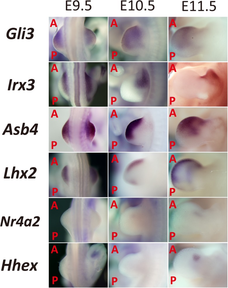
Fig 1. Expression pattern of anterior-localized genes in the limb bud based on EMBRYS.
Candidate transcription-associated factors involved in limb bud patterning were selected from EMBRYS and classified according to their anterior expression patterns. (A: Anterior; P: Posterior).
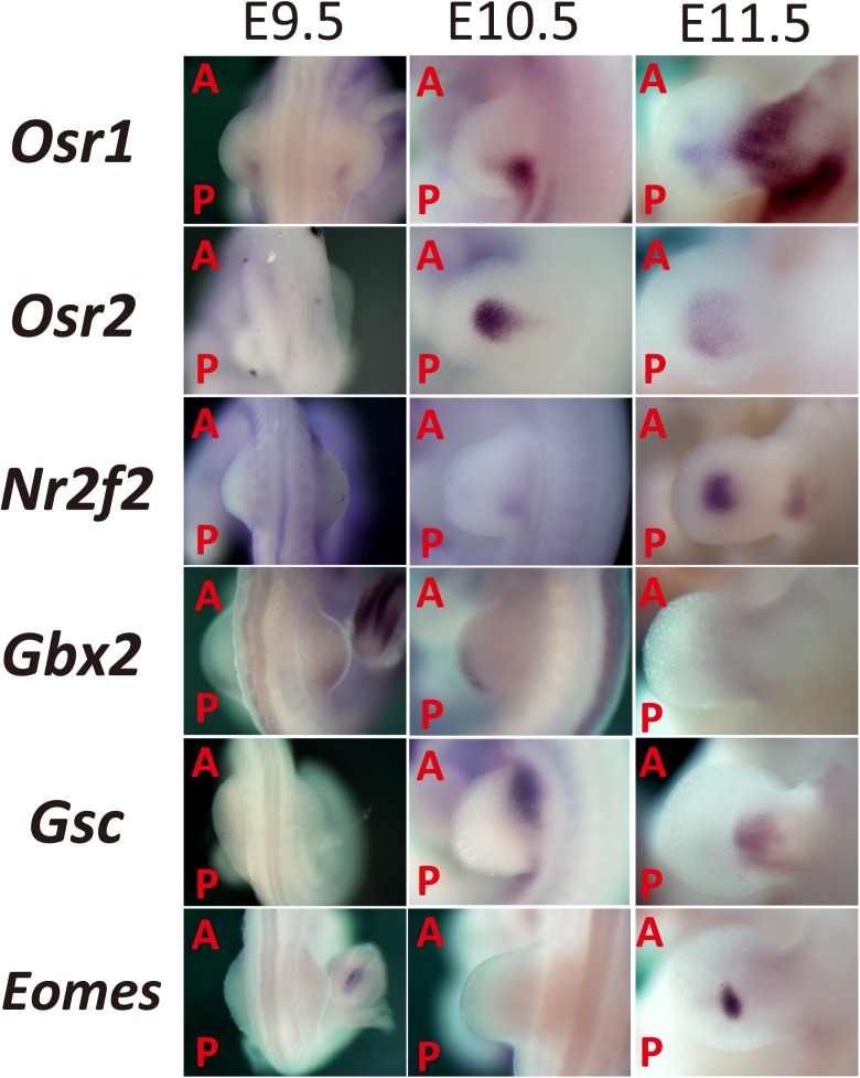
Fig 2. Expression pattern of candidate’s specifying the central part of the mouse limb bud based on EMBRYS.
Candidate transcription-associated factors involved in limb bud patterning were selected from EMBRYS and classified according to their expression pattern, particularly those showing significant localization in the limb bud mesenchyme. (A: Anterior; P: Posterior).
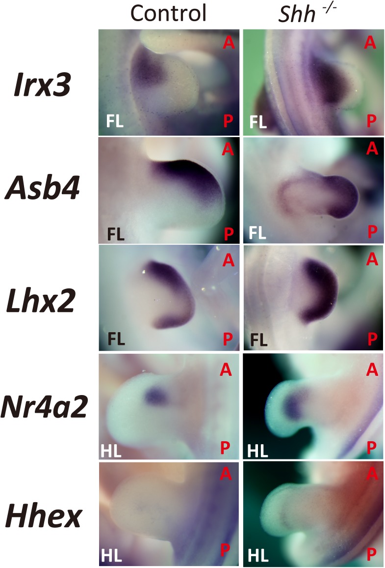
Fig 3. Expression pattern of candidates that are anterior regulators but expanded posteriorly in Shh-KO mouse limb buds.
Whole-mount in situ analysis was performed upon limbs of Shh-KO embryos (E10.5–11.5). Disruption of Shh resulted in posteriorly expanded expression of candidates in the developing limb bud (Control: WT or Shh +/-; FL: Forelimb; HL: Hind limb; A: Anterior; P: Posterior).
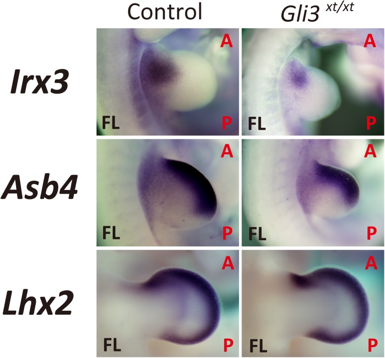
Fig 4. Reduced expression of Irx3 in Gli3-deficient limb buds.
Whole-mount in situ analysis of Gli3xt/xt embryos (E10.5–12.5). Expression of Irx3 was reduced while no significant change was seen in the expression of Asb4 and Lhx2 in Gli3-deficient limb buds. (Control: WT or Shh +/-; FL: Forelimb; HL: Hindlimb; A: Anterior; P: Posterior).
Plasmid construction
The insert sequence, CAG-loxP-CAT-polyA-loxP-polyA, [23] was amplified by PCR. The linear vector from the pT2AL200R175-CAGGS-eGFP plasmid was amplified by inverse PCR [24]. PrimeSTAR Max DNA polymerase (Takara Bio) was used to amplify both fragments. The insert fragment was cloned into the linear vector to generate the pT2AL200-CAG-loxP-CAT-polyA-loxP-polyA-R175 (pT2A-CAG-loxP) plasmid using the GeneArt Seamless Cloning and Assembly kit (Thermo Fisher Scientific) according to the manufacturer’s protocol. The targeting vector (pT2A-CAG-loxP-Irx3) for microinjection was prepared as follows: the coding DNA sequence (CDS) of Irx3 from the limb bud cDNA of an ICR mouse (a strain of albino mice) was amplified by PCR using the KAPA HiFi Ready Mix (KAPA BIOSYSTEMS) and cloned into the pT2A-CAG-loxP plasmid. The pT2A-CAG-loxP-Irx3 vector carried a floxed CAT gene and Irx3 CDS inserted between 200 bp and 175 bp of minimal Tol2 elements, respectively (Fig 5A). The inserted Irx3 sequence was validated by DNA sequencing. All primers used for PCR are shown in S1 Table.
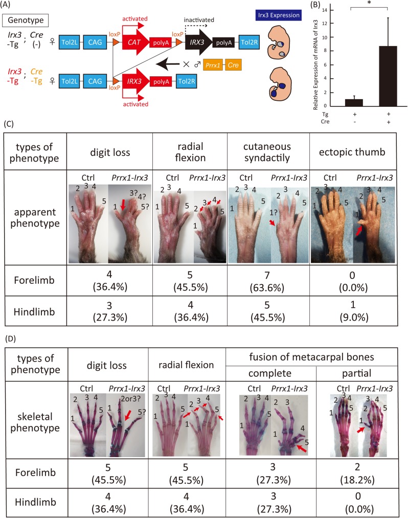
Fig 5. Generation and phenotypic analysis of transgenic mice (Prrx1-Irx3) overexpressing Irx3 in the developing mouse limb bud.
(A) Generation of transgenic mice overexpressing Irx3 in the developing limb bud. (B) Eight fold enhanced expression of Irx3 in the limb tissue of Prrx1-Irx3 than the control (Irx3-Tg/Cre -). All data were expressed as the means ± SEM, (n = 5). *P < 0.05. (C-D) Different phenotypes of Prrx1-Irx3 autopod were classified according to (C) apparent or (D) skeletal malformation (Prrx1-Irx3: Irx3-Tg and Cre-Tg; Ctrl: Irx3-Tg and Cre (-)).
Generation of transgenic Irx3 overexpressing mice
The Institutional Animal Care and Use Committee of Tokyo Medical and Dental University approved all animal experiments. The mice were sacrificed by cervical dislocation to alleviate suffering. Tol2 mRNA was transcribed from linearized pCS-mT2TP [25] using the mMessage mMachine SP6 Transcription kit (Thermo Fisher Scientific). The resulting transcripts were purified by LiCl precipitation. To generate an Irx3 transgenic mouse carrying the floxed CAT gene adjacent to the CDS of Irx3 (Fig 5A), the pT2A-CAG-loxP-Irx3 vector and Tol2 mRNA were microinjected into pronuclear-stage frozen BDF1 mouse embryos (Ark Resource) (S1A and S1B Fig). The resulting chimeric offspring (F0 mice) were crossed with C57BL/6 mice. To confirm germ-line transmission, the resulting progeny mice (F1) were genotyped by PCR analysis (S1C Fig). The Irx3 transgenic female mice were then crossed with Prrx1-Cre transgenic male mice (Jackson Laboratory) to generate Prrx1-Irx3 mice (Fig 5A). Primers used for genotyping are shown in S1 Table.
RNA isolation, reverse transcription, and quantitative real-time PCR
Total RNA was extracted from the limb epidermis of Prrx1-Irx3 or Irx3-Tg mice using ISOGEN (Nippon Gene). The extracted RNA was used as a template for reverse transcription using SuperScript II Reverse Transcriptase (Thermo Fisher Scientific). The mRNA expression of Irx3 was quantified by quantitative real-time PCR using Power SYBR Green PCR Master Mix (Thermo Fisher scientific) and were normalized to Gapdh mRNA levels (Fig 5B). Sequences of primers used for real-time PCR are shown in S1 Table.
Preparations of skeletal specimen
After observing the apparent phenotype of 3-week-old Prrx1-Irx3 or Irx3-Tg mice (Fig 5C), skeletal preparations were made and analyzed.
Skin and viscera were removed from the limbs of transgenic Prrx1-Irx3 or Irx3 mice. The specimens were then dehydrated in 100% ethanol for 120 h and degreased in acetone for 48 h. Subsequently, samples were stained with Alcian blue solution (7.5 g Alcian blue 8GX (Sigma) in 10 ml glacial acetic acid (Sigma) and 40 ml of 95% ethanol) at RT for 12 h. After washing with 95% ethanol for 10 min, the samples were treated with 2% KOH and stained with Alizarin red solution (7.5 mg Alizarin red S (Sigma) in 100 ml of 1% KOH solution). Specimens were washed with 20% glycerol in 1% KOH for 120 h and then with 20% glycerol in 20% ethanol for 12 h (Fig 5D).
Statistical analysis
The two-tailed independent Student’s t-test was used to calculate all P values. Asterisks in figures indicate differences with statistical significance as follows: *P < 0.05.
Results
Initial EMBRYS-based screening for candidate genes involved in AP patterning based on spatial localizations and temporal expression changes of 691 transcription-associated factors in the mouse limb bud
Previously, we established a WISH database of transcription factors that are present in mouse embryos termed EMBRYS [19]. To identify factors involved in limb bud patterning, the expression pattern of each factor was analyzed. According to the database, transcripts of certain transcription-associated factors displayed characteristic spatial localization and temporal expression changes in different parts of the mouse embryo at E9.5–11.5. Initially, 691 transcription-associated factors expressed in the mouse limb bud were selected and classified into categories based on their spatial expression patterns [26] (Figs 1 and 2). To our interest, some of the expression patterns were polarized towards the anterior part of the limb bud, resulting in a pattern of expression reminiscent of Gli3 expression (Fig 1). We predicted that these candidates might play a crucial role in specifying the future anterior-side of the mouse limb during development.
Formerly, Irx3 was identified as a transcription factor involved in specifying the anterior side of the limb bud [26, 27]. It is expressed on the proximal side of the primordial limb bud before E9.5. By E10.5, it localizes to the anterior-proximal region and the future radial side of the zeugopod and stylopod (Fig 1).
The SOCS box superfamily protein Asb4 mediates vascular differentiation [28]. Like Irx3, Asb4 is expressed in the anterior half of the distal part of the limb bud at E9.5. From E10.5 to 11.5, Asb4 localizes towards the anterior side of the limb bud (Fig 1).
Lhx2, a vertebrate homologue of apterous, is expressed under the confined distal mesoderm beneath the AER. Through the initiation and maintenance of the AER, Lhx2 regulates limb bud outgrowth in the chick [29, 30]. The expression pattern of Lhx2 was similar to those of Irx3 and Asb4 at E9.5. However, at E10.5, Lhx2 was also expressed at the posterior margin of the limb bud, which was consistent with previous reports (Fig 1).
Nr4a2 is a member of the nuclear receptor family of intracellular transcription factors and is involved in neuronal differentiation [31, 32]. It was found to be expressed towards the anterior end at E10.5–11.5 (Fig 1).
Hhex is a homeobox gene involved in the formation of the heart, vascular system, forebrain, thyroid, and liver [33, 34]. Additionally, it is known to regulate the AP patterning in Xenopus and mice [35]. Hhex was expressed as a spot on the anterior side of the limb bud at E11.5 (Fig 1).
Transcription factors with median-localized expression patterns were also identified in the present study. Odd-skipped related genes, such as Osr1 and Osr2, encode C2H2 zinc-finger transcription factors and are involved in the development of the embryonic heart, urogenital system [36], and the secondary palate [37]. Osr1 and Osr2 display dynamic expression patterns in the developing limb bud and are highly conserved between chicks and mice. Osr1 and Osr2 are partially co-expressed in the proximal limb bud but show mutually exclusive expression patterns in the distal autopod at E11.5–12.5 [38]. Present findings from EMBRYS indicate expression of Osr1 in the posterior-proximal part of the limb bud and in adjacent flanks at E9.5–10.5. Furthermore, the expression of Osr1 was distributed throughout the median mesenchyme of the autopod, prospective zeugopod, and stylopod at E11.5. In contrast, the expression of Osr2 was localized to the center of the limb bud at E10.5, and a shift in expression pattern was seen to the medial mesenchyme of the autopod at E11.5 (Fig 2).
Nr2f2 is a nuclear receptor subfamily member that plays a crucial role in skeletal muscle development during the limb bud outgrowth [39]. Nr2f2 was expressed in multiple developing tissues, such as the proximal region of the limb bud at E10.5 and in the center of the autopod at E11.5 (Fig 2).
Gbx2 is a homeobox gene required for the morphogenesis of the hindbrain [40]. Gbx2 was expressed along the posterior-middle part of the limb bud at E9.5–10.5 and was almost undetectable at E11.5 (Fig 2).
Goosecoid (Gsc) encodes a member of the bicoid subfamily of the paired homeobox family and plays a crucial role in craniofacial and rib cage development [41, 42]. As described previously [43], Gsc was expressed in the anterior-proximal part of the limb bud at E10.5 and was localized to the proximal region of the autopods extending posteriorly to the zeugopods and stylopods (Fig 2).
Eomes, also known as Tbr2 or Eomesodermin, is a member of the T-box family of transcription factors. It is associated with neurogenesis, cardiogenesis, and tumor immune response [44–46]. Eomes was localized to a single spot in the center of the autopod at E11.5 (Fig 2). Previous study indicated localization of Eomes expression to the prospective digits at E14.5 in autopods [47] and to the metacarpal pre-cartilage condensation at E16.5 [48].
In conclusion, the results from EMBRYS enabled selection of genes showing specific AP polarity expression in the developing limb bud. Further, we aimed at identifying distinctive genes expressed anteriorly as candidates for regulating the anterior development of the limb bud.
Candidate genes expressed towards the anterior side of the limb buds selected from EMBRYS showed posteriorly expanded expression pattern in Shh-KO limb buds
Interestingly, each selected candidate gene displayed different spatio-temporal expression patterns, suggesting their distinctive developmental roles in the specification of the anterior-side mesenchymal cells during limb bud development.
Shh is a crucial morphogen in the developing limb bud imparting posterior identity to the limb mesenchymal cells. Additionally, SHH in the ZPA mediates AP polarization [12]. Accordingly, Shh-KO mice have digit 1 and a shortened zeugopod [49]. In contrast, Gli3-deficient mouse models, such as the extra-toes (Xt) mice, exhibits symmetric polydactyly, the so-called mirror image digit duplication [21, 49, 50]. Shh expression is activated by Hand2 and retinoic acid [51]. Expression of Shh, is kept restricted to the posterior margin of the limb bud by the mutual antagonism between Gli3 and Hand2 [52]. In the present study, the identified candidate factors were further screened based on their regulation by SHH and Gli3 signaling during the development of the mouse limb bud.
To narrow down the selected candidates regulating anterior specification identified from EMBRYS, we performed WISH analysis of E10.5–11.5 limb buds from Shh-KO mouse embryos. Deletion of Shh resulted in widened posterior expression pattern of the selected genes (Fig 3).
Irx3 showed a posteriorly expanded expression pattern in Shh-deficient developing limb buds than those of the wild type, which was consistent with previous results [26], suggesting a loss of Irx3 expressional polarity caused by the absence of Shh. Similar results were observed for other candidate genes. Asb4 expression, which was originally localized to the anterior-half of the distal wild type limb bud, showed extended expression to the complete distal part and in addition was posteriorly expanded in the Shh-KO limb bud. Lhx2, which displayed a localized expression pattern along the lateral ridge in the wild type limb bud [29, 30], was expanded to the whole ridge in the Shh-KO limb bud. Furthermore, Nr4a2 and Hhex were no longer asymmetrically expressed and their expression were posteriorly expanded in Shh-KO limb buds (Fig 3).
Thus, the expression pattern of the analyzed genes indicates that they are inhibited by Shh in the posterior side of the limb bud. They typically function at the anterior side of the limb bud and are independent of SHH signaling or negative regulation by SHH.
Disruption of Gli3 affects the expression pattern of the selected candidates in the developing limb bud
Evaluation of changed expression pattern by WISH analysis of the selected candidates using Shh-KO limb buds revealed that all the candidates that displayed an anterior-localized expression pattern in wild type limb buds (Fig 1) expanded posteriorly in Shh-deficient limb buds (Fig 3). These results probably suggest their function as regulators specifying the anterior side of the developing limb bud. To further analyze these candidates, WISH analysis in Gli3-KO (extra-toes: Gli3xt/xt) embryos was performed and the change in expression pattern compared to the wild type limb bud was evaluated. We hypothesized that candidates regulated by the Gli3 pathway would show reduced expression in Gli3-deficient mouse limb buds.
Among the candidates analyzed, the expression domain of Irx3 was reduced and was localized more anterior-proximally in Gli3-deficient limb buds (Fig 4), which was consistent with previous results using Gli3/Kif7 double KO limb buds [26]. No significant changes were detected in the expression of Asb4 and Lhx2 in Gli3-deficient limb buds (Fig 4).
Collectively, through an EMBRYS-based stepwise selection method, several transcription-associated factors were narrowed down to few specific ones. Irx3 was selected for further analysis because of its crucial role in specifying the anterior mesenchymal cells in the limb bud.
Generation of a transgenic mouse model overexpressing Irx3 using the Tol2 transposon system for in vivo phenotypic analysis
To validate the screening system, in vivo experiments were designed to analyze Irx3 by generating a transgenic mice overexpressing Irx3 induced by Cre recombinase under the control of the Prrx1 regulatory element (Prrx1-Irx3 mouse) (Fig 5A). Firstly, an Irx3 transgenic mouse carrying a floxed CAT gene adjacent to the CDS of Irx3 was created. Protocols for Tol2-transposon-mediated generation of transgenic animals were as previously described [24, 25, 53, 54]. Tol2 mRNA and the Tol2 targeting vector were co-injected to facilitate the integration of large DNA fragments into the genome of pronuclear stage embryos (S1 Fig). The resultant mice were crossed with Prrx1-Cre male mouse to obtain the Prrx1-Irx3 mouse strain (Fig 5A, S1B and S1C Fig) [55]. The expression level of Irx3 in the Prrx1-Irx3 limbs was approximately 8-fold higher than in the wild type limbs (Fig 5B).
The Prrx1-Irx3 mice showed diverse limb malformations probably because of the disruption of autopod patterning. These phenotypes were highly penetrant and were classified into four categories: cutaneous fusion of digits 2–3 (63.6%), loss of digits 2–4 (45.5%), radial flexion of digits 2–4 (45.5%), and fusion of the metacarpal and metatarsal bones (45.5%) (Fig 5C and 5D). Thus, the Irx3 gain-of-function mice displayed complicated autopod phenotypes suggesting a significant role of Irx3 in regulating limb bud patterning, especially in the autopod.
In summary, the present study established an EMBRYS-based stepwise screening system for developmentally crucial transcription-associated factors. Candidates for anterior regulators, such as Irx3, were efficiently and systematically selected through multiple screening steps. In vivo analysis using transgenic mouse overexpressing Irx3 was performed to assess the screening system. Finally, to confirm the effectiveness and efficiency of the screening system, Prrx1-Irx3 mice were evaluated for complex phenotypes in the autopod.
Discussion
Morphogenesis of the developing limb bud is a highly reproducible model to study body plan formation. The process involves axis formation, patterning, outgrowth, and differentiation through interweaving gene interactions [1, 2]. Several transcriptional regulators play crucial roles in developmental and differential processes [6, 17, 18]. However, the exact mechanisms that orchestrate these processes remain to be elucidated [3].
Previously, we have established EMBRYS as a tool to identify novel transcription-associated factors that are essential for development [19]. In the present study, we established an EMBRYS-based stepwise screening system for searching candidate transcription-associated factors in a systematic and efficient manner. Firstly, we used EMBRYS to select candidates that exhibited a characteristic polarity along the AP axis. The expression pattern of several candidates showed clear localization along the anterior side of the developing limb bud mesenchyme similar to the expression pattern of Gli3 (Fig 1). To narrow down candidate transcription-associated factors that may play a role in establishing AP polarity in the limb bud under the regulation of Shh or Gli3, WISH analysis was performed in Shh- and Gli3- deficient embryos. The candidates exhibited a posteriorly expanded expression pattern in Shh-deficient compared to wild type embryos (Fig 3). Additionally, expression of Irx3 was reduced in the limb buds of Gli3-deficient embryos than in the wild type embryos (Fig 4), which was consistent with the previous studies [26, 27, 29, 30]. We therefore decided to focus on Irx3 and its role as an anterior regulator in the developing limb bud [33, 34]. A mouse model overexpressing Irx3 specifically in the limb bud was established for in vivo analysis of Irx3 (Fig 5A). The Prrx1-Irx3 mice displayed diverse malformations in the autopods due to defects in AP polarity (Fig 5C and 5D). Taken together, we validated an example of the EMBRYS-based stepwise screening system where candidate transcription-associated factors were efficiently selected, narrowed-down, and then analyzed in vivo to clarify their developmental role.
Initial screening identified five candidates showing anteriorly localized expression pattern in the developing limb bud (Fig 1). Previous reports indicate that these transcription-associated factors might play important roles during development.
Irx encodes a highly conserved three-amino acid-loop-extension (TALE) class homeoprotein at the N-terminus [56] and an IRO box at the C-terminus. Iro was first identified as a prepatterning gene for Drosophila bristles [57, 58]. Iro and Irx consist of clusters. In mammals, the Irx family consist of 6 paralogs that are classified into 2 clusters: Irx1, 2, and 4, and Irx3, 5, and 6 are classified into the IrxA and IrxB clusters, respectively [59, 60]. The characteristic genomic structures of Iro and Irx are highly conserved among Drosophila [57, 58, 60], Xenopus [60–66], zebrafish [67], chicken, [68] and mammals [27, 64, 69–78]. Furthermore, transcripts in the same clusters show almost identical expression patterns [69, 76]. Previous studies have shown that they play crucial roles in the development of sensory organs [57, 58], the nervous system [60–63, 65, 67, 71, 76, 79], the retina [63, 71], kidneys [64, 66], the heart [68, 69, 75, 77], female gonads [74], and limb buds [26, 27, 69, 71, 78].
Asb4 mediates oxygen-dependent vascular differentiation [28]. Lhx2 regulates limb bud outgrowth through the initiation and maintenance of the AER in chick embryos [29, 30]. Nr4a2 regulates the differentiation and maintenance of the dopaminergic system, [31, 32] and might also be relevant in patterning the forelimb bud based on its expression pattern [80]. Hhex is involved in the development of the heart, vascular system, forebrain, thyroid, and liver [33, 34]. To our interest, Hhex has been reported to regulate AP formation along the trunk in Xenopus and mice [35]. However, despite the characteristic expression pattern of the candidate genes in the developing limb bud, their role in patterning the limb bud along the AP axis has not been elucidated.
The initial screening of the candidate genes provided us with clues about their functions. Through EMBRYS and subsequent analysis of altered temporal expression, the list of candidate genes were further refined. Gli3, Irx3, Asb4, and Lhx2 were predicted to be expressed in significantly wide regions in the E9.5 limb bud, suggesting their activated expression in the formation of the limb bud and their probable involvement in the early specification of the AP axis. In contrast, the expression of Nr4a2 and Hhex were observed at a later stage of the developing limb bud, suggesting their involvement at a later stage of development such as late proliferation, differentiation, or digit specification.
Spatial expression patterns also suggested clues about the function of the candidates. At E11.5, Irx3 was expressed along the radial side of the zeugopods and stylopods, suggesting its involvement in the outgrowth of these structures. Asb4 was expressed in mesenchymal cells along the margin of the limb bud. Considering the previous reports [28], Asb4 might be involved in angiogenesis along the anterior side of the developing limb bud, regulating proliferation and differentiation of anterior mesenchymal cells. From this viewpoint, Lhx2 showed similar marginal expression patterns whereas Nr4a2 and Hhex showed spotty expression patterns that were not marginal but were still included along the anterior side of the limb bud.
Following the initial screening, the regulation of candidate factors by SHH or Gli3 signaling during limb bud development was further analyzed. Each of these five candidates displayed a significant posterior expression expansion in the Shh-KO limb bud. Interestingly, these expansions resulted in uniform expression patterns along the AP axis, which might be attributed to the loss of AP polarity resulting from the lack of Shh. On the other hand, it was also impressive that not all of these candidates showed significant expression reductions in Gli3-deficient limb buds. This suggests that some of the candidates might be regulated by different functions of Gli3 under SHH signaling pathway during the anterior specification of the limb bud progenitor cells.
Of the candidates that were identified during this screen, Irx3 was identified for further analysis. It has been implicated that the IrxB gene cluster, including Irx3 and 5, is involved in AP patterning of the developing limb bud [26, 27]. Previous study has indicated the regulation of Gli3, Shh, Fgf8, and their downstream target genes by Irx3 resulting in the specification of anterior progenitors in the developing limb buds [27].
We cast a focus on Irx3 as an example for in vivo analysis following the EMBRYS-based screening steps. Phenotypic analysis of Prrx1-Irx3 mice displayed 4 types of autopod malformation: cutaneous fusion of interdigits, deficit of digits 2–4, radial deviation of digits 2–4, and fusion of the metacarpal and metatarsal bones (Fig 5C and 5D). Some of these phenotypes were observed in family members with mutations in HOXD13 or HOXA13 genes [81] and in HOXD13-KO mice [82]. In addition, Prrx1-Hand2; Gli3 conditional double KO mice displayed similar phenotypes including the fusion of the metatarsals or a digit deficit [83]. These data suggests that overexpression of Irx3 affects digit patterning in the developing limb bud. Overall, the present study demonstrates proof-of-concept for our screening system in identifying transcription-associated factors that regulate AP patterning in the limb.
Additional genes, such as Gsc, Eomes, and Osr2 were clearly expressed in the middle region of the limb bud with a spotted pattern after E10.5 (Fig 2). Gsc is one of the transcription factor expressed in the Spemann’s organizer of the Xenopus embryo. When Gsc mRNA was microinjected into the ventral cells, twinned axes were induced, indicating that Gsc was sufficient to function like the Spemann's organizer [84]. Eomes is expressed in the mesoderm of Xenopus embryos, and ectopic expression of Eomes in the animal cap induces nearly all of the mesodermal genes [85]. Osr2 is initially expressed in early gastrulation in the mesoderm and endodermal region and is necessary for kidney induction [86]. Since these key transcription factors of early gastrulation are also expressed adjacently in the limb mesenchyme after E10.5, it is interesting to speculate that a key transcription network interacting with the secreted growth factors during Xenopus gastrulation was co-opted in late limb mesenchyme induction.
In summary, we established an EMBRYS-based stepwise screening system to identify candidates for transcription-associated factors that might be involved in developmental processes such as AP patterning of the limb bud. This system is widely applicable to embryonic tissues other than the limb bud or to transcription-associated factors with other functions such as differentiation, proliferation, and pluripotency. The benefit of this screening system is the easy and efficient access to 3-dimensional and spatio-temporal gene expression patterns for each gene in an initial high-throughput screen. This enables quick and efficient analysis of expressional dynamics along transcriptional hierarches and to predict their roles during development. Moreover, following the initial screen, candidates of interest under the control of developmental master genes could be effectively selected. This is then followed by in vivo experiments, performed more efficiently nowadays because of improvements in genome editing technologies. As a result, we can gain insight into the spatio-temporal regulations of body plan formation by using the limb bud as a model to study the mechanisms of specification and morphogenesis.
Supporting information
(A) Irx3 transgenic mice was generated using the Tol2 transposon system. (B) High efficiency of Irx3 transgenesis compared to a gene targeting system. (C) Genotyping strategy of transgenic Irx3 mice.
(TIF)
(XLSX)
Acknowledgments
We would like to thank all our colleagues who supported us and who were involved in helpful discussions.
We would like to thank Editage (www.editage.jp) for English language editing.
Data Availability
All relevant data are within the paper and its Supporting Information files.
Funding Statement
This research is supported by AMED-CREST (the Core Research for the Evolutionary Science and Technology) from AMED (the Japan Agency for Medical Research and Development), JSPS KAKENHI [Grant Numbers: 26113008, 15H02560, 15K15544], grants from the National Institutes of Health [Grant Numbers: AR050631, AR065379], and the Naito Foundation to H.A., grants from the National Institutes of Health [Grant Number: AR064195] to Y.K., and grants from the G. Harold and Leila Y. Mathers Charitable Foundation, The Leona M. and Harry B. Helmsley Charitable Trust and the Universidad Catolica de Murcia (UCAM) to J.C.I.B. The funders had no role in study design, data collection and analysis, decision to publish, or preparation of the manuscript.
References
- 1.Zeller R, Lopez-Rios J, Zuniga A. Vertebrate limb bud development: moving towards integrative analysis of organogenesis. Nature reviews Genetics. 2009;10(12):845–58. doi: 10.1038/nrg2681 [DOI] [PubMed] [Google Scholar]
- 2.Tabin C, Wolpert L. Rethinking the proximodistal axis of the vertebrate limb in the molecular era. Genes & development. 2007;21(12):1433–42. [DOI] [PubMed] [Google Scholar]
- 3.Benazet JD, Zeller R. Vertebrate limb development: moving from classical morphogen gradients to an integrated 4-dimensional patterning system. Cold Spring Harb Perspect Biol. 2009;1(4):a001339 doi: 10.1101/cshperspect.a001339 [DOI] [PMC free article] [PubMed] [Google Scholar]
- 4.Oliver G, Wright CV, Hardwicke J, De Robertis EM. A gradient of homeodomain protein in developing forelimbs of Xenopus and mouse embryos. Cell. 1988;55(6):1017–24. [DOI] [PubMed] [Google Scholar]
- 5.Molven A, Wright C, Bremiller R, De Robertis EM, Kimmel CB. Expression of a homeobox gene product in normal and mutant zebrafish embryos: evolution of the tetrapod body plan. Development. 1990;109(2):279–88. [DOI] [PubMed] [Google Scholar]
- 6.Burke AC, Nelson CE, Morgan BA, Tabin C. Hox genes and the evolution of vertebrate axial morphology. Development. 1995;121(2):333–46. [DOI] [PubMed] [Google Scholar]
- 7.Saunders JW. The proximo‐distal sequence of origin of the parts of the chick wing and the role of the ectoderm. Journal of Experimental Zoology. 1948;108(3):363–403. [DOI] [PubMed] [Google Scholar]
- 8.Saunders JW, Reuss C. Inductive and axial properties of prospective wing-bud mesoderm in the chick embryo. Developmental biology. 1974;38(1):41–50. [DOI] [PubMed] [Google Scholar]
- 9.Fernandez-Teran M, Ros MA. The Apical Ectodermal Ridge: morphological aspects and signaling pathways. 2008. [DOI] [PubMed]
- 10.Saunders J, Gasseling M. Ectodermal-mesenchymal interactions in the origin of limb symmetry. Epithelial-mesenchymal interactions. 1968:78–97. [Google Scholar]
- 11.Tickle C, Summerbell D, Wolpert L. Positional signalling and specification of digits in chick limb morphogenesis. Nature. 1975;254(5497):199–202. [DOI] [PubMed] [Google Scholar]
- 12.Riddle RD, Johnson RL, Laufer E, Tabin C. Sonic hedgehog mediates the polarizing activity of the ZPA. Cell. 1993;75(7):1401–16. [DOI] [PubMed] [Google Scholar]
- 13.te Welscher P, Fernandez-Teran M, Ros MA, Zeller R. Mutual genetic antagonism involving GLI3 and dHAND prepatterns the vertebrate limb bud mesenchyme prior to SHH signaling. Genes & development. 2002;16(4):421–6. [DOI] [PMC free article] [PubMed] [Google Scholar]
- 14.Yang Y, Drossopoulou G, Chuang P, Duprez D, Marti E, Bumcrot D, et al. Relationship between dose, distance and time in Sonic Hedgehog-mediated regulation of anteroposterior polarity in the chick limb. Development. 1997;124(21):4393–404. [DOI] [PubMed] [Google Scholar]
- 15.Harfe BD, Scherz PJ, Nissim S, Tian H, McMahon AP, Tabin CJ. Evidence for an expansion-based temporal Shh gradient in specifying vertebrate digit identities. Cell. 2004;118(4):517–28. doi: 10.1016/j.cell.2004.07.024 [DOI] [PubMed] [Google Scholar]
- 16.Vokes SA, Ji H, Wong WH, McMahon AP. A genome-scale analysis of the cis-regulatory circuitry underlying sonic hedgehog-mediated patterning of the mammalian limb. Genes & development. 2008;22(19):2651–63. [DOI] [PMC free article] [PubMed] [Google Scholar]
- 17.Gray PA, Fu H, Luo P, Zhao Q, Yu J, Ferrari A, et al. Mouse brain organization revealed through direct genome-scale TF expression analysis. Science. 2004;306(5705):2255–7. doi: 10.1126/science.1104935 [DOI] [PubMed] [Google Scholar]
- 18.Jessell TM. Neuronal specification in the spinal cord: inductive signals and transcriptional codes. Nature Reviews Genetics. 2000;1(1):20–9. doi: 10.1038/35049541 [DOI] [PubMed] [Google Scholar]
- 19.Yokoyama S, Ito Y, Ueno-Kudoh H, Shimizu H, Uchibe K, Albini S, et al. A systems approach reveals that the myogenesis genome network is regulated by the transcriptional repressor RP58. Dev Cell. 2009;17(6):836–48. doi: 10.1016/j.devcel.2009.10.011 [DOI] [PMC free article] [PubMed] [Google Scholar]
- 20.Chiang C, Litingtung Y, Lee E, Young KE, Corden JL, Westphal H, et al. Cyclopia and defective axial patterning in mice lacking Sonic hedgehog gene function. 1996. [DOI] [PubMed]
- 21.Hui C-c Joyner AL. A mouse model of Greig cephalo–polysyndactyly syndrome: the extra–toesJ mutation contains an intragenic deletion of the Gli3 gene. Nature genetics. 1993;3(3):241–6. doi: 10.1038/ng0393-241 [DOI] [PubMed] [Google Scholar]
- 22.Kawakami Y, Tsuda M, Takahashi S, Taniguchi N, Esteban CR, Zemmyo M, et al. Transcriptional coactivator PGC-1alpha regulates chondrogenesis via association with Sox9. Proceedings of the National Academy of Sciences of the United States of America. 2005;102(7):2414–9. doi: 10.1073/pnas.0407510102 [DOI] [PMC free article] [PubMed] [Google Scholar]
- 23.Araki K, Araki M, Miyazaki J-i, Vassalli P. Site-specific recombination of a transgene in fertilized eggs by transient expression of Cre recombinase. Proceedings of the National Academy of Sciences. 1995;92(1):160–4. [DOI] [PMC free article] [PubMed] [Google Scholar]
- 24.Urasaki A, Morvan G, Kawakami K. Functional dissection of the Tol2 transposable element identified the minimal cis-sequence and a highly repetitive sequence in the subterminal region essential for transposition. Genetics. 2006;174(2):639–49. doi: 10.1534/genetics.106.060244 [DOI] [PMC free article] [PubMed] [Google Scholar]
- 25.Kawakami K, Noda T. Transposition of the Tol2 element, an Ac-like element from the Japanese medaka fish Oryzias latipes, in mouse embryonic stem cells. Genetics. 2004;166(2):895–9. [DOI] [PMC free article] [PubMed] [Google Scholar]
- 26.Zhulyn O, Li D, Deimling S, Vakili NA, Mo R, Puviindran V, et al. A switch from low to high Shh activity regulates establishment of limb progenitors and signaling centers. Dev Cell. 2014;29(2):241–9. doi: 10.1016/j.devcel.2014.03.002 [DOI] [PubMed] [Google Scholar]
- 27.Li D, Sakuma R, Vakili NA, Mo R, Puviindran V, Deimling S, et al. Formation of proximal and anterior limb skeleton requires early function of Irx3 and Irx5 and is negatively regulated by Shh signaling. Dev Cell. 2014;29(2):233–40. doi: 10.1016/j.devcel.2014.03.001 [DOI] [PubMed] [Google Scholar]
- 28.Ferguson JE 3rd, Wu Y, Smith K, Charles P, Powers K, Wang H, et al. ASB4 is a hydroxylation substrate of FIH and promotes vascular differentiation via an oxygen-dependent mechanism. Mol Cell Biol. 2007;27(18):6407–19. doi: 10.1128/MCB.00511-07 [DOI] [PMC free article] [PubMed] [Google Scholar]
- 29.Rodriguez-Esteban C, Schwabe J, Pena J, Rincon-Limas DE, Magallón J, Botas J, et al. Lhx2, a vertebrate homologue of apterous, regulates vertebrate limb outgrowth. Development. 1998;125(20):3925–34. [DOI] [PubMed] [Google Scholar]
- 30.Kanegae Y, Tavares AT, Belmonte JCI, Verma IM. Role of Rel/NF-κB transcription factors during the outgrowth of the vertebrate limb. Nature. 1998;392(6676):611–4. doi: 10.1038/33429 [DOI] [PubMed] [Google Scholar]
- 31.Sacchetti P, Carpentier R, Segard P, Olive-Cren C, Lefebvre P. Multiple signaling pathways regulate the transcriptional activity of the orphan nuclear receptor NURR1. Nucleic Acids Res. 2006;34(19):5515–27. doi: 10.1093/nar/gkl712 [DOI] [PMC free article] [PubMed] [Google Scholar]
- 32.Kim J-Y, Koh HC, Lee J-Y, Chang M-Y, Kim Y-C, Chung H-Y, et al. Dopaminergic neuronal differentiation from rat embryonic neural precursors by Nurr1 overexpression. Journal of Neurochemistry. 2003;85(6):1443–54. [DOI] [PubMed] [Google Scholar]
- 33.Hallaq H, Pinter E, Enciso J, McGrath J, Zeiss C, Brueckner M, et al. A null mutation of Hhex results in abnormal cardiac development, defective vasculogenesis and elevated Vegfa levels. Development. 2004;131(20):5197–209. doi: 10.1242/dev.01393 [DOI] [PubMed] [Google Scholar]
- 34.Barbera JM, Clements M, Thomas P, Rodriguez T, Meloy D, Kioussis D, et al. The homeobox gene Hex is required in definitive endodermal tissues for normal forebrain, liver and thyroid formation. Development. 2000;127(11):2433–45. [DOI] [PubMed] [Google Scholar]
- 35.Zamparini AL, Watts T, Gardner CE, Tomlinson SR, Johnston GI, Brickman JM. Hex acts with beta-catenin to regulate anteroposterior patterning via a Groucho-related co-repressor and Nodal. Development. 2006;133(18):3709–22. doi: 10.1242/dev.02516 [DOI] [PubMed] [Google Scholar]
- 36.Wang Q, Lan Y, Cho ES, Maltby KM, Jiang R. Odd-skipped related 1 (Odd 1) is an essential regulator of heart and urogenital development. Dev Biol. 2005;288(2):582–94. doi: 10.1016/j.ydbio.2005.09.024 [DOI] [PMC free article] [PubMed] [Google Scholar]
- 37.Lan Y, Ovitt CE, Cho ES, Maltby KM, Wang Q, Jiang R. Odd-skipped related 2 (Osr2) encodes a key intrinsic regulator of secondary palate growth and morphogenesis. Development. 2004;131(13):3207–16. doi: 10.1242/dev.01175 [DOI] [PubMed] [Google Scholar]
- 38.Stricker S, Brieske N, Haupt J, Mundlos S. Comparative expression pattern of Odd-skipped related genes Osr1 and Osr2 in chick embryonic development. Gene Expr Patterns. 2006;6(8):826–34. doi: 10.1016/j.modgep.2006.02.003 [DOI] [PubMed] [Google Scholar]
- 39.Lee CT, Li L, Takamoto N, Martin JF, Demayo FJ, Tsai MJ, et al. The nuclear orphan receptor COUP-TFII is required for limb and skeletal muscle development. Mol Cell Biol. 2004;24(24):10835–43. doi: 10.1128/MCB.24.24.10835-10843.2004 [DOI] [PMC free article] [PubMed] [Google Scholar]
- 40.Wassarman KM, Lewandoski M, Campbell K, Joyner AL, Rubenstein J, Martinez S, et al. Specification of the anterior hindbrain and establishment of a normal mid/hindbrain organizer is dependent on Gbx2 gene function. Development. 1997;124(15):2923–34. [DOI] [PubMed] [Google Scholar]
- 41.Rivera-Pérez JA, Mallo M, Gendron-Maguire M, Gridley T, Behringer RR. Goosecoid is not an essential component of the mouse gastrula organizer but is required for craniofacial and rib development. Development. 1995;121(9):3005–12. [DOI] [PubMed] [Google Scholar]
- 42.Yamada G, Ueno K, Nakamura S, Hanamure Y, Yasui K, Uemura M, et al. Nasal and pharyngeal abnormalities caused by the mouse goosecoid gene mutation. Biochemical and biophysical research communications. 1997;233(1):161–5. doi: 10.1006/bbrc.1997.6315 [DOI] [PubMed] [Google Scholar]
- 43.Gaunt SJ, Blum M, De Robertis EM. Expression of the mouse goosecoid gene during mid-embryogenesis may mark mesenchymal cell lineages in the developing head, limbs and body wall. Development. 1993;117(2):769–78. [DOI] [PubMed] [Google Scholar]
- 44.Costello I, Pimeisl IM, Drager S, Bikoff EK, Robertson EJ, Arnold SJ. The T-box transcription factor Eomesodermin acts upstream of Mesp1 to specify cardiac mesoderm during mouse gastrulation. Nature cell biology. 2011;13(9):1084–91. doi: 10.1038/ncb2304 [DOI] [PMC free article] [PubMed] [Google Scholar]
- 45.Zhu Y, Ju S, Chen E, Dai S, Li C, Morel P, et al. T-bet and eomesodermin are required for T cell-mediated antitumor immune responses. J Immunol. 2010;185(6):3174–83. doi: 10.4049/jimmunol.1000749 [DOI] [PubMed] [Google Scholar]
- 46.Englund C, Fink A, Lau C, Pham D, Daza RA, Bulfone A, et al. Pax6, Tbr2, and Tbr1 are expressed sequentially by radial glia, intermediate progenitor cells, and postmitotic neurons in developing neocortex. J Neurosci. 2005;25(1):247–51. doi: 10.1523/JNEUROSCI.2899-04.2005 [DOI] [PMC free article] [PubMed] [Google Scholar]
- 47.Kimura N, Nakashima K, Ueno M, Kiyama H, Taga T. A novel mammalian T-box-containing gene, Tbr2, expressed in mouse developing brain. Developmental brain research. 1999;115(2):183–93. [DOI] [PubMed] [Google Scholar]
- 48.Bulfone A, Martinez S, Marigo V, Campanella M, Basile A, Quaderi N, et al. Expression pattern of the Tbr2 (Eomesodermin) gene during mouse and chick brain development. Mechanisms of development. 1999;84(1):133–8. [DOI] [PubMed] [Google Scholar]
- 49.Litingtung Y, Dahn RD, Li Y, Fallon JF, Chiang C. Shh and Gli3 are dispensable for limb skeleton formation but regulate digit number and identity. Nature. 2002;418(6901):979–83. doi: 10.1038/nature01033 [DOI] [PubMed] [Google Scholar]
- 50.te Welscher P, Zuniga A, Kuijper S, Drenth T, Goedemans HJ, Meijlink F, et al. Progression of vertebrate limb development through SHH-mediated counteraction of GLI3. Science. 2002;298(5594):827–30. doi: 10.1126/science.1075620 [DOI] [PubMed] [Google Scholar]
- 51.Niederreither K, Vermot J, Schuhbaur B, Chambon P, Dollé P. Embryonic retinoic acid synthesis is required for forelimb growth and anteroposterior patterning in the mouse. Development. 2002;129(15):3563–74. [DOI] [PubMed] [Google Scholar]
- 52.Zúñiga A, Zeller R. Gli3 (Xt) and formin (ld) participate in the positioning of the polarising region and control of posterior limb-bud identity. Development. 1999;126(1):13–21. [DOI] [PubMed] [Google Scholar]
- 53.Kawakami K. Tol2: a versatile gene transfer vector in vertebrates. Genome biology. 2007;8(1):1. [DOI] [PMC free article] [PubMed] [Google Scholar]
- 54.Kawakami K, Takeda H, Kawakami N, Kobayashi M, Matsuda N, Mishina M. A transposon-mediated gene trap approach identifies developmentally regulated genes in zebrafish. Dev Cell. 2004;7(1):133–44. doi: 10.1016/j.devcel.2004.06.005 [DOI] [PubMed] [Google Scholar]
- 55.Logan M, Martin JF, Nagy A, Lobe C, Olson EN, Tabin CJ. Expression of Cre Recombinase in the developing mouse limb bud driven by a Prxl enhancer. Genesis. 2002;33(2):77–80. doi: 10.1002/gene.10092 [DOI] [PubMed] [Google Scholar]
- 56.Bürglin TR. Analysis of TALE superclass homeobox genes (MEIS, PBC, KNOX, Iroquois, TGIF) reveals a novel domain conserved between plants and animals. Nucleic acids research. 1997;25(21):4173–80. [DOI] [PMC free article] [PubMed] [Google Scholar]
- 57.Dambly-Chaudière C, Jamet E, Burri M, Bopp D, Basler K, Hafen E, et al. The paired box gene pox neuro: A determiant of poly-innervated sense organs in Drosophila. Cell. 1992;69(1):159–72. [DOI] [PubMed] [Google Scholar]
- 58.Leyns L, Gómez-Skarmeta J-L, Dambly-Chaudière C. iroquois: a prepattern gene that controls the formation of bristles on the thorax ofDrosophila. Mechanisms of development. 1996;59(1):63–72. [DOI] [PubMed] [Google Scholar]
- 59.Peters T, Dildrop R, Ausmeier K, Rüther U. Organization of mouse Iroquois homeobox genes in two clusters suggests a conserved regulation and function in vertebrate development. Genome research. 2000;10(10):1453–62. [DOI] [PMC free article] [PubMed] [Google Scholar]
- 60.Cavodeassi F, Modolell J, Gómez-Skarmeta JL. The Iroquois family of genes: from body building to neural patterning. Development. 2001;128(15):2847–55. [DOI] [PubMed] [Google Scholar]
- 61.Bellefroid EJ, Kobbe A, Gruss P, Pieler T, Gurdon JB, Papalopulu N. Xiro3 encodes a Xenopus homolog of the Drosophila Iroquois genes and functions in neural specification. The EMBO Journal. 1998;17(1):191–203. doi: 10.1093/emboj/17.1.191 [DOI] [PMC free article] [PubMed] [Google Scholar]
- 62.Gómez‐Skarmeta JL, Glavic A, de la Calle‐Mustienes E, Modolell J, Mayor R. Xiro, a Xenopus homolog of the Drosophila Iroquois complex genes, controls development at the neural plate. The EMBO journal. 1998;17(1):181–90. doi: 10.1093/emboj/17.1.181 [DOI] [PMC free article] [PubMed] [Google Scholar]
- 63.Garriock RJ, Vokes SA, Small EM, Larson R, Krieg PA. Developmental expression of the Xenopus Iroquois-family homeobox genes, Irx4 and Irx5. Development genes and evolution. 2001;211(5):257–60. [DOI] [PubMed] [Google Scholar]
- 64.Reggiani L, Raciti D, Airik R, Kispert A, Brandli AW. The prepattern transcription factor Irx3 directs nephron segment identity. Genes & development. 2007;21(18):2358–70. [DOI] [PMC free article] [PubMed] [Google Scholar]
- 65.Rodríguez-Seguel E, Alarcón P, Gómez-Skarmeta JL. The Xenopus Irx genes are essential for neural patterning and define the border between prethalamus and thalamus through mutual antagonism with the anterior repressors Fezf and Arx. Developmental biology. 2009;329(2):258–68. doi: 10.1016/j.ydbio.2009.02.028 [DOI] [PubMed] [Google Scholar]
- 66.Alarcón P, Rodríguez-Seguel E, Fernández-González A, Rubio R, Gómez-Skarmeta JL. A dual requirement for Iroquois genes during Xenopus kidney development. Development. 2008;135(19):3197–207. doi: 10.1242/dev.023697 [DOI] [PubMed] [Google Scholar]
- 67.Lecaudey V, Anselme I, Dildrop R, Rüther U, Schneider‐Maunoury S. Expression of the zebrafish Iroquois genes during early nervous system formation and patterning. Journal of Comparative Neurology. 2005;492(3):289–302. doi: 10.1002/cne.20765 [DOI] [PubMed] [Google Scholar]
- 68.Bao Z-Z, Bruneau BG, Seidman J, Seidman CE, Cepko CL. Regulation of chamber-specific gene expression in the developing heart by Irx4. Science. 1999;283(5405):1161–4. [DOI] [PubMed] [Google Scholar]
- 69.Houweling AC, Dildrop R, Peters T, Mummenhoff J, Moorman AF, Rüther U, et al. Gene and cluster-specific expression of the Iroquois family members during mouse development. Mechanisms of development. 2001;107(1):169–74. [DOI] [PubMed] [Google Scholar]
- 70.de la Calle-Mustienes E, Feijoo CG, Manzanares M, Tena JJ, Rodriguez-Seguel E, Letizia A, et al. A functional survey of the enhancer activity of conserved non-coding sequences from vertebrate Iroquois cluster gene deserts. Genome research. 2005;15(8):1061–72. doi: 10.1101/gr.4004805 [DOI] [PMC free article] [PubMed] [Google Scholar]
- 71.Bosse A, Zülch A, Becker M-B, Torres M, Gómez-Skarmeta JL, Modolell J, et al. Identification of the vertebrate Iroquois homeobox gene family with overlapping expression during early development of the nervous system. Mechanisms of development. 1997;69(1):169–81. [DOI] [PubMed] [Google Scholar]
- 72.Bosse A, Stoykova A, Nieselt‐Struwe K, Chowdhury K, Copeland NG, Jenkins NA, et al. Identification of a novel mouse Iroquois homeobox gene, Irx5, and chromosomal localisation of all members of the mouse Iroquois gene family. Developmental Dynamics. 2000;218(1):160–74. doi: 10.1002/(SICI)1097-0177(200005)218:1<160::AID-DVDY14>3.0.CO;2-2 [DOI] [PubMed] [Google Scholar]
- 73.Cohen DR, Cheng CW, Cheng SH, Hui C-c. Expression of two novel mouse Iroquois homeobox genes during neurogenesis. Mechanisms of development. 2000;91(1):317–21. [DOI] [PubMed] [Google Scholar]
- 74.Jorgensen JS, Gao L. Irx3 is differentially up-regulated in female gonads during sex determination. Gene Expr Patterns. 2005;5(6):756–62. doi: 10.1016/j.modgep.2005.04.011 [DOI] [PubMed] [Google Scholar]
- 75.Gaborit N, Sakuma R, Wylie JN, Kim KH, Zhang SS, Hui CC, et al. Cooperative and antagonistic roles for Irx3 and Irx5 in cardiac morphogenesis and postnatal physiology. Development. 2012;139(21):4007–19. doi: 10.1242/dev.081703 [DOI] [PMC free article] [PubMed] [Google Scholar]
- 76.Gómez-Skarmeta JL, Modolell J. Iroquois genes: genomic organization and function in vertebrate neural development. Current opinion in genetics & development. 2002;12(4):403–8. [DOI] [PubMed] [Google Scholar]
- 77.Christoffels VM, Keijser AG, Houweling AC, Clout DE, Moorman AF. Patterning the embryonic heart: identification of five mouse Iroquois homeobox genes in the developing heart. Dev Biol. 2000;224(2):263–74. doi: 10.1006/dbio.2000.9801 [DOI] [PubMed] [Google Scholar]
- 78.Zülch A, Becker M-B, Gruss P. Expression pattern of Irx1 and Irx2 during mouse digit development. Mechanisms of development. 2001;106(1):159–62. [DOI] [PubMed] [Google Scholar]
- 79.Robertshaw E, Matsumoto K, Lumsden A, Kiecker C. Irx3 and Pax6 establish differential competence for Shh-mediated induction of GABAergic and glutamatergic neurons of the thalamus. Proceedings of the National Academy of Sciences of the United States of America. 2013;110(41):E3919–26. doi: 10.1073/pnas.1304311110 [DOI] [PMC free article] [PubMed] [Google Scholar]
- 80.Ahmed HA, Ibrahim LL, El Mekkawy DA, El Wakil A. Expression Pattern of the Orphan Nuclear Receptor, Nurr1, in the Developing Mouse Forelimb and its Relationship to Limb Skeletogenesis and Osteogenesis. OnLine Journal of Biological Sciences. 2015;15(3):162. [Google Scholar]
- 81.Goodman FR. Limb malformations and the human HOX genes. Am J Med Genet. 2002;112(3):256–65. doi: 10.1002/ajmg.10776 [DOI] [PubMed] [Google Scholar]
- 82.Kmita M, Fraudeau N, Hérault Y, Duboule D. Serial deletions and duplications suggest a mechanism for the collinearity of Hoxd genes in limbs. Nature. 2002;420(6912):145–50. doi: 10.1038/nature01189 [DOI] [PubMed] [Google Scholar]
- 83.Galli A, Robay D, Osterwalder M, Bao X, Benazet JD, Tariq M, et al. Distinct roles of Hand2 in initiating polarity and posterior Shh expression during the onset of mouse limb bud development. PLoS Genet. 2010;6(4):e1000901 doi: 10.1371/journal.pgen.1000901 [DOI] [PMC free article] [PubMed] [Google Scholar]
- 84.Cho KW, Blumberg B, Steinbeisser H, De Robertis EM. Molecular nature of Spemann's organizer: the role of the Xenopus homeobox gene goosecoid. Cell. 1991;67(6):1111–20. [DOI] [PMC free article] [PubMed] [Google Scholar]
- 85.Ryan K, Garrett N, Mitchell A, Gurdon J. Eomesodermin, a key early gene in Xenopus mesoderm differentiation. Cell. 1996;87(6):989–1000. [DOI] [PubMed] [Google Scholar]
- 86.Tena JJ, Neto A, de la Calle-Mustienes E, Bras-Pereira C, Casares F, Gómez-Skarmeta JL. Odd-skipped genes encode repressors that control kidney development. Developmental biology. 2007;301(2):518–31. doi: 10.1016/j.ydbio.2006.08.063 [DOI] [PubMed] [Google Scholar]
Associated Data
This section collects any data citations, data availability statements, or supplementary materials included in this article.
Supplementary Materials
(A) Irx3 transgenic mice was generated using the Tol2 transposon system. (B) High efficiency of Irx3 transgenesis compared to a gene targeting system. (C) Genotyping strategy of transgenic Irx3 mice.
(TIF)
(XLSX)
Data Availability Statement
All relevant data are within the paper and its Supporting Information files.


