Abstract
Background:
The acute kidney injury (AKI) may do damage to remote organs. Objective of the study is to investigate effect of seaweed extract (SE) on brain oxidative damage in kidney ischemia/reperfusion rats.
Material and Methods:
Animals were randomly divided into five groups. SE pre-fed to rats.
Results:
Kidney I/R may cause oxidative injury in kidneys and brains tissue in rats. SE pre-treatment can decrease lipid peroxidation levels and increase antioxidant enzymes activities in kidney and brain hippocampus of kidney I/R rats.
Conclusion:
Our results indicate that SE is useful for brain nerve function keeping in kidney I/R rats.
Keywords: Kidney, Brain nerve, Ischemia, Oxidative injury
Introduction
I/R injury indicate the injury to the tissue or organ after an ischemic tissue or organ is supplied with a blood flow or oxygen supply. The kidney is a high perfusion organ, which is more sensitive to ischemia and reperfusion. Therefore, renal I/R injuries are commoner in clinical therapy. The acute kidney injury (AKI) is a severe problem, and the disease is around 7% in hospitalized patients. The some reasons for AKI include prerenal, renal, and postrenal. AKI may lead to the decreased cardiac output, hypotension, and renal artery disease. The unattended AKI may lead to death that is generally because of nonrenal complications and involves many organ dysfunctions (Kizilbash et al, 2016). The pathological and physiological changes of renal I/R were found in the course of acute blood loss, toxic body, renal vascular surgery and renal transplantation.
Lots of oxygen free radicals were produced in tissue vascular endothelial cells and polymorphonuclear cells through many kinds of ways, e.g., xanthine oxidase, mitochondria and peanut four acids and so on. In recent years, a large number of studies have confirmed that free radical reaction is an important cause of renal ischemia reperfusion injury. This can lead to a decrease in the amount of anti-oxidant factors as well as increase in the amount of the pro-oxidant substances. Oxidative stress is probably one of the mechanisms involved in neuronal damage induced by I/R. The brain contains many antioxidant molecules that prevent and/or inhibit harmful free radical reactions (Woodman et al, 2009). Kidney I/R injury often cause damage to remote organs.
Marine algae have lots of bioactive substances, e.g. polyphenols, minerals, polysaccharides, and amino acids. They possess a large amount of biological activities, e.g. anti-bacterial, anti-asthmatic and anti-oxidant effects. Algae contain a large amount of iodine, which can significantly reduce the amount of cholesterol in the blood, which is beneficial to maintaining the function of the cardiovascular system. Leukemia algin acid and radioactive strontium can synthesize insoluble matter. Algae can prevent synthetic insoluble matter formation. Therefore, strontium may be excreted from body, So that strontium does not cause leukemia in the body.
In this study, we examine effect of SE on oxidative injury in kidney and brains in kidney I/R rats.
Experimental Design
In this study, Sprague Dawley male rats were randomly divided into five groups (8 rats in each). Group 1 was sham-operated rats administered physiological saline solution 25 days prior to sham operation; group 2 was sham-operated rats fed only SE (400mg/kg) 25 days prior to sham operation; group 3 was model rats subjected to renal I/R; and group 4 and 5 were SE-treated rats fed SE (200 and 400mg/kg) 25 days prior to ischemia and just before reperfusion. Under anesthesia (100 mg/kg ketamine and 0.75 mg/kg chlorpromazine by intraperitoneal injection), an abdominal incision was made and a right nephrectomy was performed. Following nephrectomy, left renal artery and vein were isolated and then renal pedicle was occluded for 60 min to induce ischemia. Clamps were removed, and blood flow to the kidneys was reestablished. The abdomen was closed during reperfusion. The rats were sacrificed 60 min after reperfusion.
Antioxidant Index Analysis
Kidney and brain hippocampus MDA, GSH, SOD, CAT, GPx levels were determined by using commercially available kits.
Data Analysis
Experiment values are calculated as mean ± standard error of the mean (n=8). SPSS 11.5 (SPSS Inc, Chicago, IL, USA) was used in the results analysis. The analysis of variance (one-way classification) method was used for comparing the five different groups. The significance of difference was set at P < 0.05.
Results
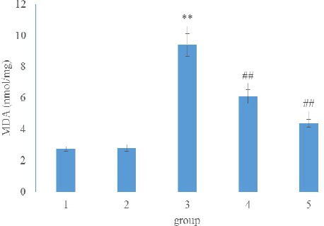
As shown in Fig. 1, induction of renal I/R in rats significantly increased kidney MDA level as compared to the values of the sham control group (p < 0.01). By contrast, rats treated with SE (200, 400 mg/kg) for 25 days prior to renal I/R showed significant reduction in the kidney MDA levels (p < 0.01) as compared to renal I/R control. There isn’t statistical difference (p>0.05) between group 1 and group 2.
Figure 1.

Effect of SE on kidney MDA level in kidney I/R rats
Fig 2 showed kidney GSH, SOD, CAT, GPx levels in renal I/R rats. A significant decreased kidney GSH, SOD, CAT, GPx levels in renal I/R rats were observed as compared to sham control group. However, administration of SE (200, 400 mg/kg) for 25 days prior to renal I/R significantly increased kidney GSH, SOD, CAT, GPx levels (p < 0.01) as compared to I/R group. SE pre-treatment didn’t cause significant effect on kidney GSH, SOD, CAT, GPx levels in sham rats.
Figure 2.

Effect of SE on kidney GSH (a), SOD (b), CAT (c) and GSH-Px (d) levels in kidney I/R rats
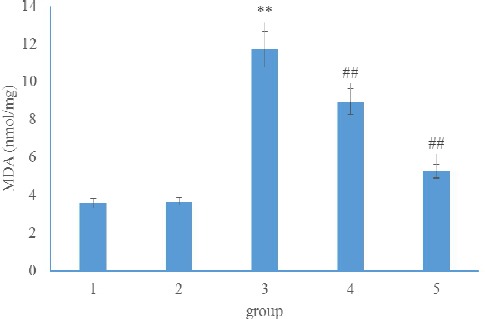
Fig 3 and 4 showed brain hippocampus MDA, GSH, SOD, CAT, GPx levels in renal I/R rats. A significant increased brain hippocampus MDA and decreased brain hippocampus GSH, SOD, CAT, GPx levels in renal I/R rats were observed as compared to sham control group. However, administration of SE (200, 400 mg/kg) for 25 days prior to renal I/R significantly decreased brain hippocampus MDA and increased brain hippocampus GSH, SOD, CAT, GPx levels (p < 0.01) as compared to I/R group. No significant change between group 1 and group 2 was detected.
Figure 3.

Effect of SE on brain hippocampus GSH level in kidney I/R rats
Figure 4.

Effect of SE on brain hippocampus SOD, CAT and GSH-Px levels in kidney I/R rats
Discussion
Kidney I/R may cause severe destructive effects to organ tissues. The some mechanisms of kidney I/R induced organ damages include microvascular dysfunctions, metabolism disorder of vaso-active substances, oxidative injury, increased endothelial cells damage, and local activation reaction of inflammation. These processes are very harmful and destroy functional and structural integrity of the organs including kidney after kidney I/R (Quoilin et al, 2014). Kidney I/R is the main pathogenesis of ischemic acute renal failure, which has high morbidity and mortality. It often accompany a series of coherent cellular events, including reactive oxygen species (ROS) release, apoptosis, necrosis, infiltration of inflammatory cells and the release of the active medium, which leads to tissue damage.
Malondialdehyde (MDA), the final product of lipid oxidation, affects the mitochondrial respiratory chain complex and the key enzyme activities in the mitochondria. In body, free radicals stimulate the lipid peroxidation reaction and cause the final oxidation product malondialdehyde which induced protein, nucleic acid and other large biological molecules cross-linking polymerization and is toxic to cells. Through analyzing MDA level, the degree of membrane lipid peroxidation can be evaluated, and the damage degree of membrane system and the stress resistance of plants can also be determined indirectly. GSH is a kind of three peptides containing the gamma amide bond and the thiol group, which is composed of glutamic acid, cysteine and glycine. It exists in almost every cell of the body. Glutathione can help maintain normal immune system function, and has the function of antioxidation and integrated detoxification. Its main physiological function is to be able to remove free radicals in the human body. As an important antioxidant in the body, GSH protects the thiol groups of many proteins and enzymes. The structure of GSH contains a reactive thiol group -SH, which is easy to be oxidized, which makes it become the main free radical scavenger in vivo. Catalase exists in the peroxide of the cells, is a marker enzyme of the enzyme, which accounts for 40% of the total amount of the enzyme. It catalyzes the decomposition of hydrogen peroxide into oxygen and water. Catalase exists in all tissues of all known animals, especially in the liver. Catalase is one of the key enzymes in the biological defense system, it free cells from H2O2’s poison. GSH-Px is a powerful reducing agent and can eliminate the damage caused by free radicals in the cells. When the free radical scavenging agent in the tissue is reduced, the active oxygen produced by the organism cannot be effectively removed, The unsaturated double bond of poly unsaturated fatty acid and fatty acid in cell membrane phospholipids is vulnerable to oxygen free radical attack. Results lead to lipid peroxidation reaction, provoke the chain reaction of free radicals and proliferation which products MDA. “MDA further decreased the membrane fluidity, increased the permeability, mitochondrial swelling, resulted in lysosomal damage and the release of lysosomal enzymes, aggravate tissue cell damage, speed up cell death (Zhang et al, 2014; Mummedy et al, 2014).”
The main objective of our current work was to investigate the protective role of SE against kidney I/R induced renal and brain oxidative injury. In the current study, kidney I/R induced severe oxidative injury (like increased MDA, decreased GSH, SOD, CAT and GPx) in rats. Free radicals are excessively produced during kidney I/R. At the same time, this can decrease the function of the body’s free radical defense system, such as SOD and other antioxidant compounds, which cannot effectively remove free radicals. This results in free radical accumulation. Free radicals attack the cell membrane which is rich in phospholipids, so that the membrane structure is damaged, and cells swell. SE treatment significantly decreased kidney MDA, increased GSH, SOD, CAT and GPx levels in kidney I/R rats. This indicates that SE can alleviate renal oxidative injury in kidney I/R rats.
Increase of oxidative stress and deterioration of systemic reactions cause remote organ dysfunction caused by I/R. Kidney I/R injury lead to oxidative stress or damage in remote organs, caused a decrease in the activities of antioxidant enzymes (superoxide dismutase, catalase and the level of glutathione). In the current study it has been also observed that there was an increase in the levels of MDA and a decrease in the levels of GSH, SOD, CAT and GPx levels in brain hippocampus tissue of renal I/R rats. These findings suggest that oxidative stress response develops after renal I/RI plays an important role in the remote organ injury. By contrast, SE pre-treatment markedly decreased brain hippocampus MDA, increased GSH, SOD, CAT and GPx levels in kidney I/R rats. This indicates that SE can alleviate brain oxidative injury in kidney I/R rats.
Conclusion
Kidney I/R may lead to remote organ brain oxidative injury and cause brain nerve dysfunction. SE treatment may alleviate brain hippocampus oxidative damage induced by kidney I/R. This is useful for brain nerve function.
References
- 1.Kizilbash S.J, Kashtan C.E, Chavers B.M, Cao Q, Smith A.R. Acute Kidney Injury and the Risk of Mortality in Children Undergoing Hematopoietic Stem Cell Transplantation. Biology of Blood and Marrow Transplantation. 2016;22(7):1264–1270. doi: 10.1016/j.bbmt.2016.03.014. [DOI] [PMC free article] [PubMed] [Google Scholar]
- 2.Mummedy S, Wan N, Wan A. Restoration of glutamine synthetase activity, nitric oxide levels and amelioration of oxidative stress by propolis in kainic acid mediated excitotoxicity. African Journal of Traditional Complementary and Alternative Medicines. 2014;11(2):458–463. doi: 10.4314/ajtcam.v11i2.33. [DOI] [PMC free article] [PubMed] [Google Scholar]
- 3.Quoilin C, Mouithys-Mickalad A, Lécart S, Fontaine-Aupart M.-P, Hoebeke M. Evidence of oxidative stress and mitochondrial respiratory chain dysfunction in an in vitro model of sepsis-induced kidney injury. BiochimicaetBiophysicaActa (BBA) - Bioenergetics. 2014;1837(10):1790–1800. doi: 10.1016/j.bbabio.2014.07.005. [DOI] [PubMed] [Google Scholar]
- 4.Woodman R, Lockette W. Alpha-methyltyrosine inhibits formation of reactive oxygen species and diminishes apoptosis in PC12 cells. Brain Research. 2009;1296:137–147. doi: 10.1016/j.brainres.2009.07.084. [DOI] [PubMed] [Google Scholar]
- 5.Zhang D.Q, Duan L.Z, Zhou N. Market survey on traditional medicine of the third month fair in dali prefecture in yunnan Province, Southwest china. African Journal of Traditional Complementary and Alternative Medicines. 2014;11(2):377–401. doi: 10.4314/ajtcam.v11i2.25. [DOI] [PMC free article] [PubMed] [Google Scholar]


