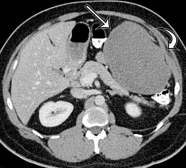
Figure 18a.

Desmoid tumor in a 50-year-old man with vague pain in the left upper quadrant. (a) Axial contrast-enhanced CT image shows a well-circumscribed mass (straight arrow), which has low attenuation compared with the adherent transversus abdominis muscle (curved arrow). (b, c) Axial T2-weighted (b) and contrast-enhanced T1-weighted fat-suppressed (c) MR images show heterogeneous intrinsic high T2 signal intensity within the mass (arrow on b), with avid enhancement of the mass (arrow on c).
