Abstract
Background
Traveling wave MR uses the far fields in signal excitation and reception, therefore its acquisition efficiency is low in contrast to the conventional near field magnetic resonance (MR). Here we show a simple and efficient method based on the local resonator to improving sensitivity of traveling wave MR technique. The proposed method utilizes a standalone or free local resonator to amplify the radio frequency magnetic fields in the interested target. The resonators have no wire connections to the MR system and thus can be conveniently placed to any place around imaging simples.
Methods
A rectangular loop L/C resonator to be used as the free local resonator was tuned to the proton Larmor frequency at 7T. Traveling wave MR experiments with and without the wireless free local resonator were performed on a living rat using a 7T whole body MR scanner. The signal-to-noise ratio (SNR) or sensitivity of the images acquired was compared and evaluated.
Results
In vivo 7T imaging results show that traveling wave MR with a wireless free local resonator placed near the head of a living rat achieves at least 10-fold SNR gain over the images acquired on the same rat using conventional traveling wave MR method, i.e. imaging with no free local resonators.
Conclusions
The proposed free local resonator technique is able to enhance the MR sensitivity and acquisition efficiency of traveling wave MR at ultrahigh fields in vivo. This method can be a simple solution to alleviating low sensitivity problem of traveling wave MRI.
Keywords: Ultrahigh field magnetic resonance (MR), magnetic resonance imaging, MR sensitivity, body imaging, microstrip resonator, traveling wave
Introduction
Because of the high operating frequency in ultrahigh field MR imaging, it is technical challenging to design large-size RF coils required for imaging large samples. Traveling wave MR developed recently utilizes the bore of the MR scanner as a waveguide to support the propagation of electromagnetic wave generated by a relatively small transceive RF coil, e.g., a patch antenna (1-6). It has demonstrated its unique capability of imaging large-size samples at ultrahigh fields. This technique alleviates the RF design difficulties in high frequency large-sized coils and simplifies large sample imaging at ultrahigh fields. Nevertheless, unlike the conventional MR method where more sensitive near-field generated by RF coils is used for signal excitation and reception, traveling wave MR technique suffers from its low signal-to-noise ratio (SNR) or sensitivity (6-8), a critical measure in MR performance. The low sensitivity problem dramatically limits the imaging applications of the traveling wave MR. Here we show a simple and efficient method to enhancing imaging sensitivity of traveling wave MR on a whole body 7T MR system. The method is based on the utilization of wireless-type free local resonators (7) that are able to amplify the radio frequency magnetic fields, i.e., B1 fields, making the MR signal excitation and reception more efficient. The local resonators tuned at the Larmor frequency of proton at 7T are free of physically connection to the MR system and are placed in the region of interest near the imaging sample. The method is experimentally demonstrated and investigated using a research-dedicated 7-Tesla whole body MR system. In vivo traveling wave MR imaging in rats is performed with and without the free local resonators for performance comparison and validation.
Methods
Traveling wave excitation and reception using microstrip resonators
To simplify the experiment, a straight-type microstrip resonator (9-22), rather than a patch antenna conventionally used in traveling wave MR imaging, was constructed to transmit and receive MR signals (23). The straight-type microstrip resonator with capacitive termination was designed and constructed to operate at 298.2 MHz, the proton Larmor frequency of our 7T whole body MR system. To help improve the transmit and receive efficiency, low loss material, Teflon, with a relatively low permittivity of 2.1 and a thickness of 0.6 cm was used for the substrate of the microstrip resonator. All the conductors in the microstrip resonator were made from back-adhesive copper tapes. The length of the microstrip resonator was 18 cm. The width of the strip conductor was 1.2 cm. The microstrip resonator was connected through an impedance matching and frequency tuning network and a T/R switch to the MR system. A sketch describing the microstrip resonator and the relative position to the magnet for the traveling-wave imaging experiment is shown in Figure 1, left inset.
Figure 1.
Schematic of the microstrip resonator as a transceive antenna, and the experiment setup of traveling wave MRI using a free local resonator for SNR enhancement on a whole body 7T MR imaging system.
Wireless free local resonator for SNR enhancement
A passive rectangular LC loop resonator with dimensions of 3.8 cm × 5 cm was constructed to resonate at 298.2 MHz. This LC loop resonator, a wireless resonator in this study, was a regular lumped element circuit. The resonant frequency of the LC loop resonator in the loaded case was determined by transmission coefficient (S21) measurements taken on a network analyzer with sniffers. This passive LC resonator does not need to have physical connections to the MR system and can be freely place to any area of interest of a MR sample for traveling wave MR imaging acquisition.
MR imaging experiment
In traveling wave MR imaging experiment, the microstrip resonator was placed in one end of the magnet bore, i.e., the patient end in this case, approximately 80 cm away from the imaging sample (or the center of the magnet). This capacitive terminated microstrip resonator was connected to the 7T MR system and served as a source generating traveling wave in the magnetic bore and also the receiver of the MR signals. The passive LC loop resonator, which had no physical connections to the MR system, was positioned near the head of a living healthy rat for amplifying the local B1 field and MR signal intensity. The experiment setup is illustrated in Figure 1.
Before the imaging experiment, the resonant frequency and input impedance of the microstrip resonator and the resonant frequency of the LC loop resonator in this experiment setup were re-measured on the network analyzer, and adjusted when needed. The animal experimental protocols were conducted under the guidelines of the National Institutes of Health and the Institutional Animal Care and Use Committee of the University of California San Francisco.
All the imaging experiments were performed on a whole body 7T MR scanner (GE Healthcare, Waukesha, WI, USA). Its magnetic bore size is large enough to support a cut-off frequency of 286 MHz, which enables traveling wave MR experiment at 7T. To simplify the experiment, no extra RF shielding was used in the magnet bore which is believed to be able to form a more efficient waveguide. In vivo traveling wave MR imaging in healthy rats was performed with and without the free passive local loop resonator. The imaging results were compared in terms of SNR and field distribution. Gradient echo sequence was used in all imaging acquisitions. The imaging parameters used in this study for imaging acquisitions with and without free passive local resonators were TR/TE =250 ms/3.3 ms, matrix size =128×128, slice thickness =3 mm (with free passive local resonator), field-of-view (FOV) = 8 cm × 8 cm (with free passive local resonator). To gain a reasonable SNR in imaging without free passive local resonator for performance comparison, another set of acquisition parameters were used in imaging acquisitions without free passive local resonator, which were TR/TE = 250 ms/3.3 ms, matrix size =128×128, slice thickness =5 mm, field-of-view (FOV) =15 cm × 15 cm.
Results and discussion
Through the scattering parameter S11 measurement taken on the network analyzer, the resonant frequency of the transmit/receive microstrip resonator for MR signal excitation and reception was tuned to 298.2 MHz, and the input impedance was matched to 50 Ohm with a S11 of −35dB or better. The resonance mode used for this imaging experiment was the primary resonance which has the lowest frequency. The high order harmonics of the microstrip resonator might be able to be used for the traveling wave MR imaging. The free passive loop resonator loaded with the rat head also resonated at 298.2 MHz. Generally, in MR imaging experiments that involve two resonators or RF coils resonating at the same frequency, mutual coupling between the two resonators is a critical parameter affecting the imaging performance. Minimized mutual coupling has to be maintained to ensure the resonators resonate at the correct frequency and well behave in RF magnetic field distribution. Due to the far field effect, as expected, mutual coupling between the two resonators (i.e., the microstrip resonator and free passive local loop resonator), both operating at 298.2 MHz, was not observed on the S11 measurement, making this two-resonator setup possible for imaging acquisition.
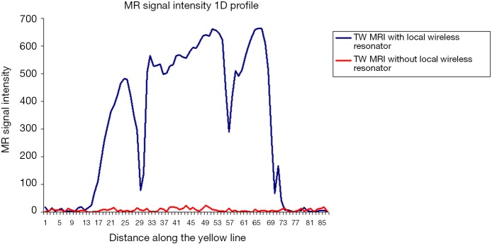
In vivo rat head images acquired using traveling wave MRI with and without free passive local resonators at 7T are shown in Figure 2. The measured highest achievable SNR was 195 for imaging with free passive local resonator (Figure 2, left inset) and 2 for imaging without free passive local resonator (Figure 2, middle inset). In the imaging acquisition with favorable acquisition parameters (i.e., thicker slice of 5 mm and larger FOV of 15 cm2), SNR of 20 was obtained in the regular traveling wave imaging without free passive local resonator. These results indicate a nearly 10-fold SNR gain of the proposed method using free passive local resonators over traditional traveling wave MR. Figure 3 shows the 1-D profiles of MR signal intensity along a line across the same slice of the rat head images acquired with and without free passive local resonators using the same acquisition parameters. The imaging method with free local resonator demonstrates substantial signal intensity gain over that of conventional traveling wave imaging method without free local resonator.
Figure 2.
In vivo rat head traveling-wave (TW) images acquired at 7T with and without the free local resonator. Gradient recalled echo (GRE) sequence was used in all imaging acquisitions. Imaging parameters were: TR/TE =250 ms/3.3 ms, slice thickness =3 mm, matrix size =128×128, field-of-view (FOV) = 8 cm × 8 cm, acquisition time 5.3 min. Image in right inset had slightly different parameters of FOV = 15 cm × 15 cm and slice thickness =5 mm. Left inset shows TW imaging with the free local resonator. Middle inset and right inset are TW images acquired without the free local resonator from the same rat. The measured highest achievable SNR is 195 (left inset), 2 (middle inset), and 20 (right inset), respectively. These image results demonstrate that the proposed free local resonator method can substantially boost SNR of traveling-wave MR.
Figure 3.
1-D profiles of MR signal intensity along the yellow dash line in the images acquired using traveling wave technique with and without free local resonators.
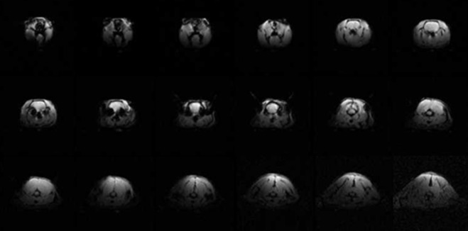
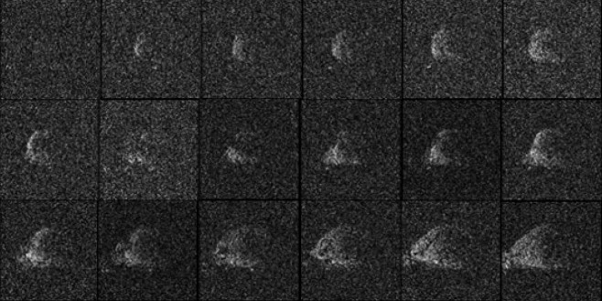
To evaluate the imaging distribution and SNR performance in the area covered by the free local resonator, multi-slice imaging in transverse orientation was acquired using conventional traveling wave MR imaging (i.e., TW MR with no free local resonator) and the proposed traveling wave MR imaging with free local resonator. The acquisition parameters for both experiments were the same. The multi-slice imaging results shown in Figures 4 and 5 indicate sufficient image coverage and typical surface coil behavior, substantiating the amplification capability in the entire area covered by the free local resonator.
Figure 4.
In vivo multiple-slice rat head traveling wave images acquired with free local wireless resonator. As described in Methods, the free local resonator used in this work was a rectangular loop surface coil. These images show the expected behavior of a surface coil.
Figure 5.
In vivo multiple-slice rat head traveling wave images acquired with no free local wireless resonators, showing much less sensitivity over the results shown in Figure 4.
In this work, only one free local resonator was utilized. If more free local resonators are applied to the imaging sample, it is possible to implement multi-point imaging experiments covering different organs or tissues in whole body ultrahigh field MR, as long as the organs or tissues interested are in the linear range of the gradient coils used in the imaging experiments. On the type of free local resonator, it might be also possible to use a multi-element array, instead of a single local resonator, to further enhance SNR for the area of interest in the traveling wave MRI.
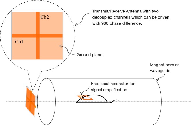
As described in Methods and Materials, signal excitation and reception in this traveling wave MR imaging experiment were achieved by using a single straight-type microstrip resonator. In the RF field generation and signal pick-up of traveling wave MR, more microstrip resonators can be employed to form a multichannel transceive array. It can be another way to further enhancement of SNR in traveling wave MRI. Figure 6 demonstrates the simplest case of multichannel transceive array with only two array elements. If the two transceive elements are well decoupled electromagnetically, approximately 40% SNR gain can be expected over the traveling wave MRI with only one transceive element as demonstrated in this work. Unlike the element layout shown in Figure 6 where the two elements were placed orthogonally and thus intrinsically decoupled, when more resonant elements are involved, electromagnetic coupling among elements may become an issue. This coupling issue can be possibly addressed by using the magnetic wall or ICE decoupling technology reported in literatures (24-35).
Figure 6.
Experiment setup and schematic of a microstrip transceive antenna with two resonant elements or channels for further improvement of detection sensitivity and transmit efficiency in traveling wave MR imaging. If the two transceive channels are sufficiently decoupled electromagnetically, additional ~40% SNR gain can be expected.
Conclusions
A method of enhancing signal-to-noise ratio or sensitivity of traveling wave MR imaging is proposed and experimentally demonstrated on a whole body ultrahigh field 7T MR system. By positioning a free local resonator, i.e. a standalone wireless resonator, near an imaging sample, SNR of traveling wave MR can be amplified significantly in the area covered by the free local resonator. This method provides a simple and practical approach to increasing SNR in traveling wave MR imaging.
Acknowledgements
Funding: This work was funded by the National Institutes of Health under Award Number EB008699, EB020283 and P41EB013598, and a Cohn Fund Award.
Disclaimer: The content is solely the responsibility of the author and does not necessarily represent the official views of the National Institutes of Health or Cohn Fund.
Footnotes
Conflicts of Interest: The author has no conflicts of interest to declare.
References
- 1.Brunner DO, De Zanche N, Frohlich J, Paska J, Pruessmann KP. Travelling-wave nuclear magnetic resonance. Nature 2009; 457:994-8. 10.1038/nature07752 [DOI] [PubMed] [Google Scholar]
- 2.Webb AG, Collins CM, Versluis MJ, Kan HE, Smith NB. MRI and localized proton spectroscopy in human leg muscle at 7 Tesla using longitudinal traveling waves. Magn Reson Med 2010;63:297-302. 10.1002/mrm.22262 [DOI] [PMC free article] [PubMed] [Google Scholar]
- 3.Zhang B, Wiggins G, Duan Q, Lattanzi R, Sodickson DK. Whole-body travelling-wave imaging at 7 tesla: simulations and early in-vivo experience. In: Proceedings of the 17th Scientific Meeting and Exhibition of ISMRM, Honolulu, HI, 2009:499. [Google Scholar]
- 4.Pang Y, Wang C, Vigneron D, Zhang X. Parallel traveling-wave MRI: antenna array approach to traveling-wave MRI for parallel transmission and acquisition. In: Proceedings of the 18th Scientific Meeting and Exhibition of ISMRM, Stockholm, 2010:3794. [Google Scholar]
- 5.Pang Y, Vigneron D, Zhang X. Parallel traveling-wave MRI: A feasibility study. Magn Reson Med 2012;67:965-78. 10.1002/mrm.23073 [DOI] [PMC free article] [PubMed] [Google Scholar]
- 6.Kroeze H, van de Bank BL, Visser F, Lagendijk JJ, Luijten P, Klomp DW, van den Berg CA. Carotids imaging at 7 Tesla using traveling wave excitation and local receiver coils. ISMRM 2009:1320.
- 7.Zhang X, Pang Y, Vigneron D. SNR Enhancement by Free Local Resonators for Traveling Wave MRI. In: Proceedings of the 22nd Scientific Meeting and Exhibition of ISMRM, Milan, 2014:1357. [Google Scholar]
- 8.Yan X, Zhang X. Theoretical and simulation verification of SNR enhancement in traveling wave MRI using free local resonators. In: Proceedings of the 24th Scientific Meeting and Exhibition of ISMRM, Singapore, 2016:3535. [Google Scholar]
- 9.Zhang X, Ugurbil K, Chen W. Microstrip RF surface coil design for extremely high-field MRI and spectroscopy. Magn Reson Med 2001;46:443-50. 10.1002/mrm.1212 [DOI] [PubMed] [Google Scholar]
- 10.Zhang X, Ugurbil K, Chen W. A microstrip transmission line volume coil for human head MR imaging at 4T. J Magn Reson 2003;161:242-51. 10.1016/S1090-7807(03)00004-1 [DOI] [PubMed] [Google Scholar]
- 11.Zhang X, Ugurbil K, Sainati R, Chen W. An inverted-microstrip resonator for human head proton MR imaging at 7 tesla. IEEE Trans Biomed Eng 2005;52:495-504. 10.1109/TBME.2004.842968 [DOI] [PubMed] [Google Scholar]
- 12.Zhang X, Zhu X, Chen W. Higher-order harmonic transmission-line RF coil design for MR applications. Magn Reson Med 2005;53:1234-9. 10.1002/mrm.20462 [DOI] [PubMed] [Google Scholar]
- 13.Adriany G, Van de Moortele PF, Wiesinger F, Moeller S, Strupp J, Andersen P, Snyder C, Zhang X, Chen W, Pruessmann K, Boesiger P, Vaughan T, Ugurbil K. Transmit and receive transmission line arrays for 7 Tesla parallel imaging. Magn Reson Med 2005;53:434-45. 10.1002/mrm.20321 [DOI] [PubMed] [Google Scholar]
- 14.Wu B, Wang C, Krug R, Kelley DA, Xu D, Pang Y, Banerjee S, Vigneron D, Nelson S, Majumdar S, Zhang X. 7T human spine imaging arrays with adjustable inductive decoupling. IEEE Trans Biomed Eng 2010;57:397-403. 10.1109/TBME.2009.2030170 [DOI] [PMC free article] [PubMed] [Google Scholar]
- 15.Wu B, Wang C, Kelley DA, Xu D, Vigneron D, Nelson S, Zhang X. Shielded microstrip array for 7T human MR imaging. IEEE Trans Med Imaging 2010; 29: 179-84. 10.1109/TMI.2009.2033597 [DOI] [PMC free article] [PubMed] [Google Scholar]
- 16.Wu B, Wang C, Lu J, Pang Y, Nelson S, Vigneron D, Zhang X. Multi-channel Microstrip Transceiver Arrays using Harmonics for High Field MR Imaging in Humans. IEEE Trans Med Imaging 2012;31:183-91. 10.1109/TMI.2011.2166273 [DOI] [PMC free article] [PubMed] [Google Scholar]
- 17.Zhang X, Ugurbil K, Chen W. Method and apparatus for magnetic resonance imaging and spectroscopy using microstrip transmission line coils. U.S. Patent 7023209, University of Minnesota, 2006.
- 18.Jasinski K, Mlynarczyk A, Latta P, Volotovskyy V, Weglarz W, Tomanek B. A volume microstrip RF coil for MRI microscopy. Magn Reson Imaging 2012;30: 70-7. 10.1016/j.mri.2011.07.010 [DOI] [PubMed] [Google Scholar]
- 19.Klomp D, Raaijmakers A, Arteaga de Castro C, Boer V, Kroeze H, Luijten P, Lagendijk J, van den Berg C. Uniform prostate imaging and spectroscopy at 7 T: comparison between a microstrip array and an endorectal coil. NMR Biomed 2011;24:358-65. [DOI] [PubMed] [Google Scholar]
- 20.Brunner D, Zanche N, Froehlich J, Baumann D, Pruessmann K. A symmetrically fed microstrip coil array for 7T. In: Proceedings of the 15th Scientific Meeting and Exhibition of ISMRM, Honolulu, HI, 2007:448. [Google Scholar]
- 21.Yan X, Wei L, Xue R, Zhang X. Closely-spaced double-row microstrip RF arrays for parallel MR imaging at ultrahigh fields. Applied Magnetic Resonance 2015;46:1239-48. 10.1007/s00723-015-0712-1 [DOI] [PMC free article] [PubMed] [Google Scholar]
- 22.Rutledge O, Kwak T, Cao P, Zhang X. Design and test of a double-nuclear RF coil for 1H MRI and 13C MRSI at 7T. J Magn Reson 2016;267:15-21. 10.1016/j.jmr.2016.04.001 [DOI] [PMC free article] [PubMed] [Google Scholar]
- 23.Vareth M, Flynn A, Bian W, Li Y, Vigneron D, Nelson S, Zhang X. Accelerated Parallel Traveling Wave MR and Compressed Sensing Using a 2-Channel Transceiver Array. In: Proceedings of the 21st Scientific Meeting and Exhibition of ISMRM, Salt Lake City, 2013:122. [Google Scholar]
- 24.Xie Z, Zhang X. A novel decoupling technique for non-overlapped microstrip array coil at 7T MR imaging. In: Proceedings of the 16th Scientific Meeting and Exhibition of ISMRM, Toronto, 2008:1068. [Google Scholar]
- 25.Xie Z, Zhang X. An 8-channel microstrip array coil for mouse parallel MR imaging at 7T by using magnetic wall decoupling technique. In: Proceedings of the 16th Scientific Meeting and Exhibition of ISMRM, Toronto, 2973. [Google Scholar]
- 26.Xie Z, Zhang X. An 8-channel non-overlapped spinal cord array coil for 7T MR imaging. In: Proceedings of the 16th Scientific Meeting and Exhibition of ISMRM, Toronto, p2974. [Google Scholar]
- 27.Xie Z, Zhang X. An eigenvalue/eigenvector analysis of decoupling methods and its application at 7T MR imaging. In: Proceedings of the 16th Scientific Meeting and Exhibition of ISMRM, Toronto, p2972. [Google Scholar]
- 28.Li Y, Xie Z, Pang Y, Vigneron D, Zhang X. ICE Decoupling Technique for RF Coil Array Designs. Medical Physics 2011;38:4086- 93. 10.1118/1.3598112 [DOI] [PMC free article] [PubMed] [Google Scholar]
- 29.Yan X, Zhang X, Feng B, Ma C, Wei L, Xue R. 7T transmit/receive arrays using ICE decoupling for human head MR imaging. IEEE Trans Med Imaging 2014;33:1781-7. 10.1109/TMI.2014.2313879 [DOI] [PubMed] [Google Scholar]
- 30.Yan X, Zhang X, Wei L, Xue R. Magnetic wall decoupling method for monopole coil array in ultrahigh field MRI: a feasibility test. Quant Imaging Med Surg 2014;4:79-86. [DOI] [PMC free article] [PubMed] [Google Scholar]
- 31.Yan X, Zhang X, Wei L, Xue R. Hybrid monopole/loop coil array for human head MR imaging at 7T. Applied Magnetic Resonance 2015;46:541-50. 10.1007/s00723-015-0656-5 [DOI] [PMC free article] [PubMed] [Google Scholar]
- 32.Yan X, Zhang X. Decoupling and matching network for monopole antenna arrays in ultrahigh field MRI. Quant Imaging Med Surg 2015;5:546-51. [DOI] [PMC free article] [PubMed] [Google Scholar]
- 33.Yan X, Zhang X, Wei L, Xue R. Theoretical analysis of magnetic wall decoupling method for radiative antenna arrays in ultrahigh magnetic field MRI. Concept in Magn Reson Part B 2015;45B:183-90. 10.1002/cmr.b.21312 [DOI] [Google Scholar]
- 34.Yan X, Cao Z, Zhang X. Simulation verification of SNR and parallel imaging improvements by ICE-decoupled loop array in MRI. Appl Magn Reson 2016;47: 395-403. 10.1007/s00723-016-0764-x [DOI] [PMC free article] [PubMed] [Google Scholar]
- 35.Yan X, Zhang X, Wei L, Xue R. Eight-channel monopole array using ICE decoupling for human head MR imaging at 7 T. Appl Magn Reson 2016;47:527-38. 10.1007/s00723-016-0775-7 [DOI] [PMC free article] [PubMed] [Google Scholar]


