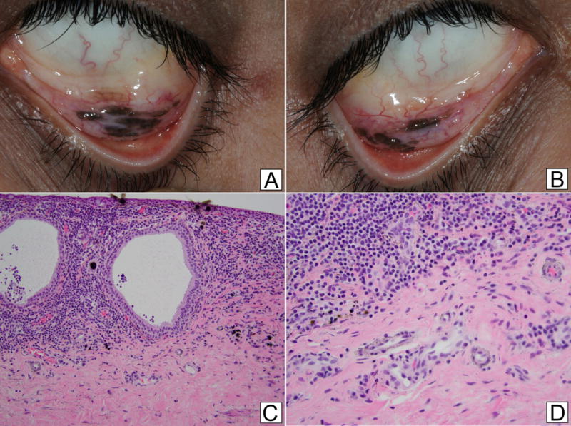
Figure 1. Photographs of Inferior Fornices and Histopathology.

a.b. External photographs of the left (a) and right (b) inferior fornices showing elevated pigmented lesions with surrounding hypervascularity
c. A dense lichenoid lymphocytic infiltrate surrounds cystic invaginations of the mucosa, consistent with reactive lymphoid hyperplasia. Within surrounding submucosa, small rounded black deposits are noted (Hematoxylin and Eosin, 200×)
d. Higher power view shows fine to coarse black granules of silver embedded within the submucosa. A few rounded larger elements are also noted. (Hematoxylin and Eosin, 400×)
