Abstract
Introduction: The aim of this study was to evaluate the effect of various concentrations of NaOCl on shear bond strength of composite resin to dentin of primary teeth, prepared with laser and bur.
Methods: In this in vitro study, 48 primary molars were sectioned at mesiodistal direction and were randomly divided into 6 groups; G1: bur, G2: bur + NaOCl 2.5%, G3: bur + NaOCl 5.25%, G4: laser, G5: laser + NaOCl 2.5%, G6: laser + NaOCl 5.25%. One-Step Plus adhesive was applied after phosphoric acid gel and NaOCl over the dentin surfaces for all groups, and composite resin cylinders were bonded to the samples. After thermocycling, shear bond strengths of composite resin to dentin were measured and statistical analyses were done by means of t test and analysis of variance (ANOVA).
Results: The mean shear bond strength showed no significant difference between the groups prepared with bur (13.82 ± 3.49) and laser (14.18 ± 3.65) (P > 0.05). The mean difference of shear bond strength between three groups G1, G2 and G3 and between G4, G5 and G6 were not statistically significant (P > 0.05). Scanning electron microscopy (SEM) figures showed an irregular surface in laser groups and fairly complete removal of smear layer from the orifices of the dentinal tubules, in the group in which NaOCl was used.
Conclusion: The application of different concentrations of NaOCl does not significantly improve the bond strength in dentin surfaces prepared with laser or bur
Keywords: Er:YAG Lasers, Primary teeth, Electron scanning microscopy, Hypochlorite, Sodium, Shear bond strength
Introduction
Recently, laser is being increasingly used in dentistry.1 Among all types of lasers, Erbium-Doped Yttrium Aluminum Garnet laser (Er:YAG( is commonly used in dental hard tissues2 and its ability for removing enamel and dentin is comparable to conventional methods.1
Cavity preparation with Er:YAG laser has advantages such as removing dental hard tissues with minimal pulp injury,3,4 creating a rough and irregular surface,2 less side effects due to severe heat produced by conventional methods e.g. cracking, melting or charring in the remaining tissue,3,4 lowering the pain and vibration during preparation, which increases the patient’s comfort and is a key factor in pediatric dentistry.5,6
Despite appropriate morphologic characteristics of surfaces prepared by this laser for bonding, some studies have reported lower bond strength compared to conventional methods. A probable reason may be the denaturation and alteration in collagen network, which may prevent the penetration of adhesive into dentin tubules7-9 and therefore lead to the penetration of bacteria and oral fluids, hydrolysis and destruction of collagen fibers, which may decrease bond strength and increase microleakage.10,11 The presence of subsurface fissures due to heat during irradiation might also be a destructive factor for bonding process.7-9
After etching, collagen network of demineralized dentin creates a smooth surface with low surface energy.12 If collagen is dissolved in this surface, penetrability increases and resin will penetrate more efficiently, while a layer of minerals remains on the relatively demineralized dentin. This probably creates a more reliable bond directly to the dentin hydroxy apatite crystals from which collagen fibers have been removed.13
Sodium hypochlorite is a non-specific proteolytic agent which can remove organic material, carbonate and magnesium ions and may change the composition of dentin.14,15 Some studies have proposed this agent to be used after dentin etching, in order to affect the remaining collagen layer, thus increase the adhesion.16-18
It seems that the efficacy of this method is related to the type of solvent solution contained in the bonding system.19 In a study it was shown that when any variable such as hypochlorite is added to the conventional bonding process, the acetone-based adhesives become less sensitive than water-based adhesives.20
Considering the increase in the use of adhesives and laser technology in pediatric dentistry, the evaluation of the interaction between adhesive systems and lased primary dentin is beneficial.21 Besides, there is a considerable difference between primary and permanent teeth regarding structure and composition, so the results of the studies on permanent teeth cannot be generalized to primary teeth,22 and further researches are required to assess the factors affecting the bond strength using Er:YAG laser in primary teeth.
The aim of this study was to assess the effect of laser irradiation and different concentrations of sodium hypochlorite on shear bond strength between composite resin and dentin of primary teeth.
Methods
A total of 48 extracted primary human molars with sound buccal and lingual surfaces were used in this study. For disinfection, the teeth were immersed in 0.5% chloramine T solution for 7 days.23 Prior to use, the specimens were polished and cleansed by water/pumice slurry with prophylaxis rubber cups at low speed. In case there was any crack or other dental abnormalities in buccal or lingual surfaces, the specimen was excluded from the study.
To prepare the specimens, at first the roots were sectioned 2 mm below the cemento-enamel junction (CEJ) and then the crown was divided into buccal and lingual parts in mesiodistal direction, vertical to the longitudinal axis by a diamond bur (D&Z, Germany). Overall, 96 specimens were assessed.
All specimens (except for 12, which should have been assessed by electron scanning microscopy [SEM]) were mounted in an acrylic self-cure resin (Pattern resin, GC, Tokyo, Japan). The specimens were randomly divided into two groups (n = 42), for surface preparation by Er:YAG laser or bur. Each group was then divided into 3 subgroups (n = 14), according to the type of sodium hypochlorite: without sodium hypochlorite, with sodium hypochlorite 2.5% and 5.25%. Two specimens for SEM analysis were added to each group (n = 16).
Dentinal surfaces of the groups 1,2, and 3 were prepared using the diamond bur (Tizkavan, Iran) on a high-speed hand-piece (NSK, Japan) with water spray cooling; in groups 4, 5, and 6 the surfaces were prepared by Er:YAG laser (Fotona, Fidelis Plus III, Slovenia), with 2.94 µm wavelength, 200 mJ energy, pulse repetition rate of 10 Hz, and micro-short pulse (MSP) along with air-water cooling of 7 mL/min, in non-contact mode from 17 mm distance with a RO2-C-919 hand-piece.24,25
In order to expose the dentinal surface, 1mm of enamel was removed by a diamond bur and Er:YAG laser in the first to third groups and fourth to sixth groups, respectively. The dentinal surface was demarcated by attaching a piece of insulating tape with a central hole of 1.5 mm diameter, in order to limit the border of irradiation and dentinal bonding.
All specimens were etched with a 35% phosphoric acid gel (Bisco, Schamburg, USA) for 15 seconds, rinsed and dried according to the manufacturers’ instructions. Then 2 concentrations of sodium hypochlorite were applied on the dentinal surface for 30 seconds: 2.5% in second and fifth groups, and 5.25% in third and sixth groups. The specimens were then washed for 30 seconds and were gently dried.26
In the next step, One-Step Plus bonding (Bisco, Schamburg, USA), an acetone-based adhesive was applied on the dentinal surface of almost all specimens (except for those selected to be analyzed by SEM) according to the manufacturer’s instructions, and all specimens were cured for 30 seconds. AELITE composite (Bisco, Schamburg, USA) was bonded to the surface of all specimens by a plastic cylinder (2 mm diameter and height) and then was cured for 40 seconds with a light-cured unit (Arialux, ApadanataK, Iran), at a light intensity of 500 mW/cm2.
The specimens were immersed in distilled water at 37˚C and then thermocycled for1000 cycles from 5 ± 2˚C to 5 5± 2˚C, with 30 seconds dwell time and 15 seconds transfer time (Vafaei, Iran).
Finally, in order to determine the composite bond strength to dentin in all groups, an instron testing machine (Dartec series, HC 10, Strobridge, England) with a cross head speed of 0.5 mm/min parallel to the tooth surface in the interface of resin and tooth was used. The composite bond strength was calculated in MPa by dividing the force applied to the composite at the time of failure to the cross-section of the specimens.
Electron Scanning Microscopy Study
Dentinal specimens (2 samples of each group) were prepared by the same method as other specimens to assess morphologic changes in the surface. Specimens were kept dry in separate containers (in order to assess dentinal surface under field emission -SEM, vacuum is required and specimens should be dry). Specimens were mounted in a specific plate and were gold-coated and then assessed by FE-SEM (Hitachi 54160, Japan); and appropriate pictures with ×1000 and ×5000 magnifications were obtained. The morphologic characteristics of dentinal surface were evaluated for surface roughness, presence of smear layer, appearance of dentinal tubules and presence of melting or crack. Data were analyzed by SPSS (version 17) using Student’s t test, one-way analysis of variance (ANOVA) and two-way ANOVA tests. Shapiro-Wilk test was used to test the normal distribution of the subjects.
Results
The study was performed on 84 primary teeth divided into 2 groups based on the method of surface preparation (laser or bur). Each group was then divided into 3 subgroups (n = 14), regarding the use of sodium hypochlorite: without sodium hypochlorite, with sodium hypochlorite 2.5% and 5.25%.
The mean of shear bond strength to dentin was a little higher in laser group than bur group, although not statistically significant (P = 0.65). Table 1 shows shear bond strength (means and standard deviations) obtained using different concentrations of sodium hypochlorite.
Table 1. Comparison of Shear Bond Strength (Means ± SD, MPa) Regarding Sodium Hypochlorite Use .
| Sodium Hypochlorite Use | Number | Mean ± SD (MPa) |
| No | 28 | 13.47 ± 3.18 |
| Yes (2.5%) | 28 | 14.08 ± 3.76 |
| Yes (5.25%) | 28 | 14.44 ± 3.76 |
One-way analysis of variance (ANOVA), P value = 0.59
The shear bond strength means were slightly higher after using sodium hypochlorite 5.25% without statistical significance (P = 0.78) in the 3 bur-prepared groups. The mean shear bond strength among laser-prepared groups was slightly higher after using sodium hypochlorite 2.5% and 5.25%, but the difference was not statistically significant (P = 0.62). Table 2 compares shear bond strength regarding the preparation method and sodium hypochlorite in all groups.
Table 2. Shear Bond Strength (Means ± SD, MPa) Regarding the Preparation Method and Sodium Hypochlorite Use .
| Preparation Method | Sodium Hypochlorite Use | Number | Strength, Mean ± SD, MPa |
| Bur | No | 14 | 13.56 ± 3.36 |
| 2.5% | 14 | 13.53 ± 3.64 | |
| 5.25% | 14 | 14.36 ± 3.64 | |
| Laser | No | 14 | 13.39 ± 3.1 |
| 2.5% | 14 | 14.63 ± 3.93 | |
| 5.25% | 14 | 14.51 ± 4 |
Results of two-way ANOVA test:
Preparation method: F = 0.2, P = 0.65.
Sodium hypochlorite: F = 0.5, P = 0.6.
Preparation method and sodium hypochlorite: F = 0.23, P = 0.79.
Electron Scanning Microscopy Results
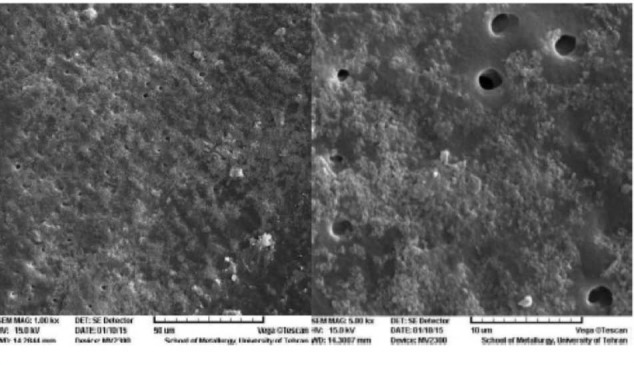
In the SEM figures of the first group, a smooth dentinal surface was observed which was the opening of dentinal tubules in some parts, but in other parts a smear layer with irregular debris covered the tubules (Figure 1).
Figure 1.

SEM Figures of Group 1 With ×1000 (Left) and ×5000 (Right) Magnification.
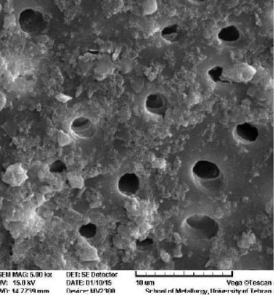
The results of SEM analysis for the second group showed a relatively clean and smooth dentinal surface and lower amount of smear layer compared to the first group. In this group, there were more exposed dentinal tubules with larger diameters and obvious appearance (Figure 2).
Figure 2.

SEM Figures of the Group 2 With ×5000 Magnification.
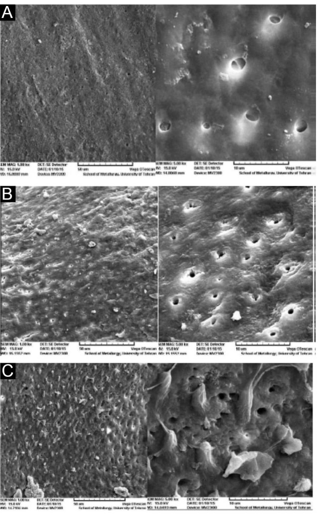
The SEM figures of the third group showed a smooth, clean dentinal surface with no smear layer compared to the first and second groups. The orifice of the dentinal tubules were completely open and obvious (Figure 3A).
Figure 3.

(A) SEM Figures of Group 3 With ×1000 (Left) and ×5000 (Right) Magnification. (B) SEM Figures of Group 4 With ×1000 (Left) and ×5000 (Right) Magnification. (C) SEM Figures of Group 5 With ×1000 (Left) and ×5000 (Right) Magnification.
The forth group SEM figures showed a highly irregular and flaky surface with smaller amount of smear layer compared to the teeth prepared by bur. Inter-tubular dentin was better eliminated by laser than peri-tubular dentin, due to its water rich property. Therefore a cuff-like high density circle could be seen around the dentinal tubules (Figure 3B).
The SEM figures of the fifth group showed a highly irregular and flaky surface with more dentinal tubules without smear layer. In addition, inter-tubular dentin and tubules were clearly observed. This group in comparison with the previous one (without sodium hypochlorite), has a smaller amount of smear layer and better exposure of inter-tubular dentin (Figure 3A).
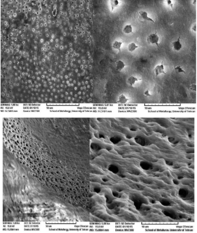
The SEM figures of the sixth group showed a smoother surface with large amount of exposed, open dentinal tubules. In this Figure, no smear layer and debris are seen (Figure 4).
Figure 4.

(A) SEM Figures of Group 6 With ×1000 (Left) and ×5000 (Right) Magnification.
Discussion
The desirable bond of resin to dentin is obtained after complete penetration of the monomer into the demineralized dentin. The incomplete penetration of resin leaves the collagen fibers unsupported, so they will be exposed to oral environment, which may lead to hydrolytic damage and nano-leakage in the hybrid layer.13
Some studies have shown that the application of sodium hypochlorite, as a deproteinizing substance on the dentinal surface, can remove organic materials and a part of smear layer, however it cannot completely open dentinal tubules, and it seems that it does not affect inorganic substances.27 In contrast, some other studies have shown complete removal of collagen fibers by different concentrations of NaOCl used in different applications.28,29
After using NaOCl, a hydrophilic surface is created and wettability of the surface will be increased through a chemical reaction between the primer and the deproteinized dentin, which leads to a better spread of bonding on the surface.30
NaOCl removes collagen fibers from demineralized surface of dentin and makes it similar to enamel.31 After deproteinization, dentin gains a porous structure with surface roughness and irregularity, which creates a micromechanical bond and makes a thicker hybrid layer that accelerates the penetration of acid and bonding agents, and eventually increases bond strength.30
Therefore, some studies has recommended to use NaOCl in concentrations varying from 2.5% to 13% after dentin etching, to remove the remained collagenous layer and to increase the adhesion.16-18
Using sodium hypochlorite at a concentration of 5% removes the organic materials without any change in the mechanical properties of the dentin, compared to higher concentration (10%), which increases micro-leakage.29,32 For all these reasons, 2.5% and 5.25% concentrations of sodium hypochlorite were used in this study.
It seems as well that the efficiency of treatment with NaOCl in improving bond strength depends on the type of solvent solution contained in the adhesive system.19 Some studies have shown higher bond strength with acetone-based adhesive compared with alcohol-based adhesive.17-19 NaOCl is able to promote the rate of acetone diffusion; moreover, it has an ability to substitute water, which improves the interaction between acetone monomer and inter-tubular dentin exposed via sodium hypochlorite.19 Therefore, in the current study an acetone-based adhesive (One-Step Plus) was used.
According to the results of the current study, the mean shear bond strength in the groups prepared by laser was slightly higher than the groups prepared by bur, although the difference was not statistically significant and was in agreement with the results of the previous studies.33,34 This little increase in shear bond strength in groups prepared by laser is probably due to the morphologic properties of the surface after preparation by laser.35
In the present study, the application of sodium hypochlorite with different concentrations did not show a statistically significant increase in mean shear bond strength compared with the other groups without application of sodium hypochlorite, which was consistent with the results of the study conducted by Gowda et al. They reported that using 10% sodium hypochlorite can completely remove the smear layer, and higher concentrations can decrease the application time.36 In the current study, sodium hypochlorite was applied for 30 seconds on the specimens’ surface. The increase in exposure time (60 seconds or more) may lead to a statistically significant difference.
The results of the current study showed that after removing collagen from dentin of the primary teeth by sodium hypochlorite followed by the use of acetone-based adhesive in groups prepared either by laser or bur, the difference in shear bond strength was negligible. The results were in agreement with the study conducted by Correr et al.29 The structural difference of primary teeth dentin with permanent ones is probably the reason for this insignificant difference, despite the complete removal of collagen. Primary teeth are less calcified than permanent teeth, and contain more water and organic substances. The dentinal tubules are less aligned and intertubular dentin contains more water than peritubular dentin.37 Therefore, primary teeth may need different application time and concentration of sodium hypochlorite in order to improve the bond strength compared to permanent teeth; thus the need for more research in this field.
The results of the current study were consistent with the studies which used the same bonding38,39; but there was inconsistency with the results of some other studies that used alcohol-based adhesive or other acetone-based adhesive.17,19,40
Perdigão et al,17 Saboia et al19 and Prati et al40 showed that using sodium hypochlorite for removal of organic materials from demineralized dentin before the application of Single Bond (water/alcohol-based) significantly decreases the shear bond strength. This lower strength is probably due to the slower diffusion of alcoholic solvents compared to acetone solvents. Single Bond (water/alcohol-based) has lower capacity to permeate the nano-metric spaces produced by NaOCl and eventually some void spaces in the bonding interface can weaken the bonding strength. Although this defect may be compensated by increasing the time of application of water/alcohol systems.41 Additionally, inter-tubular spaces created by sodium hypochlorite cannot be filled by relatively large polyalkenoic acid molecules in the Single Bond, which can be a reason for the decrease in bond strength.42
Pioch et al18 and Saboia et al19 used Prime & Bond 2-1 and all 2 bond (acetone-based adhesive), and found a positive effect of sodium hypochlorite on shear bond strength. The increase in bond strength after using acetone-based systems is probably due to the high ability of acetone to penetrate and push away water, which in turn improves wettability and penetration of adhesive systems with acetone solvents into the inter-tubular demineralized dentin treated by sodium hypochlorite.43
The insignificant difference between groups regarding shear bond strength in the current study, despite using one-step-plus acetone bonding compared to other acetone bondings can be explained as following: acidic monomers in the bonding systems with low pH (e.g. Prime & Bond 2-1) may re-etch the inorganic phase of dentin surface which is free from collagen in the depth of 0.3-0.5 µm, so a hybrid layer may be created, which is enough for a high bond strength,40,44 One-Step Plus bonding is mildly acidic (pH = 4-5) to re-etch deproteinized dentin surface. On the other hand, pH can affect polymerization as well.45 Sodium hypochlorite has alkaline pH and if the bonding system pH is not highly acidic, it cannot neutralize the alkaline pH, and dentin surface remains alkaline which may interfere with the polymerization of monomers.
Sodium hypochlorite, despite its advantages, has some negative effects on bond strength which can explain the results of the present study. According to the recent studies, 10% sodium hypochlorite can contract demineralized dentin and decrease N/Ca ratio in dentin, which results in dentin cohesive failure and a decrease in bond strength.46
NaOCl is an oxidizing agent and interferes with the polymerization of bonding’s monomer. Some researchers have used acid ascorbic (as a reductive agent) after dentin surface treatment with NaOCl.47
It seems that the combination of beneficial and destructive effects of this substance (NaOCl) on the bond strength may neutralize each other, so no change is observed in mean bond strength with or without NaOCl use. In general, considering different and controversial results of different studies on the effect of NaOCl use on demineralized dentin, it can be concluded that this technique is very sensitive, and different factors such as bonding system composition, depth of dentin (superficial or deep dentin), the type of dentin (human or cow), and its wettability (being dry or wet), may affect the results.48
As a limitation of this study, the type of failure (cohesive or adhesive) in the interface between dentin and restoration was not determined. Further investigations on using NaOCl with different application durations and with other acetone-based adhesive need to be conducted.
Clinical Relevance
According to the results of the current study, the use of NaOCl with different concentrations is not able to increase the mean shear bond strength of composite restorations on dentin surface prepared by laser or bur, in primary teeth.
Conflict of Interests
The authors declare no conflict of interest, financial or other, exists
Ethical Considerations
This study was accomplished on hopeless teeth that were extracted due to poor prognosis.
Acknowledgments
The authors thankfully acknowledge the support from the Laser Research Center of Dentistry, School of Dentistry, Yazd University of Medical Sciences.
Please cite this article as follows: Bahrololoomi Z, Dadkhah A, Alemrajabi M. The effect of Er:YAG laser irradiation and different concentrations of sodium hypochlorite on shear bond strength of composite to primary teeth’s dentin. J Lasers Med Sci. 2017;8(1):29-35. doi:10.15171/jlms.2017.06.
References
- 1.Hosain M, Yamada Y, Nakamura Y, Murakami Y, Tamaki Y, Matsumoto K. A study on surface roughnes and microleakage test in cavities prepared by Er:YAG laser irradiation and etched bur cavities. Lasers Med Sci. 2003;18:25–31. doi: 10.1007/s10103-002-0235-5. [DOI] [PubMed] [Google Scholar]
- 2.Navarro RS, Gouw-Soares S, Cassoni A, Haypek P, Zezell DM, de Paula Eduardo C. The influence of erbium:yttrium–aluminum–garnet laser ablation with variable pulse width on morphology and microleakage of composite restorations. Lasers Med Sci. 2010;25(6):881–889. doi: 10.1007/s10103-009-0736-6. [DOI] [PubMed] [Google Scholar]
- 3.Hibst R, Keller U. Experimental studies of the application of the Er:YAG laser on dental hard substances: I Measurement of the ablation rate. Lasers Surg Med. 1989;9:338–344. doi: 10.1002/lsm.1900090405. [DOI] [PubMed] [Google Scholar]
- 4.Tokonabe H, Kouji R, Watanabe H, Nakamura Y, Matsumoto K. Morphological changes of human teeth with Er:YAG laser irradiation. J Clin Laser Med Surg. 1999;17:7–12. doi: 10.1089/clm.1999.17.7. [DOI] [PubMed] [Google Scholar]
- 5.Apel C, Schafer C, Gutknecht N. Demineralization of Er:YAG and Er,Cr:YSGG laser-prepared enamel cavities in vitro. Caries Res. 2003;37(1):34–37. doi: 10.1159/000068228. [DOI] [PubMed] [Google Scholar]
- 6.Sungurtekin E, Oztaş N. The effect of erbium, chromium:yttrium-scandium-galliumgarnet laser etching on marginal integrity of a resin-based fissure sealant in primary teeth. Lasers Med Sci. 2010;25(6):841–847. doi: 10.1007/s10103-009-0720-1. [DOI] [PubMed] [Google Scholar]
- 7.Dunn WJ, Davis JT, Bush AC. Shear bond strength and SEM evaluation of composite bonded to Er:YAG laser-prepared dentin and enamel. J Dent Mater. 2005;21:616–624. doi: 10.1016/j.dental.2004.11.003. [DOI] [PubMed] [Google Scholar]
- 8.Firat E, Gurgan S, Gutknecht N. Microtensile bond strength of an etch-and-rinse adhesive to enamel and dentin after Er:YAG laser pretreatment with different pulse durations. Lasers Med Sci. 2012;27(1):15–21. doi: 10.1007/s10103-010-0830-9. [DOI] [PubMed] [Google Scholar]
- 9.Giachetti L, Scaminaci Ruso D, Scarpelli F, Vitale M. SEM analysis of dentin treated with the Er:YAG laser: a pilot study of the consequences resulting from laser use on adhesion mechanisms. J Clin Laser Med Surg. 2004;22(1):35–41. doi: 10.1089/104454704773660958. [DOI] [PubMed] [Google Scholar]
- 10. Summitt JB, Robbins WJ, Schwartz RS. Fundamental of Operative Dentistry. 2nd ed. Chicago: Quintesence; 2001.
- 11.Gwinnett AJ. Altered tisue contribution to interfacial bond strength with acid conditioned dentin. Am J Dent. 1994;7:24324–6. [PubMed] [Google Scholar]
- 12. Roberson T, Heymann HO, Swife EJ. Sturdevants Art & Science of Operative Dentistry. 4thed. St Louis: Mosby; 2012:240.
- 13.Vargas MA, Cobb DS, Armstrong SR. Resin-dentin shear bond strength and interfacial ultrastructure with and without a hybrid layer. Oper Dent. 1997;22:159–166. [PubMed] [Google Scholar]
- 14.Kunawarote S, Nakajima M, Foxton RM, Tagami J. Effect of pretreatment with mildly acidic hypochlorous acid on adhesion to caries-affected dentin using a self-etch adhesive. Eur J Oral Sci. 2011;119:86–92. doi: 10.1111/j.1600-0722.2010.00788.x. [DOI] [PubMed] [Google Scholar]
- 15.Taniguchi G, Nakajima M, Hosaka K. et al. Improving the effect of NaOCl pretreatment on bonding tocaries-affected dentin using self-etch adhesives. J Adhes Dent. 2009;37:769–775. doi: 10.1016/j.jdent.2009.06.005. [DOI] [PubMed] [Google Scholar]
- 16.Monticelli F, Toledano M, Silva AS, Osorio E, Osorio R. Sealing effectiveness of etch-and-rinse vs self-etching adhesives after water aging: influence of acid etching and NaOCl dentin pretreatment. J Adhes Dent. 2008;10(3):183–188. [PubMed] [Google Scholar]
- 17.Perdigão J, Lopes M, Geraldeli S, Lopes GC, García-Godoy F. Effect of ahypochlorite gel on dentin bonding. Dent Mater. 2000;16:311–323. doi: 10.1016/s0109-5641(00)00021-x. [DOI] [PubMed] [Google Scholar]
- 18.Pioch T, Kobaslija S, Huseinbegović A, Müller K, Dörfer CE. The effect of NaOCl dentin treatment on nanoleakage formation. J Biomed Mater Res. 2001;56:578–583. doi: 10.1002/1097-4636(20010915)56:4<578::aid-jbm1130>3.0.co;2-#. [DOI] [PubMed] [Google Scholar]
- 19.Saboia VPA, Pimenta LAF, Ambrosiano GMB. Effect of collagen removal on microleakage of resin composite restorations. Oper Dent. 2002;27:38–43. [PubMed] [Google Scholar]
- 20.Saraceni CH, Liberti E, Navarro RS, Casoni A, Kodama R, Oda M. Er:YAG-laser and sodium hypochlorite influence on bond to dentin. Microsc Res Tech. 2013;76(1):72–78. doi: 10.1002/jemt.22138. [DOI] [PubMed] [Google Scholar]
- 21.Angker I, Swain MV, Kilpatrick N. Micro-mechanical characterization of the properties of primary tooth dentin. J Dent. 2003;31:261–267. doi: 10.1016/s0300-5712(03)00045-9. [DOI] [PubMed] [Google Scholar]
- 22.Kornblit R, Bosù M, Mari D, Rocca JP, Polimeni A. Enamel and dentine of deciduous teeth Er:YAG laser prepared: a SEM study. Eur J Paediatr Dent. 2009;10:75–82. [PubMed] [Google Scholar]
- 23.Baghalian A, Nakhjavani Y, Hooshmand T, Motahhary P, Bahramian H. Microleakage of Er:YAG laser and dental bur prepared cavities in primary teeth restored with different adhesive restorative materials. Laser Med Sci. 2013;28:1453–1460. doi: 10.1007/s10103-012-1222-0. [DOI] [PubMed] [Google Scholar]
- 24.Jaberi Ansari Z, Fekrazad R, Feizi S, Younesi F. The effect of an Er,Cr:YSGG laser on micro – shear bond strength of composite to the enamel and dentin of human permanent teeth. Laser Med Sci. 2011;27(4):761–765. doi: 10.1007/s10103-011-0961-7. [DOI] [PubMed] [Google Scholar]
- 25.Wanderley RL, Monghini EM, Pecora JD, Palma-Dibb RG, Borsatto MC. Shear bond strength to enamel of primary teeth irradiated with varying Er:YAG laser energies and SEM examination of the surface morphology: an in vitro study. Photomed Laser Surg. 2005;23(3):260–267. doi: 10.1089/pho.2005.23.260. [DOI] [PubMed] [Google Scholar]
- 26.Lahmouzi J, Farache M, Umana M, Compere P, Nysen-Behets C, Samir N. Influence of sodium hypochlorite on Er:YAG laser-irradiated dentin and its effect on the quality of adaptation of the composite restoration margins. Photomed Laser Surg. 2012;30(11):655–662. doi: 10.1089/pho.2012.3249. [DOI] [PubMed] [Google Scholar]
- 27.Hawkins CL, Davis MJ. Hypochorite-induced oxidation of protein in plasma: formation of chloramines and nitrogen-centred radical and their role in protein fragmentation. Biochem J. 1999;340:539–548. [PMC free article] [PubMed] [Google Scholar]
- 28.Marshall GW, Yucel N, Blooch M, Kinney JH Hablitz S, Marshall SJ. Sodium hypochlorite alteration of dentin and dentin collagen. Surf Sci. 2001;491:444–455. [Google Scholar]
- 29.Correr GM, Puppin-Rontani RM, Correr L, CoelhoSinhoreti MA, Consani S. Effect of sodium hypochlorite on dentin bonding in primary teeth. J Adhes Dent. 2004;6:307–312. [PubMed] [Google Scholar]
- 30.Toledano M, Osorio R, Perdigao J, Rosales JI, Thompson JY, CabrerizoVilchez MA. Effect of acid etching and collagen removal on dentin wettability and roughnes. J Biomed Mater Res. 1999;47(2):198–203. doi: 10.1002/(sici)1097-4636(199911)47:2<198::aid-jbm9>3.0.co;2-l. [DOI] [PubMed] [Google Scholar]
- 31.Kim J, Uchiyama T, Carrilho M. et al. Chlorhexidine binding to mineralized versus demineralized dentin powder. Dent Mater. 2010;26(8):771–778. doi: 10.1016/j.dental.2010.04.001. [DOI] [PMC free article] [PubMed] [Google Scholar]
- 32.Beseggio W, Consolmagno EC, Carvalho FL. et al. Effect of deproteinization and tubular occlusion on microtensile bond strength and microleakage resin composite restoration. J Appl Oral Sci. 2009;17:462–466. doi: 10.1590/S1678-77572009000500021. [DOI] [PMC free article] [PubMed] [Google Scholar]
- 33.da Silva MP, Barceleiro MO, Dias KR, Zanin F. Shear bond strength of two adhesive systems bonded to Er:YAG laser-prepared dentin. Gen Dent. 2011;59(3):96–100. [PubMed] [Google Scholar]
- 34.Flury S, Koch T, Peutzfeldt A, Lusi A. Micromorphology and adhesive performance of Er:YAG laser-treated dentin of primary teeth. Lasers Med Sci. 2012;27(3):529–535. doi: 10.1007/s10103-011-0882-5. [DOI] [PubMed] [Google Scholar]
- 35.Keller U, Hibts R. Effect of Er:YAG laser on enamel bonding of composite material. Proc SPIE. 1993;1880:127–133. [Google Scholar]
- 36.Gowda L, Das UM. Effect of various concentrations of sodium hypochlorite on primary dentin: an in vitro scanning electron microscopic study. J Clin Pediatr Dent. 2012;37(1):37–43. doi: 10.17796/jcpd.37.1.285706488224hq84. [DOI] [PubMed] [Google Scholar]
- 37.Zhang S, Chen T, Ge LH. Scanning electron microscopy study of cavity preparation in deciduous teeth using the Er:YAG laser with different powers. Lasers Med Sci. 2012;27(1):141–144. doi: 10.1007/s10103-010-0854-1. [DOI] [PubMed] [Google Scholar]
- 38.Inai N, Kanemura N, Tagami J, Watanabe LG, Marshall SJ, Marshall GW. Adhesion between collagen depleted dentin and dentin adhesives. Am J Dent. 1998;11(3):123–127. [PubMed] [Google Scholar]
- 39.Kanca J, Kanemula N, Tagami j, Watanaba LG. Clues to mechanism of adhesion. Am J of Dent. 1998;4:213–215. [Google Scholar]
- 40.Prati C, Chersoni S, Pashely DII. Effect of removal of surface collagen fibrils on resin-dentin bonding. Dent Mater. 1999;15:323–331. doi: 10.1016/s0109-5641(99)00052-4. [DOI] [PubMed] [Google Scholar]
- 41.Kasraie S, Ataie M, Mani Kashani K, Ghasemi A. The effect of collagen removal on shear bond strength of four single bottle adhesive systems. J Dent Med. 2006;19(2):17–27. [Google Scholar]
- 42.Pishevar LMS, Jafari M, Omidi K. Effect of collagen fibrils removal on shear bond strength of total etch and self etch adhesive systems. J Dent Med. 2009;22(3):86–92. [Google Scholar]
- 43.Sano H, Yoshikawa T, Pereira PRN. et al. Long term durability of dentin bonds made with a self etching primer, in vivo. J Dent Res. 1999;7:906–911. doi: 10.1177/00220345990780041101. [DOI] [PubMed] [Google Scholar]
- 44.Montes M, Goes MF, Ambrosano GMB, Duarte RM, Sobrinho LC. The effect of collagen removal and use of a low-viscosity resin liner on marginal adaptation of resin composite restoration with margin in dentin. Oper Dent. 2003;28:378–387. [PubMed] [Google Scholar]
- 45.Sanares A, Etthagarum A, Pashley DH. Adverse surface interaction between one-bottle light cured adhesive and chemical cured composite. Dent Mater. 2001;76:542–556. doi: 10.1016/s0109-5641(01)00016-1. [DOI] [PubMed] [Google Scholar]
- 46.Phrukkanon S, Burrow MF, Hartley PG, Tyas MJ. The influence of modification of etched hovinc dentin on bond strength. Dent Mater. 2000;16:255–265. doi: 10.1016/s0109-5641(00)00015-4. [DOI] [PubMed] [Google Scholar]
- 47.Morris MD, Lee KW, Agee KA, Bouillaguet S, Pashely DH. Effect of sodium hypochlorite and RC-Prep on bond strength of resin cement to endodontic surface. J Endod. 2001;27:753–757. doi: 10.1097/00004770-200112000-00010. [DOI] [PubMed] [Google Scholar]
- 48.Sauro S, Mannocci F, Tay FR. et al. Deproteinization effect of NaOCl acid etched in clinically relevant vs prolonged period of application A confocal and environmental scanning electron microscopy study. Oper Dent. 2009;34:166–73. doi: 10.2341/08-56. [DOI] [PubMed] [Google Scholar]


