Abstract
Patient: Female, 63
Final Diagnosis: Extramedullary involvement of multiple myeloma
Symptoms: Right shoulder/upper arm, neuropathic pain
Medication: High-dose dexamethasone therapy
Clinical Procedure: FDG PET/CT
Specialty: Hematology • Nuclear Medicine
Objective:
Rare disease
Background:
Peripheral or cranial nerve root dysfunction secondary to invasion of the CNS in multiple myeloma is a rare clinical event that is frequently mistaken for other diagnoses. We describe the clinical utility of 18F-fluorodeoxyglucose positron emission tomography (FDG-PET)/CT scanning for diagnosing neuro-myelomatosis.
Case Report:
A 63-year-old woman whose chief complaints were right shoulder and upper extremity pain underwent MRI and 18F-FDG PET/CT scan. MRI revealed a non-specific brachial plexus tumor. 18F-FDG PET/CT demonstrated intense FDG uptake in multiple intramedullary lesions and in the adjacent right brachial plexus, indicating extra-medullary neural involvement associated with multiple myeloma, which was confirmed later by a bone marrow biopsy.
Conclusions:
This is the first reported case of neuro-myelomatosis of the brachial plexus. It highlights the utility of the 18F-FDG PET/CT scan as a valuable diagnostic modality.
MeSH Keywords: Brachial Plexus Neuropathies, Central Nervous System, Fluorodeoxyglucose F18, Multiple Myeloma, Neoplasm Invasiveness, Positron-Emission Tomography
Background
Neurologic manifestations often complicate the course of patients with multiple myeloma (MM) and the peripheral neuropathies are usually related to amyloidosis or compression by tumors [1]. The pathogenesis of extramedullary involvement in MM is speculated to be as follows: 1) direct extension from MM skeletal lesions with disruption of the cortical bone; or 2) hematogenous metastatic spread to any tissue or organ, the most frequent being the skin, liver, kidney, or central nervous system [2]. The reported incidence of extramedullary involvement in newly diagnosed MM ranges from 7% to 18% [3]. Although several imaging techniques can aid in the assessment of extramedullary involvement in MM, the International Myeloma Working Group published a consensus statement indicating that PET/CT imaging should be performed in all patients in whom extramedullary involvement is suspected [4].
Here, we present a case of neuro-myelomatosis of the brachial plexus diagnosed using 18F-FDG PET/CT. To the best of our knowledge, it is the first documented case of neuro-myelomatosis of the brachial plexus.
Case Report
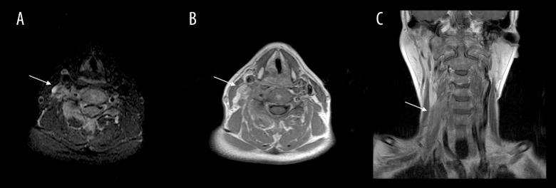
A 63-year-old Japanese woman visited a general practitioner with chief complaints of right shoulder and upper extremity pain. Although the patient’s physical examination was unremarkable, the transverse T2-weighted MRI images (T2WI) of head and neck (Figure 1A) and fat-saturated T2WI (Figure 1B) revealed a mild, high-intensity lesion along the right brachial plexus. Coronal gadolinium-enhanced T1-weighted images (Figure 1C) revealed mild, diffuse contrast-enhancement in the lesion, which is a non-specific signal pattern of brachial plexus lesions such as metastatic tumors, benign neurogenic tumors, malignant nerve sheath tumors, and Ewing sarcomas [5].
Figure 1.
Head and neck MRI images. Transverse T2-weighted image (T2WI) (A), fat-saturated T2WI (B), and coronal gadolinium-enhanced T1-weighted image (C).
One week after the MRI findings, the patient presented with unexpected thrombocytopenia (3.2×10⁴/μL), high serum level of LDH (16320 U/L), and IgD (197 mg/dL). Then, the serum immunoelectrophoresis and bone marrow biopsy were quickly performed for advanced diagnostic purposes. These results were as follows: M-protein of the IgD-lambda type, and infiltration of clonal plasma cells with CD3 (−), CD4 (−), CD7 (+), CD10 (−), CD20 (−), CD38(+), CD56 (−), CD138 (−), Bcl-2 (−), Bcl-6 (−), c-Myc (+), MUM-1 (+), PAX5(−), OCT2(+), bob1(+), kappa(−) and lambda(+). Thus, physicians strongly suspected MM from the patient’s clinical characteristics.
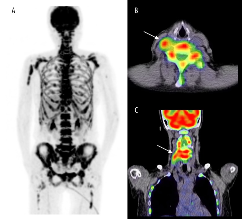
As shown in Figure 2, The 18F-FDG PET/CT fusion images and maximum intensity projections of her whole-body scan revealed high-intensity FDG uptake in multiple intramedullary lesions [6], and similar uptake was observed along the right brachial plexus, where the mass lesion had been detected previously via MRI. The 18F-FDG PET/CT images revealed neither disruption of cortical bone adjacent to the medullary lesions nor remodeling/destruction of trabecular bone, consistent with neuro-myelomatosis of the brachial plexus, which is defined as extramedullary neural involvement associated with MM.
Figure 2.
18F-FDG PET/CT images. Maximum intensity projections of the whole-body scan (A) and fusion images (B, C).
The neuropathic pain was improved with high-dose dexamethasone therapy. In addition, after the combination chemotherapy with etoposide, prednisolone, vincristine, Adriamycin, and cyclophosphamide, the plasma cells in the bone marrow almost disappeared. The 18F-FDG PET/CT images confirmed complete metabolic remission of the intramedullary lesions and the right brachial plexus lesion.
Discussion
One of the clinical features of IgD MM is the common occur-rence of cytogenetic abnormalities as well as extramedullary involvement [7]; thus, this condition may present with variable symptoms caused by the invasion of a variety of organs, including neuro-myelomatosis. Although neurological manifestations frequently complicate the course of patients with MM [8], peripheral neuropathy is also a common complication of many of the systemic amyloidoses [9]. Neuropathies related with neuro-myelomatosis are treatable pathological conditions; therefore, differentiating neuro-myelomatosis from neuro-amyloidosis is of clinical importance.
Conclusions
Although neuro-myelomatosis are difficult to diagnose, this case establishes 18F-FDG PET/CT as a potentially useful imaging modality for the diagnosis of extramedullary lesions associated with MM.
Footnotes
Conflicts of interest
The authors declare that they have no conflicts of interest.
References:
- 1.Marchettini P, Lacerenza M, Mauri E, et al. Painful peripheral neuropathies. Curr Neuropharmacol. 2006;4(3):175–81. doi: 10.2174/157015906778019536. [DOI] [PMC free article] [PubMed] [Google Scholar]
- 2.Blade J, Fernandez de Larrea C, Rosinol L, et al. Soft-tissue plasmacytomas in multiple myeloma: Incidence, mechanisms of extramedullary spread, and treatment approach. J Clin Oncol. 2011;29(28):3805–12. doi: 10.1200/JCO.2011.34.9290. [DOI] [PubMed] [Google Scholar]
- 3.Blade J, de Larrea CF, Rosinol L. Extramedullary involvement in multiple myeloma. Haematologica. 2012;97(11):1618–19. doi: 10.3324/haematol.2012.078519. [DOI] [PMC free article] [PubMed] [Google Scholar]
- 4.Dimopoulos M, Terpos E, Comenzo RL, et al. International myeloma working group consensus statement and guidelines regarding the current role of imaging techniques in the diagnosis and monitoring of multiple myeloma. Leukemia. 2009;23(9):1545–56. doi: 10.1038/leu.2009.89. [DOI] [PubMed] [Google Scholar]
- 5.Wittenberg KH, Adkins MC. MR imaging of nontraumatic brachial plexopathies: Frequency and spectrum of findings. Radiographics. 2000;20(4):1023–32. doi: 10.1148/radiographics.20.4.g00jl091023. [DOI] [PubMed] [Google Scholar]
- 6.Hanrahan CJ, Christensen CR, Crim JR. Current concepts in the evaluation of multiple myeloma with mr imaging and fdg pet/ct. Radiographics. 2010;30(1):127–42. doi: 10.1148/rg.301095066. [DOI] [PubMed] [Google Scholar]
- 7.Pandey S, Kyle RA. Unusual myelomas: A review of igd and ige variants. Oncology (Williston Park) 2013;27(8):798–803. [PubMed] [Google Scholar]
- 8.Fassas AB, Muwalla F, Berryman T, et al. Myeloma of the central nervous system: Association with high-risk chromosomal abnormalities, plasmablastic morphology and extramedullary manifestations. Br J Haematol. 2002;117(1):103–8. doi: 10.1046/j.1365-2141.2002.03401.x. [DOI] [PubMed] [Google Scholar]
- 9.Shin SC, Robinson-Papp J. Amyloid neuropathies. Mt Sinai J Med. 2012;79(6):733–48. doi: 10.1002/msj.21352. [DOI] [PMC free article] [PubMed] [Google Scholar]


