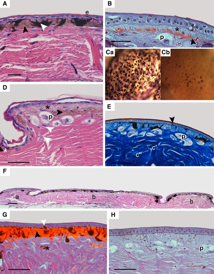
Fig. 2.
Distribution of chromatophores in tokay gecko skin; scalebar 50 µm. A, D, and F—H&E, Ca, Cb—micrograph of the skin in toto in transmitted light, E—Mallory’s stain, B, G and H—DIC (Nomarski contrast). A Blue pigmented area from dorsal part of body; epidermis (e), single melanophores with processes (white arrowhead), numerous iridophores (black arrowhead). B Orange/red pigmented area from dorsal part of head with clearly visible layer of iridophores (black arrowhead) and erythrophores (black asterisk). Parenchymatous cells (p) are visible in dermis under chromatophores. Ca, b. Micrographs of the skin in toto in transmitted light from blue area (Ca), with high number of melanophores with melanin-filled processes and from orange/red area (Cb) where melanophores are visible in smaller numbers with melanosomes in perinuclear part of these cells (Cb). D Orange/red pigmented area from back where erythrophores are more numerous (black asterisk) than in blue pigmented area, a single layer of iridophores (black arrowhead), isolated melanophores (white asterisk), fibrocytes and fibroblasts visible in dermis (white arrowhead). Parenchymatous cells (p) are visible in dermis under chromatophores. E. Orange/red area from back with single melanocyte in epidermis (black arrowhead), characteristic of tuberculate scales bundles of collagen fibres with different orientation (c’, c”), and parenchymatous cells (p) under chromatophores. F Two types of scales from dorsal part of back: overlapping scale (a) and tuberculate scales (b). G Blue area from dorsal part of tail with layer of iridophores (black arrowhead). Isolated melanocyte in epidermis (white arrowhead) and frequently deeper located melanophores (black asterisk). H Orange/red area from ventral part of trunk without iridophores (lack of contrasted cells). Parenchymatous cells (p) are visible under chromatophores

