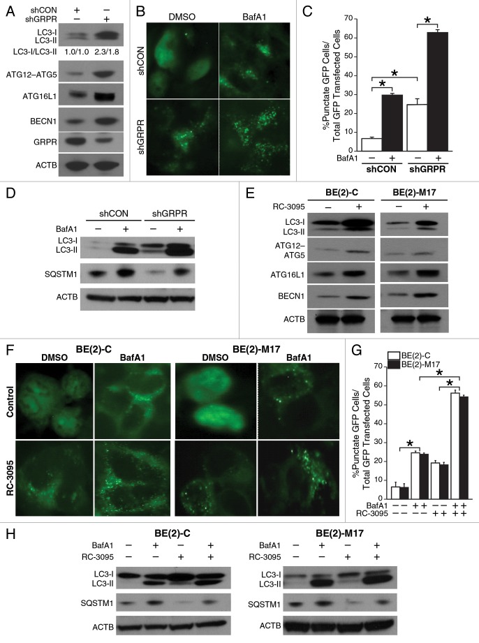
Figure 2. Inhibition of GRPR enhanced the expression of key autophagic markers in human neuroblastoma cells. (A) Immunoblotting demonstrated increased expression of ATG12–ATG5 conjugate, ATG16L1, BECN1 and enhanced conversion of LC3-I to LC3-II in GRPR-silenced (shGRPR) BE(2)-C cells when compared with control cells (shCON). (B) BE(2)-C cells silenced for GRPR (shGRPR) or controls (shCON) were transfected with EGFP-LC3 plasmid and treated with BafA1 (200 μM) for 24 h until assessed for autophagy. Representative fluorescence microscopy photos are shown. (C) The percentage of cells with punctate EGFP-LC3 fluorescence was calculated relative to all EGFP-positive cells in BE(2)-C cells (*P < 0.05). (D) Western blots for LC3-I and -II, and SQSTM1 in BE(2)-C cells silenced for GRPR (shGRPR) or control (shCON) and treated with or without BafA1 (200 μM) for 24 h. (E) Conversion of LC3-I to LC3-II, ATG12–ATG5 conjugate, ATG16L1, and BECN1 were determined by immunoblotting using lysates from EGFP-LC3-transfected BE(2)-C and BE(2)-M17 cells treated with DMSO or RC-3095 (1 μM) for 48 h. ACTB was probed to assess equal protein loading. (F) BE(2)-C and BE(2)-M17 cells were transfected with EGFP-LC3 plasmid, treated with or without BafA1 (200 μM) for 24 h and then assessed for autophagy after 48 h. Representative fluorescence microscopy photos are shown. (G) The percentage of cells with punctate EGFP-LC3 fluorescence was calculated relative to all EGFP positive cells in BE(2)-C and BE(2)-M17 cells treated with DMSO or RC-3095 (1 μM). Values shown are mean ± SEM of three separate experiments (*P < 0.05). (H) Expression of LC3-I or -II, and SQSTM1 in BE(2)-C and BE(2)-M17 cells pretreated with DMSO or RC-3095 (1 μM) and then treated with or without BafA1 (200 μM) for 24 h.

An official website of the United States government
Here's how you know
Official websites use .gov
A
.gov website belongs to an official
government organization in the United States.
Secure .gov websites use HTTPS
A lock (
) or https:// means you've safely
connected to the .gov website. Share sensitive
information only on official, secure websites.
