

Abstract
An attempt has been made to use lactoperoxidase-catalyzed iodination of excised Cucurbita hypocotyl hooks to monitor the distribution of plasma membrane fragments relative to that of phytochrome in particulate fractions from this tissue. Upon fractionation, the iodinated tissue yields a 20,000g pellet which contains 58% of the trichloroacetic acid-precipitable 125I at a specific radioactivity 12 times that of the proteins in the supernatant. On sucrose gradients, the labeled fraction has a mean isopycnic density of 1.15 g · cm−3. The distribution profile is distinct from that of the total particulate protein and does not coincide with either mitochondrial or endoplasmic reticulum markers. These observations satisfy operational criteria commonly accepted in other systems as indices of selective labeling of the cell surface. The sucrose gradient profiles of the phytochrome and 125I in the 20,000g pellets are noncoincident. In the absence of more direct evidence, this is readily interpreted to indicate a lack of association of the pigment with the plasma membrane.
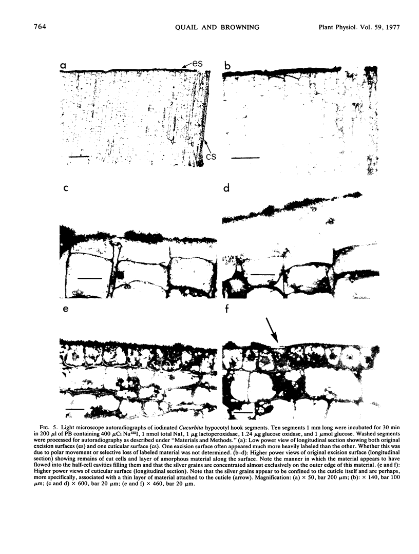
Autoradiographic analysis indicates, however, that the 125I is almost exclusively associated with an amorphous film (possibly phloem-exudate protein) overlying the cut cells at the point of prelabeling excision and along the outer physical surface of the hypocotyl cuticle. No evidence of plasma membrane labeling is apparent. The observed membrane-like behavior of the iodinated material upon cell fractionation is attributed to the preferential posthomogenization association of this material with a particular membrane fraction(s). These data indicate that in addition to the well recognized potential for spurious labeling of the internal cytoplasmic proteins of leaky cells, a further source of ambiguity in surface-labeling experiments should be considered. That is, the potential for labeling extracellular proteins of nonplasma membrane origin but with a capacity to become associated with membranes upon homogenization.
Full text
PDF


Images in this article
Selected References
These references are in PubMed. This may not be the complete list of references from this article.
- Carraway K. L. Covalent labeling of membranes. Biochim Biophys Acta. 1975 Dec 29;415(4):379–410. doi: 10.1016/0304-4157(75)90005-2. [DOI] [PubMed] [Google Scholar]
- Gadowska M., Miks B., Kawalec M., Kawiak J. Radioiodination of L 1210 cells. Folia Histochem Cytochem (Krakow) 1975;13(1-2):11–20. [PubMed] [Google Scholar]
- Hodges T. K., Leonard R. T., Bracker C. E., Keenan T. W. Purification of an ion-stimulated adenosine triphosphatase from plant roots: association with plasma membranes. Proc Natl Acad Sci U S A. 1972 Nov;69(11):3307–3311. doi: 10.1073/pnas.69.11.3307. [DOI] [PMC free article] [PubMed] [Google Scholar]
- Hubbard A. L., Cohn Z. A. The enzymatic iodination of the red cell membrane. J Cell Biol. 1972 Nov;55(2):390–405. doi: 10.1083/jcb.55.2.390. [DOI] [PMC free article] [PubMed] [Google Scholar]
- Juliano R. L., Behar-Bannelier M. An evaluation of techniques for labelling the surface proteins of cultured mammalian cells. Biochim Biophys Acta. 1975 Jan 28;375(2):249–267. doi: 10.1016/0005-2736(75)90193-5. [DOI] [PubMed] [Google Scholar]
- LOWRY O. H., ROSEBROUGH N. J., FARR A. L., RANDALL R. J. Protein measurement with the Folin phenol reagent. J Biol Chem. 1951 Nov;193(1):265–275. [PubMed] [Google Scholar]
- Lewis B. A., Elkin A., Michell R. H., Coleman R. Basolateral plasma membranes of intestinal epithelial cells. Identification by lactoperoxidase-catalysed iodination and isolation after density perturbation with digitonin. Biochem J. 1975 Oct;152(1):71–84. doi: 10.1042/bj1520071. [DOI] [PMC free article] [PubMed] [Google Scholar]
- Lord J. M., Kagawa T., Moore T. S., Beevers H. Endoplasmic reticulum as the site of lecithin formation in castor bean endosperm. J Cell Biol. 1973 Jun;57(3):659–667. doi: 10.1083/jcb.57.3.659. [DOI] [PMC free article] [PubMed] [Google Scholar]
- Morrison M. The determination of the exposed proteins on membranes by the use of lactoperoxidase. Methods Enzymol. 1974;32:103–109. doi: 10.1016/0076-6879(74)32013-7. [DOI] [PubMed] [Google Scholar]
- Spurr A. R. A low-viscosity epoxy resin embedding medium for electron microscopy. J Ultrastruct Res. 1969 Jan;26(1):31–43. doi: 10.1016/s0022-5320(69)90033-1. [DOI] [PubMed] [Google Scholar]
- Strobel G. A., Hess W. M. Evidence for the presence of the toxin-binding protein on the plasma membrane of sugarcane cells. Proc Natl Acad Sci U S A. 1974 Apr;71(4):1413–1417. doi: 10.1073/pnas.71.4.1413. [DOI] [PMC free article] [PubMed] [Google Scholar]
- Van Der Woude W. J., Lembi C. A., Morré D. J. beta-Glucan Synthetases of Plasma Membrane and Golgi Apparatus from Onion Stem. Plant Physiol. 1974 Sep;54(3):333–340. doi: 10.1104/pp.54.3.333. [DOI] [PMC free article] [PubMed] [Google Scholar]