Abstract
Fluorescent probes play an important role in the development of fluorescence-based imaging techniques for life sciences research. Gold nanoclusters (AuNCs) are a novel type of fluorescent nanomaterials which have attracted great interest in recent years. Composed of only a few atoms, these ultrasmall AuNCs exhibit quantum confinement effects and molecule-like properties. Fluorescent AuNCs have an attractive set of features including ultrasmall size, good biocompatibility and photostability, and tunable emission in the red to near-infrared spectral region, which make them promising as fluorescent labels for biological imaging. Examples of their application include live cell labeling, cancer cell targeting, cellular apoptosis monitoring, and in vivo tumor imaging. Here, we present a brief overview of recent advances in utilizing these emissive ultrasmall AuNCs as optical probes for in vitro and in vivo fluorescence imaging.
Keywords: Gold nanoclusters, Fluorescence probes, Live cell imaging, In vitro imaging, In vivo imaging, Cytotoxicity
Introduction
Among the established imaging techniques, fluorescence microscopy offers unique advantages for biophysical studies at the molecular level, for example, to analyze the folding and function of individual biomolecules (Nienhaus 2009; Helm et al. 2009) and to visualize biomolecular processes in living cells with high spatial resolution (Hedde and Nienhaus 2010). The quality of fluorescence imaging crucially relies on the performance of the fluorophores attached to the biological structures of interest to visualize them, so many researchers keep on pursuing new, robust fluorescence probes (Zhang et al. 2002; Koo et al. 2011). Ideally, fluorescence probes suitable for imaging applications should meet the following requirements: (1) they should be non-toxic to cells and other organisms; (2) they should be smaller than the biomolecules of interest, so that normal biological functions such as biomolecular interactions are not disturbed; (3) they should have optimal photophysical properties including low photobleaching, absence of blinking, and high quantum yield; and, finally, (4) they should be simple to synthesize and use for labeling biomolecules. Each of these attributes is essential for marker application in advanced imaging techniques aimed at unraveling the molecular details underlying biological processes (Fernandez-Suarez and Ting 2008).
Organic dyes have been employed as powerful and bright fluorescence markers for many years (Giepmans et al. 2006; Fernandez-Suarez and Ting 2008). However, their limited photostability and small Stokes shift restrict their use in some applications, e.g., long-term tracking and multicolor imaging applications (Resch-Genger et al. 2008). GFP-like fluorescent proteins are even less photostable than excellent organic dyes, but their key advantage for biological imaging applications is that they can be genetically encoded so that the labels are produced by the cells themselves (Wiedenmann and Nienhaus 2006; Nienhaus 2008; Nienhaus and Wiedenmann 2009). In contrast, semiconductor quantum dots have excellent brightness and photostability and are, from the photophysics point of view, very attractive labels for biological imaging (Michalet et al. 2005). Due to bulky layers for water solubilization and biofunctionalization, many currently available quantum dots have an overall diameter above 5 nm, potentially causing major kinetic or steric hindrance problems when utilized as fluorescent biomarkers for studying biomolecule interactions or tracking biological processes (Baker 2010).
In recent years, novel fluorescent biomarkers have been developed based on metal nanocrystals, known as fluorescent metal nanoclusters (NCs) (Zheng et al. 2007; Shang et al. 2011c). Metal NCs consist of a few atoms and have a core diameter of less than 2 nm. With cluster dimensions approaching the Fermi wavelength of electrons, the continuous density of states breaks up into discrete energy levels leading to the observation of dramatically different optical, electrical, and chemical properties as compared to large metal nanoparticles (Zheng et al. 2004) and, most importantly, they can exhibit strong photoluminescence. Although these luminescent metal NCs have not yet found as widespread application as conventional organic dyes, they show great promise as markers for biological imaging due to their ultrasmall size and excellent photophysical property. Among various metal NCs (e.g., Au, Ag, Cu, and Pt), AuNCs are currently most intensely studied, owing to their good biocompatibility, extraordinary stability, and facile synthesis (Muhammed and Pradeep 2010). In this review, we focus on recent developments in utilizing AuNCs as novel optical probes for fluorescence bioimaging.
Optical properties and synthesis of gold nanoclusters
The ultra-small size of AuNCs results in quantum confinement effects, which give rise to discrete electronic energy levels, molecule-like electronic transitions, enhanced photoluminescence, intrinsic magnetism, and other effects (Jin 2010). In contrast to larger Au nanoparticles, AuNCs are too small to possess the continuous density of states necessary to support a plasmon. While the photoluminescence from bulk gold is extremely low, with a quantum yield of only 10−10 (Mooradian 1969), AuNCs can have a much enhanced quantum yield in the range 10−3–10−1. Therefore, they can be sufficiently bright for many fluorescence marker applications. The as yet highest reported quantum yield of AuNCs was 70 % for dendrimer-encapsulated Au5 NCs by the Dickson group (Zheng et al. 2004). By controlling size and chemical composition, the fluorescence emission of AuNCs can be easily tuned from the visible into the near-infrared region (Zheng et al. 2004; Huang et al. 2009b).
With regard to their nonlinear optical properties, Ramakrishna et al. ((2008) demonstrated that AuNCs can be used as efficient two-photon absorbers. The two-photon absorption cross-section of AuNCs was measured to be as high as 105 Göppert-Mayer units (Polavarapu et al. 2011), higher than the values reported for typical organic dyes and quantum dots (Larson et al. 2003; Pan et al. 2007a; Feng et al. 2008). Such large two-photon absorption cross-sections make AuNCs efficient absorbers for multi-photon biological imaging. Furthermore, AuNCs have been observed to possess better photostability than organic dyes (Polavarapu et al. 2011; Wu et al. 2010; Lin et al. 2009). For instance, lipoic acid-coated AuNCs exhibited a much slower photobleaching rate than organic fluorophores such as fluorescein and rhodamine 6G (Lin et al. 2009). The photophysical merits of AuNCs mentioned above are beneficial to their biological application, giving the possibility of multiplexed detection of molecular targets, and continuous, real-time imaging of single molecules or cells over long periods.
The reliable synthesis of AuNCs with excellent optical properties is a fundamental requirement for their application. A wide variety of methods for the synthesis of water-soluble, fluorescent AuNCs have been described. These fall into two main categories. The first route involves the production of small clusters from large Au nanoparticles by etching with thiols (Huang et al. 2007; Lin et al. 2009; Muhammed et al. 2009), biomolecules (Zhou et al. 2009; Muhammed et al. 2010), or multivalent polymers (Duan and Nie 2007). Although this strategy proved to be efficient for synthesizing AuNCs with different ligands, it involves a multi-step process that makes their synthesis rather complicated and tedious. By contrast, a one-step strategy, in which fluorescent AuNCs are produced directly by reducing gold salt with a suitable reductant such as sodium borohydride (Link et al. 2002; Schaeffer et al. 2008; Shang et al. 2011a), tetrakis(hydroxymethyl)phosphonium chloride (Shang et al. 2011b), or even proteins (Xie et al. 2009; Xavier et al. 2010; Wen et al. 2011; Yan et al. 2012), is more favorable and has lately been widely adopted.
Fluorescence imaging of gold nanoclusters in vitro
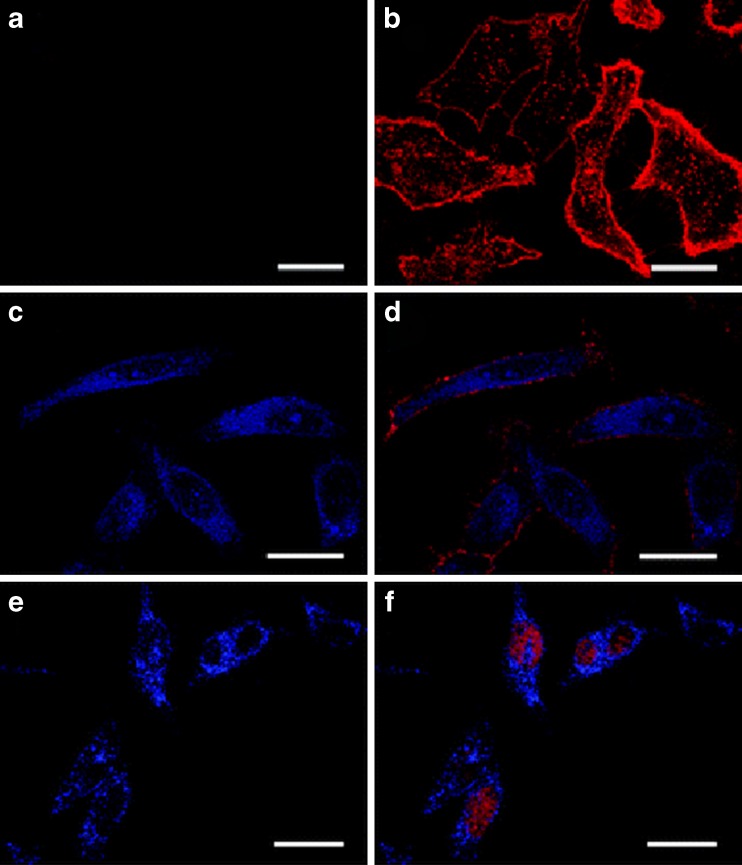
With the rapid advance of synthesis strategies, AuNCs have been successfully employed as fluorescence labels for a variety of biological purposes, such as biomolecule detection (Huang et al. 2009a; Shiang et al. 2011; Jin et al. 2011; Tian et al. 2012), intracellular metal ion sensing (Pu et al. 2011; Shang et al. 2012b), live cell labeling (Lin et al. 2009; Huang et al. 2011; Liu et al. 2011), cellular apoptosis studies (Lin et al. 2010), and targeting notorious pathogenic bacteria (Chen et al. 2010). Among the various applications of AuNCs, fluorescence imaging has made the most progress and attracted the greatest interest. In an early report, Lin et al. (2008) explored the possibility of utilizing AuNCs as fluorescence probes for nuclear targeting and intracellular imaging. Confocal microscopy images failed to show fluorescence from cells treated with 11-mercaptoundecanonic acid-capped AuNCs, suggesting a low cellular uptake efficiency for these nanoparticles (Fig. 1a). By contrast, upon functionalization of AuNCs with a site-specific targeting peptide, denoted SV40 nuclear-localization signal, intense blue PL from AuNCs was observed inside the cells (Fig. 1c, e). Colocalization experiments revealed that these peptide-coated AuNCs were distributed within both the cytoplasm (Fig. 1d) and the nucleus (Fig. 1f). Jao et al. (2010) further reported that positively charged, dendrimer-encapsulated Au8 NCs could penetrate the cell membrane and predominantly localized in the cytoplasm. Moreover, recent studies revealed that AuNCs also enter cells via endocytosis (Iversen et al. 2011; Polavarapu et al. 2011; Shang et al. 2011b). These very recent reports demonstrate the great potential of AuNCs as optical probes for intracellular imaging and subcellular targeting.
Fig. 1.
Confocal microscopy images of intracellular delivery of AuNCs. HeLa cells were treated with 11-mercaptoundecanonic acid-capped AuNCs (a, b) and peptide–functionalized AuNCs (c–f) for 1.5 h. The left panel shows fluorescence images of AuNCs only (blue), while the right panel shows colocalization images of AuNCs-treated cells that were counterstained with a membrance dye (wheat germ agglutinin-conjugated Alexa 594; b, d, red) and a nuclear dye (SYTO 59; f, red). Scale bar 25 μm. Reprinted with permission from the Royal Society of Chemistry (Lin et al. 2008)
Compared with visible light, the near-infrared region provides several advantages for cellular imaging, such as weak autofluorescence, minimal photobleaching, and low phototoxicity. Therefore, imaging agents with near-infrared fluorescence emission are particularly attractive for high-sensitivity fluorescence bioimaging (Frangioni 2003; Gao et al. 2010). Pradeep and coworkers (Muhammed et al. 2009) presented a successful example of producing near-infrared-emitting Au23 NCs with an etching-based strategy. With streptavidin bound to the particle surface, these AuNCs were observed to selectively stain human hepatoma cells (HepG2). These cells contain a large amount of biotin, which binds to streptavidin with high specificity and affinity. Recently, receptor-targeted near-infrared imaging of folate receptor-positive oral carcinoma cells was reported using folic acid-conjugated fluorescent Au25 NCs (Retnakumari et al. 2010). Receptor-targeted cancer detection was demonstrated on FR+ve oral carcinoma KB cells, where the folic acid-conjugated AuNCs were found to become internalized in significantly higher concentration compared with the negative control cell lines. The characteristic near-infrared emission from AuNCs made it possible to image the clusters under 700 – 800 nm emission, where the optical properties of blood and tissue are highly favorable for biomedical imaging. Furthermore, bright aqueous near-infrared fluorescent AuNCs capped with multidentate polymer were used as biomarkers to label cells of the hematopoietic system (Huang et al. 2011). The cancer cells incorporated more AuNCs than normal cells, as compared with the control group labeled with quantum dots.
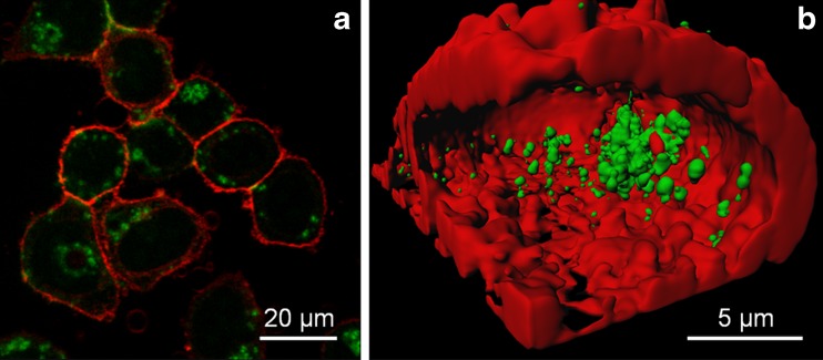
Two-photon excitation is advantageous for deep-tissue imaging owing to its ability of better tissue penetration and the often quoted reduced phototoxicity of near-infrared light excitation (Diaspro and Robello 2000). Note, however, that the latter effect can be counterbalanced by the enormous intensities required for two-photon absorption. The outstanding two-photon absorption cross-sections of AuNCs make them promising markers for two-photon cellular imaging. Chou and coworkers (Liu et al. 2009) explored two-photon imaging of human mesenchymal stem cells (hMSCs) using dextran-encapsulated AuNCs as probes. Upon two-photon excitation with 800-nm laser pulses in a confocal microscope, bright luminescence from AuNCs was observed in the cells. Glutathione-capped AuNCs with large two-photon absorption cross-section and extraordinary photostability have also shown considerable promise for two-photon excitation in live cell imaging (Polavarapu et al. 2011). Recently, we (Shang et al. 2011b) reported the internalization of D-penicillamine-coated AuNCs by imaging live HeLa cells with two-photon fluorescence excitation at 810 nm. Figure 2 shows bright luminescence from AuNCs inside the cells. Cells were also imaged at different z-positions for 3D reconstruction, confirming the presence of AuNCs inside the cells as well as adhering to the plasma membrane. Elucidating the details of the uptake mechanism of these ultrasmall NCs still requires further elaboration. However, the finding that most particles inside the cells existed in the form of large aggregates suggests that they are internalized by specific endocytosis pathways that package many tiny particles into endosomal vesicles (Jiang et al. 2010a, b; Lunov et al. 2011).
Fig. 2.
a Fluorescence images of HeLa cells after incubation with D-penicillamine-coated AuNCs (100 μg/ml) for 2 h, taken with 810-nm two-photon excitation. b Cross-section of a 3D image reconstruction, showing internalized AuNCs. AuNC fluorescence was recorded in a spectral window from 557 to 607 nm (green channel), whereas the emission of the membrane stain DiD was detected in a band from 647 to 703 nm (red channel)
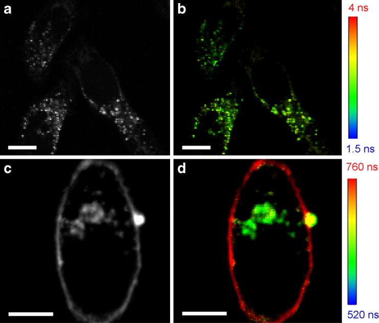
In contrast to fluorescence intensity imaging, lifetime-based imaging is independent of fluorophore concentration and laser excitation intensity. Importantly, however, the fluorescence lifetime of the fluorophores can be exquisitely sensitive to the local environment. Consequently, fluorescence lifetime imaging may provide contrast due to spatial variations of the lifetime and, thus, may yield valuable additional information (Borst and Visser 2010). We have recently introduced a facile strategy of synthesizing near-infrared-emitting lipoic acid-coated AuNCs (Shang et al. 2011a). Besides ultrasmall size, good colloidal stability, and biocompatibility, these AuNCs possess a long fluorescence lifetime (>100 ns), much longer than that of cellular autofluorescence and the fluorescence of organic dyes, so that, by using time-gated detection, their emission can be detected absolutely background-free. After exposing HeLa cells to AuNCs for 1 h, luminescent emitters with long fluorescence lifetimes (500–800 ns) were observed inside the cells, suggesting that the AuNCs had been internalized (Fig. 3). Note that the fluorescence decay of lipoic acid-coated AuNCs in the cells is slower than in aqueous solution, reflecting the modified NC environment during the uptake process including the formation of a protein corona around the particles (Röcker et al. 2009; Jiang et al. 2010b; Maffre et al. 2011). Furthermore, the fluorescence lifetime images clearly show an interesting contrast among AuNCs in different locations: NCs near the cell membrane display longer lifetimes than those inside the cells. Thus, fluorescence lifetime imaging not only reveals the cellular uptake of AuNCs but also provides information on changes in their local environment. All these results indicate that AuNCs have a great potential as robust fluorophores in biomedical imaging, especially in combination with advanced microscopy techniques (Su et al. 2010; Quan et al. 2010; Jiang et al. 2011).
Fig. 3.
Confocal microscopy images of HeLa cells without (a, b) and with (c, d) incubation with 100 μg/ml lipoic acid-coated AuNCs for 1 h. Fluorescence intensity (a, c) and lifetime (b, d) images were taken with pulsed diode laser (470 nm) excitation and a band-pass emission filter 690/70 (center wavelength/width), using time correlated single photon counting. Lifetime maps were calculated by determining the lifetime from the fluorescence decays of all the photon counts registered for each pixel. Scale bar 10 μm
Fluorescence imaging of gold nanoclusters in vivo
Compared with cellular imaging, in vivo imaging of a multicellular organism faces different challenges caused by the increase in sample complexity and the poor transmission of visible light through biological tissue (Rao et al. 2007; Wang et al. 2010). Correspondingly, ultrasmall AuNCs with emission in the near-infrared region, excellent stability, and good photophysical properties would be ideal markers for live animal imaging.
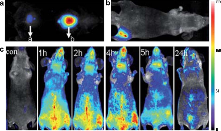
Wu et al. (2010) presented the first application of AuNCs-based in vivo fluorescence imaging; they explored the possibility of using near-infrared-emitting bovine serum albumin-stabilized AuNCs as imaging agents for tumor fluorescence imaging in vivo. The fluorescence of the AuNCs was easily visualized upon injection into the muscle up to a few millimeters owing to the enhanced tissue penetration afforded by near-infrared fluorescence (Fig. 4). AuNCs were also intravenously injected into mice for whole-body real-time in vivo imaging. Immediately after tail vein injection of AuNCs, bright fluorescence from the superficial vasculature of the whole body could easily be visualized. Fluorescence was visible in the circulation even 5 h post-injection, and decreased noticeably within a day. Further in vivo and ex vivo studies showed that these ultrasmall AuNCs accumulated predominantly in tumor sites resulting from the enhanced permeability and retention effect. The authors also presented a quantitative report on the biodistribution of AuNCs in different organs. They found that uptake of AuNCs by the reticule-endothelial system (e.g., liver, spleen) is relatively small in comparison with other nanoparticle-based contrast imaging agents due to their ultrasmall hydrodynamic size (ca. 2.7 nm).
Fig. 4.
In vivo fluorescence images of mice injected with 100 μl AuNCs (a) subcutaneously (a 0.235 mg ml−1, b 2.35 mg ml−1) and (b) intramuscularly (2.35 mg ml−1). c Real-time in vivo abdomen images taken upon intravenous injection with 200 μl of AuNCs (2.35 mg ml−−1) at different time points post-injection. Reprinted with permission from the Royal Society of Chemistry (Wu et al. 2010)
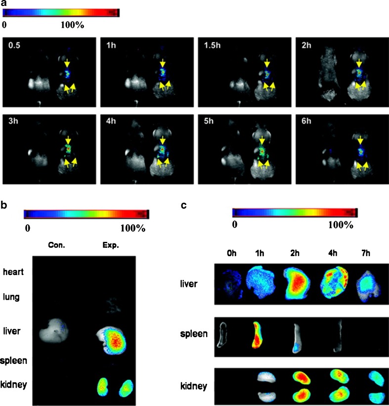
In a similar study, Sun et al. (2011) evaluated red-emitting ferritin-encapsulated AuNCs as fluorescent probes for in vivo imaging, with the ferritin providing specificity to target certain cells and tissues. They injected these AuNCs via the lateral tail vein into (nude) female mice, which were then subjected to whole-body fluorescence imaging. Fluorescent regions with a kidney-like shape were observed on either side of the spine 30 min post-injection and remained visible for at least 7 h (Fig. 5). Such a significant accumulation of nanoparticles in kidneys was assumed to indicate the involvement of a specific uptake mechanism related to the existence of certain ferritin receptors in kidney. In addition to kidney fluorescence, strong signals were also observed in the central dorsal region from liver and spleen, as further confirmed by ex vivo imaging experiments. Quantitation of the distribution of AuNCs using inductively coupled plasma mass spectrometry revealed that liver, kidney, and spleen were the major target tissues of ferritin-encapsulated AuNCs 2 h post-injection, compared with very low Au level in lung and heart. The biodistribution results are consistent with previous work on bovine serum albumin-stabilized AuNCs (Wu et al. 2010). These new results are important milestones for the development of AuNCs as contrast agents for targeted imaging and in vivo applications.
Fig. 5.
In vivo and ex vivo imaging female nude mice injected with ferritin-encapsulated AuNCs. a Whole body dorsal fluorescence images at different time points after Au NC injection. For each panel, the Au NC-injected mouse is shown on the right; a saline-injected control is shown on the left. b Fluorescence images of mouse organs 6 h after AuNCs injection. Control mice are shown on the left. c Fluorescence images of mouse organs at different time points after injection. The final concentration of AuNCs was 0.8 nmol/g body weight. The excitation filter transmitted from 576 to 621 nm; the emission was collected through a 635-nm long-pass filter. Reprinted with permission from American Chemical Society (Sun et al. 2011)
For in vivo biomedical applications, renal clearance is of fundamental importance to ensure that the contrast agents can be effectively cleared from the body, thereby avoiding accumulation in organs and interference with other diagnostic tests (Linkov et al. 2008; Schipper et al. 2009). Recently, Zheng et al. (2011) studied renal clearance of glutathione-coated luminescent AuNCs and revealed that only 3.7 % of the clusters accumulated in the liver; over 50 % of the clusters were found in urine within 24 h after intravenous injection, which is comparable to the quantum dots with the best renal clearance efficiency (Soo Choi et al. 2007). In addition, renal clearance of glutathione-coated AuNCs was at least 10 times better than that of similarly sized AuNCs coated with cysteine, a ligand known to significantly enhance renal clearance of quantum dots in vivo (Soo Choi et al. 2007). The small size in combination with suitable surface ligands not only enables the majority of the luminescent AuNCs to be cleared out of the body through kidney filtration but also stabilizes the luminescent AuNCs during blood circulation. Based on these applications and rapid progress in synthesizing near-infrared luminescent AuNCs, we anticipate their widespread use in in vivo biomedical imaging.
Biocompatibility of gold nanoclusters
Cytotoxicity and the potential influence of imaging agents on the cellular processes are primary issues in any live cell or whole animal imaging experiment (Feliu and Fadeel 2010; Soenen et al. 2011). Ultrasmall AuNCs are generally considered non-toxic, similar to bulk gold, which is chemically inert and biocompatible. It appears that toxicity should be even further alleviated due to their minimal metal content. However, recent studies reported adverse effects of Au nanoparticles on the cytoskeletal structure and cell viability by interacting with DNA and inducing oxidative stress (Pan et al. 2009; Chompoosor et al. 2010; Khlebtsov and Dykman 2011). While these toxicity issues are complicated by the wide variety of parameters, for example, cell types and physicochemical properties of the nanoparticles including size, colloidal stability, and ligand chemistry (Pan et al. 2007b; Sohaebuddin et al. 2010; Schaeublin et al. 2011), the majority of reports on luminescent AuNCs did not find adverse effects on cell viability or morphology at the concentrations used for imaging experiments. However, in light of the key relevance of nanotoxicity for biomedical applications, further inquiry into these issues is highly desirable.
Measurement of mitochondrial damage using the methyl thiazolyl tetrazolium (MTT) assay revealed negligible toxicity of AuNCs for a variety of cell lines, including human neuroblastoma cells (Polavarapu et al. 2011), HeLa cells (Shang et al. 2011a), and human endothelial cells (Retnakumari et al. 2010; Wang et al. 2011), for AuNC concentrations below 500 μg/ml. In addition, the possible occurrence of reactive oxygen stress caused by the interaction of cells with AuNCs has been evaluated (Retnakumari et al. 2010). Practically no reactive oxygen species stress was observed for folic acid-conjugated Au NC-treated cells. The cell membrane integrity upon the treatment of D-penicillamine-coated AuNCs has also been tested with a trypan blue exclusion test (Shang et al. 2011b), which also indicated that these AuNCs do not cause adverse effects. Moreover, the in vitro experiments on cultured cells did not observe morphological changes that would hint at adverse effects of AuNCs exposure within a reasonable concentration range. Particularly, Wang et al. (2011) compared the viability of endothelial cells treated with AuNCs and quantum dots and reported a better biocompatibility of AuNCs.
To assess toxicity in vivo, the body weight of AuNCs-treated mice was monitored and compared with control mice injected with phosphate buffer saline (Wu et al. 2010). Over a period of 1 month, the body weight of mice injected with bovine serum albumin-stabilized AuNCs changed only slightly, while the mice were observed to live normally without any sign of acute toxic responses or long-term toxic effects, suggesting the non-toxic nature of the AuNCs administered to the mice.
Outlook
Despite impressive progress on the biological application of fluorescent AuNCs in recent years, knowledge of their interactions within the complex biological environment is still limited. It would be important to understand the surface interactions of biomacromolecules with these ultrasmall particles and to see how these interactions affect the biological activity (Jiang et al. 2010b; Walczyk et al. 2010; Stark 2011). Moreover, we also need to better understand the mechanisms by which AuNCs are taken up by the cells (Iversen et al. 2011). In view of the large surface-to-volume ratio of these ultrasmall NCs, surface modifications, e.g., due to binding of proteins, are likely to alter their photophysical properties, which may in turn affect their performance as optical markers. Recently, we investigated the interactions of AuNCs with four different proteins (human serum albumin, apotransferrin, lysozyme, and apolipoprotein E4) and the effects on their fluorescence (Shang et al. 2012a). All proteins were observed to bind with roughly micromolar affinities to lipoic acid-coated AuNCs. Upon protein association, the fluorescence of AuNCs was significantly enhanced and, concomitantly, their luminescence lifetime was prolonged. These results provided clear evidence that protein binding to the surfaces of ultrasmall fluorescent AuNCs has a significant influence on their photophysical properties.
With the development of nanoscale contrast agents for the fields of diagnostics and whole-body imaging, integration of multiple functionalities within one nanoparticle would easily allow their detection with several imaging techniques or to include therapeutic qualities (Mulder et al. 2007). For example, in clinical applications requiring in vivo imaging in living subjects, imaging agents geared towards both fluorescence and magnetic resonance imaging can be particularly advantageous because optical and magnetic detection can be employed in a complementary fashion (Koktysh et al. 2011). In this regard, great opportunities and challenges remain for materials chemists to produce high quality, AuNCs-based multifunctional probes for advanced biomedical imaging application (Muhammed and Pradeep 2011; Durgadas et al. 2011).
Conjugation of imaging agents with biomolecules such as antibodies, enzymes, DNA, or oligosaccharides is a prerequisite to their use as biological probes for specific fluorescence imaging (Erathodiyil and Ying 2011), because it enables researchers to target desired locations within cells, tissues, and organs, reduce overall toxicity, and boost the efficiency of the imaging probes (Lin et al. 2009; Retnakumari et al. 2010). This research has as yet seen rather slow progress for AuNCs. In this regard, the presence of reactive moieties on the surface of AuNCs, allowing further mild, selective, and stable ligation with biomolecules, is of crucial importance. Many nanoparticle functionalization techniques have been reported in the literature; however, chemical engineering on the surface of AuNCs can pose particular challenges in view of their extraordinarily high surface-volume ratio, polydispersed nature, and sensitivity of the fluorescence response to the environment (Wu and Jin 2010; Shang et al. 2012a). Anyhow, engineering of luminescent AuNCs with biorecognition capabilities would be of great value for a wide range of life sciences applications.
Yet another area in which further progress will be highly appreciated is a thorough study on the toxicity of AuNCs. Although many reports have indicated a good biocompatibility, toxicity of these ultrasmall particles has so far only been explored to a limited extent. Much more needs to be done so that they can be used with confidence in biomedical applications on humans. A comprehensive examination of biocompatibility will be necessary by analyzing cellular responses, e.g., apoptosis, DNA damage, and oxidative stress (Marquis et al. 2009; Zhao et al. 2011). Moreover, extensive studies of the effects of AuNCs on cell growth and living organisms over extended periods of time need to be performed.
Acknowledgements
This work was supported by the Deutsche Forschungsgemeinschaft (DFG) through the Center for Functional Nanostructures (CFN) and the Priority Program SPP1313. L. Shang gratefully acknowledges support from the Alexander von Humboldt (AvH) Foundation.
Conflict of interest
None.
References
- Baker M. Nanotechnology imaging probes: smaller and more stable. Nat Methods. 2010;7:957–962. doi: 10.1038/nmeth1210-957. [DOI] [Google Scholar]
- Borst JW, Visser AJWG. Fluorescence lifetime imaging microscopy in life sciences. Meas Sci Technol. 2010;21:102002. doi: 10.1088/0957-0233/21/10/102002. [DOI] [Google Scholar]
- Chen W-Y, Lin J-Y, Chen W-J, Luo L, Wei-Guang Diau E, Chen Y-C. Functional gold nanoclusters as antimicrobial agents for antibiotic-resistant bacteria. Nanomedicine. 2010;5:755–764. doi: 10.2217/nnm.10.43. [DOI] [PubMed] [Google Scholar]
- Chompoosor A, Saha K, Ghosh PS, Macarthy DJ, Miranda OR, Zhu Z-J, Arcaro KF, Rotello VM. The role of surface functionality on acute cytotoxicity, ROS generation and DNA damage by cationic gold nanoparticles. Small. 2010;6:2246–2249. doi: 10.1002/smll.201000463. [DOI] [PMC free article] [PubMed] [Google Scholar]
- Diaspro A, Robello M. Two-photon excitation of fluorescence for three-dimensional optical imaging of biological structures. J Photochem Photobiol B. 2000;55:1–8. doi: 10.1016/S1011-1344(00)00028-2. [DOI] [PubMed] [Google Scholar]
- Duan H, Nie S. Etching colloidal gold nanocrystals with hyperbranched and multivalent polymers: a new route to fluorescent and water-soluble atomic clusters. J Am Chem Soc. 2007;129:2412–2413. doi: 10.1021/ja067727t. [DOI] [PubMed] [Google Scholar]
- Durgadas CV, Sharma CP, Sreenivasan K. Fluorescent and superparamagnetic hybrid quantum clusters for magnetic separation and imaging of cancer cells from blood. Nanoscale. 2011;3:4780–4787. doi: 10.1039/c1nr10900f. [DOI] [PubMed] [Google Scholar]
- Erathodiyil N, Ying JY. Functionalization of inorganic nanoparticles for bioimaging applications. Acc Chem Res. 2011;44:925–935. doi: 10.1021/ar2000327. [DOI] [PubMed] [Google Scholar]
- Feliu N, Fadeel B. Nanotoxicology: no small matter. Nanoscale. 2010;2:2514–2520. doi: 10.1039/c0nr00535e. [DOI] [PubMed] [Google Scholar]
- Feng W, Wei T, Li-Na M, Wen-Ju C, Gui-Lan Z, Guo-Feng Z, Shi-Dong C, Wei X. Optical nonlinear properties of CdSeS/ZnS core/Shell quantum dots. Chin Phys Lett. 2008;25:1461–1464. doi: 10.1088/0256-307X/25/4/080. [DOI] [Google Scholar]
- Fernandez-Suarez M, Ting A. Fluorescent probes for super-resolution imaging in living cells. Nat Rev Mol Cell Bio. 2008;9:929–943. doi: 10.1038/nrm2531. [DOI] [PubMed] [Google Scholar]
- Frangioni JV. In vivo near-infrared fluorescence imaging. Curr Opin Chem Biol. 2003;7:626–634. doi: 10.1016/j.cbpa.2003.08.007. [DOI] [PubMed] [Google Scholar]
- Gao J, Chen K, Xie R, Xie J, Lee S, Cheng Z, Peng X, Chen X. Ultrasmall near-Infrared non-cadmium quantum dots for in vivo tumor imaging. Small. 2010;6:256–261. doi: 10.1002/smll.200901672. [DOI] [PMC free article] [PubMed] [Google Scholar]
- Giepmans BNG, Adams SR, Ellisman MH, Tsien RY. The fluorescent toolbox for assessing protein location and function. Science. 2006;312:217–224. doi: 10.1126/science.1124618. [DOI] [PubMed] [Google Scholar]
- Hedde PN, Nienhaus GU. Optical imaging of nanoscale cellular structures. Biophys Rev. 2010;2:147–158. doi: 10.1007/s12551-010-0037-0. [DOI] [PMC free article] [PubMed] [Google Scholar]
- Helm M, Kobitski A, Nienhaus GU. Single-molecule Förster resonance energy transfer studies of RNA structure, dynamics and function. Biophys Rev. 2009;1:161–176. doi: 10.1007/s12551-009-0018-3. [DOI] [PMC free article] [PubMed] [Google Scholar]
- Huang CC, Yang Z, Lee KH, Chang HT. Synthesis of highly fluorescent gold nanoparticles for sensing mercury(II) Angew Chem Int Ed. 2007;46:6824–6828. doi: 10.1002/anie.200700803. [DOI] [PubMed] [Google Scholar]
- Huang C-C, Chen C-T, Shiang Y-C, Lin Z-H, Chang H-T. Synthesis of fluorescent carbohydrate-protected Au nanodots for detection of concanavalin A and escherichia coli. Anal Chem. 2009;81:875–882. doi: 10.1021/ac8010654. [DOI] [PubMed] [Google Scholar]
- Huang C, Liao H, Shiang Y, Lin Z, Yang Z, Chang H. Synthesis of wavelength-tunable luminescent gold and gold/silver nanodots. J Mater Chem. 2009;19:755–759. doi: 10.1039/b808594c. [DOI] [Google Scholar]
- Huang X, Luo Y, Li Z, Li B, Zhang H, Li L, Majeed I, Zou P, Tan B. Biolabeling hematopoietic system cells using near-Infrared fluorescent Gold nanoclusters. J Phys Chem C. 2011;115:16753–16763. doi: 10.1021/jp202612p. [DOI] [Google Scholar]
- Iversen T-G, Skotland T, Sandvig K. Endocytosis and intracellular transport of nanoparticles: Present knowledge and need for future studies. Nano Today. 2011;6:176–185. doi: 10.1016/j.nantod.2011.02.003. [DOI] [Google Scholar]
- Jao Y-C, Chen M-K, Lin S-Y. Enhanced quantum yield of dendrimer-entrapped gold nanodots by a specific ion-pair association and microwave irradiation for bioimaging. Chem Commun. 2010;46:2626–2628. doi: 10.1039/b926364k. [DOI] [PubMed] [Google Scholar]
- Jiang X, Röcker C, Hafner M, Brandholt S, Dörlich RM, Nienhaus GU. Endo- and exocytosis of zwitterionic quantum dot nanoparticles by live HeLa cells. ACS Nano. 2010;4:6787–6797. doi: 10.1021/nn101277w. [DOI] [PubMed] [Google Scholar]
- Jiang X, Weise S, Hafner M, Röcker C, Zhang F, Parak WJ, Nienhaus GU. Quantitative analysis of the protein corona on FePt nanoparticles formed by transferrin binding. J R Soc Interface. 2010;7:S5–S13. doi: 10.1098/rsif.2009.0272.focus. [DOI] [PMC free article] [PubMed] [Google Scholar]
- Jiang X, Musyanovych A, Rocker C, Landfester K, Mailander V, Nienhaus GU. Specific effects of surface carboxyl groups on anionic polystyrene particles in their interactions with mesenchymal stem cells. Nanoscale. 2011;3:2028–2035. doi: 10.1039/c0nr00944j. [DOI] [PubMed] [Google Scholar]
- Jin R. Quantum sized, thiolate-protected gold nanoclusters. Nanoscale. 2010;2:343–362. doi: 10.1039/b9nr00160c. [DOI] [PubMed] [Google Scholar]
- Jin L, Shang L, Guo S, Fang Y, Wen D, Wang L, Yin J, Dong S. Biomolecule-stabilized Au nanoclusters as a fluorescence probe for sensitive detection of glucose. Biosens Bioelectron. 2011;26:1965–1969. doi: 10.1016/j.bios.2010.08.019. [DOI] [PubMed] [Google Scholar]
- Khlebtsov N, Dykman L. Biodistribution and toxicity of engineered gold nanoparticles: a review of in vitro and in vivo studies. Chem Soc Rev. 2011;40:1647–1671. doi: 10.1039/c0cs00018c. [DOI] [PubMed] [Google Scholar]
- Koktysh D, Bright V, Pham W. Fluorescent magnetic hybrid nanoprobe for multimodal bioimaging. Nanotechnology. 2011;22:275606. doi: 10.1088/0957-4484/22/27/275606. [DOI] [PMC free article] [PubMed] [Google Scholar]
- Koo H, Huh MS, Ryu JH, Lee D-E, Sun I-C, Choi K, Kim K, Kwon IC. Nanoprobes for biomedical imaging in living systems. Nano Today. 2011;6:204–220. doi: 10.1016/j.nantod.2011.02.007. [DOI] [Google Scholar]
- Larson DR, Zipfel WR, Williams RM, Clark SW, Bruchez MP, Wise FW, Webb WW. Water-soluble quantum dots for multiphoton fluorescence imaging in vivo. Science. 2003;300:1434–1436. doi: 10.1126/science.1083780. [DOI] [PubMed] [Google Scholar]
- Lin S-Y, Chen N-T, Sum S-P, Lo L-W, Yang C-S (2008) Ligand exchanged photoluminescent gold quantum dots functionalized with leading peptides for nuclear targeting and intracellular imaging. Chem Commun:4762–4764 [DOI] [PubMed]
- Lin CA, Yang TY, Lee CH, Huang SH, Sperling RA, Zanella M, Li JK, Shen JL, Wang HH, Yeh HI, Parak WJ, Chang WH. Synthesis, characterization, and bioconjugation of fluorescent gold nanoclusters toward biological labeling applications. ACS Nano. 2009;3:395–401. doi: 10.1021/nn800632j. [DOI] [PubMed] [Google Scholar]
- Lin S-Y, Chen N-T, Sun S-P, Chang JC, Wang Y-C, Yang C-S, Lo L-W. The Protease-mediated nucleus shuttles of subnanometer gold quantum dots for real-time monitoring of apoptotic cell death. J Am Chem Soc. 2010;132:8309–8315. doi: 10.1021/ja100561k. [DOI] [PubMed] [Google Scholar]
- Link S, Beeby A, FitzGerald S, El-Sayed M, Schaaff T, Whetten R. Visible to infrared luminescence from a 28-atom gold cluster. J Phys Chem B. 2002;106:3410–3415. doi: 10.1021/jp014259v. [DOI] [Google Scholar]
- Linkov I, Satterstrom FK, Corey LM. Nanotoxicology and nanomedicine: making hard decisions. Nanomedicine. 2008;4:167–171. doi: 10.1016/j.nano.2008.01.001. [DOI] [PubMed] [Google Scholar]
- Liu C, Ho M, Chen Y, Hsieh C, Lin Y, Wang Y, Yang M, Duan H, Chen B, Lee J. Thiol-functionalized gold nanodots: two-photon absorption property and imaging in vitro. J Phys Chem C. 2009;113:21082–21089. doi: 10.1021/jp9080492. [DOI] [Google Scholar]
- Liu C-L, Wu H-T, Hsiao Y-H, Lai C-W, Shih C-W, Peng Y-K, Tang K-C, Chang H-W, Chien Y-C, Hsiao J-K, Cheng J-T, Chou P-T. Insulin-directed synthesis of fluorescent gold nanoclusters: preservation of insulin bioactivity and versatility in cell imaging. Angew Chem Int Ed. 2011;50:7056–7060. doi: 10.1002/anie.201100299. [DOI] [PubMed] [Google Scholar]
- Lunov O, Zablotskii V, Syrovets T, Röcker C, Tron K, Nienhaus GU, Simmet T. Modeling receptor-mediated endocytosis of polymer-functionalized iron oxide nanoparticles by human macrophages. Biomaterials. 2011;32:547–555. doi: 10.1016/j.biomaterials.2010.08.111. [DOI] [PubMed] [Google Scholar]
- Maffre P, Nienhaus K, Amin F, Parak WJ, Nienhaus GU. Characterization of protein adsorption onto FePt nanoparticles using dual-focus fluorescence correlation spectroscopy. Beilstein J Nanotechnol. 2011;2:374–383. doi: 10.3762/bjnano.2.43. [DOI] [PMC free article] [PubMed] [Google Scholar]
- Marquis BJ, Love SA, Braun KL, Haynes CL. Analytical methods to assess nanoparticle toxicity. Analyst. 2009;134:425–439. doi: 10.1039/b818082b. [DOI] [PubMed] [Google Scholar]
- Michalet X, Pinaud F, Bentolila L, Tsay J, Doose S, Li J, Sundaresan G, Wu A, Gambhir S, Weiss S. Quantum dots for live cells, in vivo imaging, and diagnostics. Science. 2005;307:538–544. doi: 10.1126/science.1104274. [DOI] [PMC free article] [PubMed] [Google Scholar]
- Mooradian A. Photoluminescence of metals. Phys Rev Lett. 1969;22:185–187. doi: 10.1103/PhysRevLett.22.185. [DOI] [Google Scholar]
- Muhammed HMA, Pradeep T (2010) Luminescent quantum clusters of gold as bio-Labels. In: Demchenko AP (ed) Advanced fluorescence reporters in chemistry and biology II, vol 9. Springer series on fluorescence. Springer, Berlin, pp 333–353
- Muhammed HMA, Pradeep T. Au25@SiO2: quantum clusters of gold embedded in silica. Small. 2011;7:204–208. doi: 10.1002/smll.201001332. [DOI] [PubMed] [Google Scholar]
- Muhammed HMA, Verma P, Pal S, Kumar R, Paul S, Omkumar R, Pradeep T. Bright, NIR emitting Au23 from Au25: characterization and applications including biolabeling. Chem Eur J. 2009;15:10110–10120. doi: 10.1002/chem.200901425. [DOI] [PubMed] [Google Scholar]
- Muhammed HMA, Verma PK, Pal SK, Retnakumari A, Koyakutty M, Nair S, Pradeep T. Luminescent quantum clusters of gold in bulk by albumin-induced core etching of nanoparticles: metal ion sensing, metal-enhanced luminescence, and biolabeling. Chem Eur J. 2010;16:10103–10112. doi: 10.1002/chem.201000841. [DOI] [PubMed] [Google Scholar]
- Mulder WJM, Griffioen AW, Strijkers GJ, Cormode DP, Nicolay K, Fayad ZA. Magnetic and fluorescent nanoparticles for multimodality imaging. Nanomedicine. 2007;2:307–324. doi: 10.2217/17435889.2.3.307. [DOI] [PubMed] [Google Scholar]
- Nienhaus GU. The green fluorescent protein: a key tool to study chemical processes in living cells. Angew Chem Int Ed. 2008;47:8992–8994. doi: 10.1002/anie.200804998. [DOI] [PubMed] [Google Scholar]
- Nienhaus GU. Single-molecule fluorescence studies of protein folding. Methods Mol Biol. 2009;490:311–337. doi: 10.1007/978-1-59745-367-7_13. [DOI] [PubMed] [Google Scholar]
- Nienhaus GU, Wiedenmann J. Structure, dynamics and optical properties of fluorescent proteins: perspectives for marker development. Chem Phys Chem. 2009;10:1369–1379. doi: 10.1002/cphc.200800839. [DOI] [PubMed] [Google Scholar]
- Pan L, Tamai N, Kamada K, Deki S. Nonlinear optical properties of thiol-capped CdTe quantum dots in nonresonant region. Appl Phys Lett. 2007;91:051902–051903. doi: 10.1063/1.2761494. [DOI] [Google Scholar]
- Pan Y, Neuss S, Leifert A, Fischler M, Wen F, Simon U, Schmid G, Brandau W, Jahnen-Dechent W. Size-dependent cytotoxicity of gold nanoparticles. Small. 2007;3:1941–1949. doi: 10.1002/smll.200700378. [DOI] [PubMed] [Google Scholar]
- Pan Y, Leifert A, Ruau D, Neuss S, Bornemann J, Schmid G, Brandau W, Simon U, Jahnen-Dechent W. Gold nanoparticles of diameter 1.4 nm trigger necrosis by oxidative stress and mitochondrial damage. Small. 2009;5:2067–2076. doi: 10.1002/smll.200900466. [DOI] [PubMed] [Google Scholar]
- Polavarapu L, Manna M, Xu Q-H. Biocompatible glutathione capped gold clusters as one- and two-photon excitation fluorescence contrast agents for live cells imaging. Nanoscale. 2011;3:429–434. doi: 10.1039/c0nr00458h. [DOI] [PubMed] [Google Scholar]
- Pu K-Y, Luo Z, Li K, Xie J, Liu B. Energy transfer between conjugated-oligoelectrolyte-substituted POSS and gold nanocluster for multicolor intracellular detection of mercury ion. J Phys Chem C. 2011;115:13069–13075. doi: 10.1021/jp203133t. [DOI] [Google Scholar]
- Quan T, Li P, Long F, Zeng S, Luo Q, Hedde PN, Nienhaus GU, Huang ZL. Ultra-fast, high-precision image analysis for localization-based super resolution microscopy. Opt Express. 2010;18:11867–11876. doi: 10.1364/OE.18.011867. [DOI] [PubMed] [Google Scholar]
- Ramakrishna G, Varnavski O, Kim J, Lee D, Goodson T. Quantum-sized gold clusters as efficient two-photon absorbers. J Am Chem Soc. 2008;130:5032–5033. doi: 10.1021/ja800341v. [DOI] [PubMed] [Google Scholar]
- Rao J, Dragulescu-Andrasi A, Yao H. Fluorescence imaging in vivo: recent advances. Curr Opin Biotechnol. 2007;18:17–25. doi: 10.1016/j.copbio.2007.01.003. [DOI] [PubMed] [Google Scholar]
- Resch-Genger U, Grabolle M, Cavaliere-Jaricot S, Nitschke R, Nann T. Quantum dots versus organic dyes as fluorescent labels. Nat Methods. 2008;5:763–775. doi: 10.1038/nmeth.1248. [DOI] [PubMed] [Google Scholar]
- Retnakumari A, Setua S, Menon D, Ravindran P, Muhammed H, Pradeep T, Nair S, Koyakutty M. Molecular-receptor-specific, non-toxic, near-infrared-emitting Au cluster-protein nanoconjugates for targeted cancer imaging. Nanotechnology. 2010;21:055103. doi: 10.1088/0957-4484/21/5/055103. [DOI] [PubMed] [Google Scholar]
- Röcker C, Pötzl M, Zhang F, Parak WJ, Nienhaus GU. A quantitative fluorescence study of protein monolayer formation on colloidal nanoparticles. Nat Nanotechnol. 2009;4:577–580. doi: 10.1038/nnano.2009.195. [DOI] [PubMed] [Google Scholar]
- Schaeffer N, Tan B, Dickinson C, Rosseinsky MJ, Laromaine A, McComb DW, Stevens MM, Wang Y, Petit L, Barentin C, Spiller DG, Cooper AI, Levy R (2008) Fluorescent or not? Size-dependent fluorescence switching for polymer-stabilized gold clusters in the 1.1-1.7 nm size range. Chem Commun:3986–3988 [DOI] [PubMed]
- Schaeublin NM, Braydich-Stolle LK, Schrand AM, Miller JM, Hutchison J, Schlager JJ, Hussain SM. Surface charge of gold nanoparticles mediates mechanism of toxicity. Nanoscale. 2011;3:410–420. doi: 10.1039/c0nr00478b. [DOI] [PubMed] [Google Scholar]
- Schipper ML, Iyer G, Koh AL, Cheng Z, Ebenstein Y, Aharoni A, Keren S, Bentolila LA, Li J, Rao J, Chen X, Banin U, Wu AM, Sinclair R, Weiss S, Gambhir SS. Particle size, surface coating, and PEGylation influence the biodistribution of quantum dots in living mice. Small. 2009;5:126–134. doi: 10.1002/smll.200800003. [DOI] [PMC free article] [PubMed] [Google Scholar]
- Shang L, Azadfar N, Stockmar F, Send W, Trouillet V, Bruns M, Gerthsen D, Nienhaus GU. One-pot synthesis of near-infrared fluorescent gold clusters for cellular fluorescence lifetime imaging. Small. 2011;7:2614–2620. doi: 10.1002/smll.201100746. [DOI] [PubMed] [Google Scholar]
- Shang L, Dörlich RM, Brandholt S, Schneider R, Trouillet V, Bruns M, Gerthsen D, Nienhaus GU. Facile preparation of water-soluble fluorescent gold nanoclusters for cellular imaging applications. Nanoscale. 2011;3:2009–2014. doi: 10.1039/c0nr00947d. [DOI] [PubMed] [Google Scholar]
- Shang L, Dong S, Nienhaus GU. Ultra-small fluorescent metal nanoclusters: synthesis and biological applications. Nano Today. 2011;6:401–418. doi: 10.1016/j.nantod.2011.06.004. [DOI] [Google Scholar]
- Shang L, Brandholt S, Stockmar F, Trouillet V, Bruns M, Nienhaus GU. Effect of protein adsorption on the fluorescence of ultrasmall gold nanoclusters. Small. 2012;8:661–665. doi: 10.1002/smll.201101353. [DOI] [PubMed] [Google Scholar]
- Shang L, Yang L, Stockmar F, Popescu R, Trouillet V, Bruns M, Gerthsen D, Nienhaus GU (2012b) Microwave-assisted rapid synthesis of luminescent gold nanoclusters for sensing Hg2+ in living cells using fluorescence imaging. Nanoscale. doi:10.1039/C1032NR30219E [DOI] [PubMed]
- Shiang Y-C, Lin C-A, Huang C-C, Chang H-T. Protein A-conjugated luminescent gold nanodots as a label-free assay for immunoglobulin G in plasma. Analyst. 2011;136:1177–1182. doi: 10.1039/c0an00889c. [DOI] [PubMed] [Google Scholar]
- Soenen SJ, Rivera-Gil P, Montenegro J-M, Parak WJ, De Smedt SC, Braeckmans K. Cellular toxicity of inorganic nanoparticles: Common aspects and guidelines for improved nanotoxicity evaluation. Nano Today. 2011;6:446–465. doi: 10.1016/j.nantod.2011.08.001. [DOI] [Google Scholar]
- Sohaebuddin S, Thevenot P, Baker D, Eaton J, Tang L. Nanomaterial cytotoxicity is composition, size, and cell type dependent. Part Fibre Toxicol. 2010;7:22. doi: 10.1186/1743-8977-7-22. [DOI] [PMC free article] [PubMed] [Google Scholar]
- Soo Choi H, Liu W, Misra P, Tanaka E, Zimmer JP, Itty Ipe B, Bawendi MG, Frangioni JV. Renal clearance of quantum dots. Nat Biotech. 2007;25:1165–1170. doi: 10.1038/nbt1340. [DOI] [PMC free article] [PubMed] [Google Scholar]
- Stark WJ. Nanoparticles in biological systems. Angew Chem Int Ed. 2011;50:1242–1258. doi: 10.1002/anie.200906684. [DOI] [PubMed] [Google Scholar]
- Su Y, Nykanen M, Jahn K, Whan R, Cantrill L, Soon L, Ratinac K, Braet F. Multi-dimensional correlative imaging of subcellular events: combining the strengths of light and electron microscopy. Biophys Rev. 2010;2:121–135. doi: 10.1007/s12551-010-0035-2. [DOI] [PMC free article] [PubMed] [Google Scholar]
- Sun C, Yang H, Yuan Y, Tian X, Wang L, Guo Y, Xu L, Lei J, Gao N, Anderson GJ, Liang X-J, Chen C, Zhao Y, Nie G. Controlling assembly of paired gold clusters within apoferritin nanoreactor for in vivo kidney targeting and biomedical imaging. J Am Chem Soc. 2011;133:8617–8624. doi: 10.1021/ja200746p. [DOI] [PubMed] [Google Scholar]
- Tian D, Qian Z, Xia Y, Zhu C. Gold nanocluster-based fluorescent probes for near-infrared and turn-on sensing of glutathione in living cells. Langmuir. 2012;28:3945–3951. doi: 10.1021/la204380a. [DOI] [PubMed] [Google Scholar]
- Walczyk D, Bombelli FB, Monopoli MP, Lynch I, Dawson KA. What the cell “sees” in bionanoscience. J Am Chem Soc. 2010;132:5761–5768. doi: 10.1021/ja910675v. [DOI] [PubMed] [Google Scholar]
- Wang C, Gao X, Su X. In vitro and in vivo imaging with quantum dots. Anal Bioanal Chem. 2010;397:1397–1415. doi: 10.1007/s00216-010-3481-6. [DOI] [PubMed] [Google Scholar]
- Wang H-H, Lin C-AJ, Lee C-H, Lin Y-C, Tseng Y-M, Hsieh C-L, Chen C-H, Tsai C-H, Hsieh C-T, Shen J-L, Chan W-H, Chang WH, Yeh H-I. Fluorescent gold nanoclusters as a biocompatible marker for in vitro and in vivo tracking of endothelial cells. ACS Nano. 2011;5:4337–4344. doi: 10.1021/nn102752a. [DOI] [PubMed] [Google Scholar]
- Wen F, Dong Y, Feng L, Wang S, Zhang S, Zhang X. Horseradish peroxidase functionalized fluorescent gold nanoclusters for hydrogen peroxide sensing. Anal Chem. 2011;83:1193–1196. doi: 10.1021/ac1031447. [DOI] [PubMed] [Google Scholar]
- Wiedenmann J, Nienhaus GU. Live-cell imaging with EosFP and other photoactivatable marker proteins of the GFP family. Expert Rev Proteomics. 2006;3:361–374. doi: 10.1586/14789450.3.3.361. [DOI] [PubMed] [Google Scholar]
- Wu Z, Jin R. On the ligand’s role in the fluorescence of gold nanoclusters. Nano Lett. 2010;10:2568–2573. doi: 10.1021/nl101225f. [DOI] [PubMed] [Google Scholar]
- Wu X, He X, Wang K, Xie C, Zhou B, Qing Z. Ultrasmall near-infrared gold nanoclusters for tumor fluorescence imaging in vivo. Nanoscale. 2010;2:2244–2249. doi: 10.1039/c0nr00359j. [DOI] [PubMed] [Google Scholar]
- Xavier PL, Chaudhari K, Verma PK, Pal SK, Pradeep T. Luminescent quantum clusters of gold in transferrin family protein, lactoferrin exhibiting FRET. Nanoscale. 2010;2:2769–2776. doi: 10.1039/c0nr00377h. [DOI] [PubMed] [Google Scholar]
- Xie J, Zheng Y, Ying JY. Protein-directed synthesis of highly fluorescent gold nanoclusters. J Am Chem Soc. 2009;131:888–889. doi: 10.1021/ja806804u. [DOI] [PubMed] [Google Scholar]
- Yan L, Cai Y, Zheng B, Yuan H, Guo Y, Xiao D, Choi MMF. Microwave-assisted synthesis of BSA-stabilized and HSA-protected gold nanoclusters with red emission. J Mater Chem. 2012;22:1000–1005. doi: 10.1039/c1jm13457d. [DOI] [Google Scholar]
- Zhang J, Campbell RE, Ting AY, Tsien RY. Creating new fluorescent probes for cell biology. Nat Rev Mol Cell Biol. 2002;3:906–918. doi: 10.1038/nrm976. [DOI] [PubMed] [Google Scholar]
- Zhao F, Zhao Y, Liu Y, Chang X, Chen C, Zhao Y. Cellular uptake, intracellular trafficking, and cytotoxicity of nanomaterials. Small. 2011;7:1322–1337. doi: 10.1002/smll.201100001. [DOI] [PubMed] [Google Scholar]
- Zheng J, Zhang C, Dickson RM. Highly fluorescent, water-soluble, size-tunable gold quantum dots. Phys Rev Lett. 2004;93:077402. doi: 10.1103/PhysRevLett.93.077402. [DOI] [PubMed] [Google Scholar]
- Zheng J, Nicovich PR, Dickson RM. Highly fluorescent noble-metal quantum dots. Ann Rev Phys Chem. 2007;58:409–431. doi: 10.1146/annurev.physchem.58.032806.104546. [DOI] [PMC free article] [PubMed] [Google Scholar]
- Zhou R, Shi M, Chen X, Wang M, Chen H. Atomically monodispersed and fluorescent sub-nanometer gold clusters created by biomolecule-assisted etching of nanometer-sized gold particles and rods. Chem Eur J. 2009;15:4944–4951. doi: 10.1002/chem.200802743. [DOI] [PubMed] [Google Scholar]
- Zhou C, Long M, Qin Y, Sun X, Zheng J. Luminescent gold nanoparticles with efficient renal clearance. Angew Chem Int Ed. 2011;50:3168–3172. doi: 10.1002/anie.201007321. [DOI] [PMC free article] [PubMed] [Google Scholar]


