Abstract
Background:
The purpose of this study was to compare the clinical and radiographic outcomes of fresh Aloe vera barbadensis plant extract and mineral trioxide aggregate (MTA) as pulpotomy agents in primary molar teeth.
Materials and Methods:
Pulpotomy procedure was performed in sixty primary molar teeth which were randomly allocated to two groups, i.e., Aloe vera pulpotomy (Group A) and MTA pulpotomy (Group B). All the pulpotomized teeth were evaluated clinically and radiographically at 1, 3, 6, 9, and 12 months of time interval using predetermined criteria.
Results:
The success rates between Groups A and B at the end of the 1st month were 24.1% and 96.4%, at the end of 3rd month were 57.1% and 100%, at the end of 6th month were 75% and 100%, at the end of 9th month were 66.6% and 100%, and at the end of 12 months were 100% and 100% respectively. The overall success rates at the end of 12-month follow-up period were 6.9% and 71.4%, respectively, after taking dropout patients into consideration, and the difference was statistically significant (P < 0.001).
Conclusions:
MTA pulpotomy was found to be superior when compared to fresh A. barbadensis plant extract pulpotomy in primary molars.
Keywords: Aloe vera, mineral trioxide aggregate, pulpotomy
Introduction
Preservation of primary teeth before the eruption of permanent teeth is desirable since they help to determine the shape of dental arches, act as a natural space maintainer between teeth, prevent detrimental tongue and speech habits, conserve esthetics, and maintain chewing function. Apart from that, they play an important role in growth, development, and maturation of the entire facioskeletal complex.[1] Over the years, formocresol (FC) has remained as the gold standard for pulpotomy procedure due to its very high and consistent results that date back to more than a century. Despite FC's high success rate and its position as “gold standard” in pulpotomy, a substantial shift has been seen with the use of this medicament because of certain reasons. It contains formaldehyde which is regarded to be a potential carcinogenic and mutagenic compound and is thus very toxic and its use in dentistry is of great concern.[2] In 2004, the disquiet among the dental professionals began after the classification of formaldehyde as a potent carcinogen for humans by the International Agency for Research on Cancer.[2] There lies a sufficient evidence of cancer of the nasopharynx, nasal cavity, and paranasal air sinuses as well as leukemia.[2] Studies began from 1975 when Gravenmade's glutaraldehyde[2] was introduced till the recent being the extract of Ankaferd blood stopper plant[3] in 2012, but none of the materials were regarded as ideal pulpotomy agent.
On the contrary, mineral trioxide aggregate (MTA) introduced by Torabinejad et al.[4] was found to be a very successful pulpotomy agent due to its excellent biocompatibility, property of dentin bridge formation, and less intake of procedural time with 100% clinical and radiographic success rate. In addition, it demonstrates superior long-term treatment outcomes in pulpotomy of primary molars than ferric sulfate.[5] However, it has certain drawbacks such as it is not cost-effective and thus out of reach for some of the masses of Indian population.[6] Furthermore, it exhibits a short shelf-life since it is sensitive to moisture. So as to subjugate with the shortcomings of MTA, a shift toward Mother Nature was given a thought, and various ayurvedic and medicinal plants were extensively studied.[7] One such plant falling under this category is Aloe vera (Aloe barbadensis Mill-family Liliaceae).[8] Aloe, native to Africa, is also known as “lily of the desert,” the “plant of immortality,” and the “medicinal plant.” Around 2000 years ago, the Greek scientists regarded A. vera as the universal panacea.[9] It contains 75 potentially active constituents such as vitamins, enzymes, minerals, sugars, anthraquinones, fatty acids, hormones, and other useful substances. Due to these constituents, it is very much popular for its anti-inflammatory, anti-bacterial, anti-fungal, anti-viral as well as protective nature against a broad range of microorganisms. It also exhibits excellent antioxidative property. It is also implicated in the treatment of healing of skin burns and wounds.[9]
In an animal model, Gala-Garcia et al.[10] evaluated the effect of A. vera on rat pulp tissue and found to have acceptable biocompatibility and can also lead to tertiary dentinal bridge formation. In addition, in an another in vivo human model, Gupta et al.[1] evaluated the effect of freshly extracted A. vera gel from its leaves as a pulpotomy medicament in primary molar teeth. Clinical and radiographic evaluation of all the pulpotomized teeth was carried out for about 1 month followed by histopathological evaluation. They concluded that freshly extracted A. vera gel can be used as a successful pulpotomy agent. To the best of our knowledge till date, there are no studies available in literature comparing the efficacy of fresh A. barbadensis plant extract and MTA as pulpotomy agents in primary teeth. Hence, this study was undertaken to evaluate and compare the clinical and radiographic outcomes of fresh A. barbadensis plant extract and MTA as pulpotomy agents in primary molars. The objective of this study was to introduce a natural alternative to commercially available pulpotomy agents for primary teeth which may be potent, economical, and with least side effects.
Materials and Methods
The research protocol was reviewed and approved by the Institutional Ethics Committee prior to the beginning of this study. Parents or legally responsible persons of the selected participants received detailed information about the study, materials being used along with their advantages, disadvantages, limitations, and drawbacks, and signed a free informed consent form, permitting the participation of their children. A total of 48 children who met the inclusion criteria requiring pulpotomy procedure in sixty teeth involving primary molars were included in the study after screening 127 children in the age group of 4–10 years who reported to the department of pedodontics and preventive dentistry. The sample size was calculated after power analysis which was more than 80% for this study.
The selected tooth in each patient was isolated with the help of a rubber dam, followed by application of caries detector dye (Reveal Caries Indicator, Prevest Denpro Limited, Jammu, Jammu and Kashmir, India) with an applicator tip. After 1 min, the caries detector dye was washed away with distilled water, and the stained dentin was removed with the help of a No. 557 round bur to evaluate the depth of the carious lesion and the condition of the deepest layer of carious dentin. If the deepest layer of carious dentin was darkly stained, shiny and hard on exploration, indicating arrested caries, the caries was left, a permanent restoration was carried out and the tooth was excluded from the study. In teeth where carious dentin was lightly stained, dull and soft on exploration, the carious dentin was slowly removed, and in case of an accidental exposure of the pulp, the tooth was considered for pulpotomy. In cases where carious pulp exposure was already preexisting, the tooth was directly considered for pulpotomy.
Once the tooth was selected for pulpotomy, local anesthesia (Lignox 2% adrenaline 1:80,000, Warren Indoco Remedies Ltd., Mumbai, India) was administered after removal of rubber dam, which was later reapplied for the pulpotomy procedure. The roof of the pulp chamber was removed with the help of a No. 558 straight fissure bur, and the coronal pulp was amputated with the help of a sharp sterile spoon excavator. Then, the pulp chamber was irrigated with the help of normal saline. The initial pulpal bleeding was arrested by application of pressure over the root canal orifices with a cotton pellet moistened in saline for 4 min. Upon removal of cotton pellet, if hemostasis was achieved, the tooth was indicated for either A. vera (Group A) or MTA (Group B) pulpotomy through randomization protocol performed by the parent selecting a color-coded stick out from an opaque bag mentioning the medicament with an allocation ratio of 1:1.[11] In cases where no hemostasis was achieved at the end of 4 min, the tooth was indicated for pulpectomy procedure and excluded from the study.
A healthy plant of pure A. barbadensis Mill, approximately 4 years old,[10] certified by the Indian Agricultural Research Institute, New Delhi, India, was procured at regular intervals from this institute throughout the study period. From the whole plant, a healthy leaf was selected and cut from its stem base, cleaned with 70% ethyl alcohol, and stored in distilled water for 1 h to eliminate aloin.[10] After 1 h, with the help of a sterile Bard-Parker blade, the outer green rind portion was removed, and the knife was introduced inside the inner mucilage layer as described by Ramachandran and Rao.[12] The mucilage or the inner clear jelly-like substance, approximately 10 mm, was removed and washed again. The mucilage was cut half and placed onto the pulp stumps of the tooth. The A. vera gel was further covered with a layer of collagen sponge (Abgel, Sri Gopal Krishna Labs Pvt. Ltd., Mumbai, India) followed by placement of glass ionomer cement (type 2; Ketac Molar Easymix, 3M ESPE, Germany) restoration. A stainless steel crown was cemented onto the pulpotomized tooth as a final restoration in the same appointment.
In the control group, MTA (Angelus Industries Limited, Brazil) powder was mixed with distilled water as per manufacturer's instructions into a thick paste and was placed onto the pulp stumps. After the initial setting, it was followed by the placement of glass ionomer cement (type 2; Ketac Molar Easymix, 3M ESPE, Germany) restoration. The pulpotomized tooth was prepared to receive a stainless steel crown and cemented in the same appointment. All the pulpotomized teeth were evaluated clinically and radiographically using predetermined criteria[13] (clinical: Pain, tenderness, mobility, swelling, sinus; radiographic: Widening of periodontal ligament space, radiolucency, root resorption, pulp obliteration) after 1, 3, 6, 9, and 12 months. The data obtained were tabulated and subjected to statistical analysis using Statistical Package for Social Sciences software version 17 (SPSS Inc., Chicago, IL) for Windows. Pearson's Chi-square test and Fisher's exact test were used to compare the success between the groups. The significance level of all the statistical tests utilized in this study was set at P ≤ 0.05 (5%).
Results
The mean age of the children participated in this study was observed to be 6.5 ± 1.2 years with 33 boys and 15 girls in both the groups. Table 1 shows the distribution of teeth available at various follow-up intervals. Tables 2 and 3 represent the summary of distribution of the number of teeth with clinical and radiographic success or failure [Figures 1–4] at various follow-up periods and overall success rate (clinical and radiographic) observed between Group A and in Group B. All the teeth that had experienced clinical failure were excluded from the study immediately whereas those teeth presented with only radiographic failure were kept under observation and carried[14] to the next follow-up period. It is evident from these tables that the combined (clinical and radiographic) success rates of A. vera pulpotomy and MTA pulpotomy were 6.9% and 71.4%, respectively, after taking the dropout teeth into consideration, and the difference was statistically significant (P < 0.001).
Table 1.
Distribution of teeth available at various follow-up intervals (1, 3, 6, 9, and 12 months)
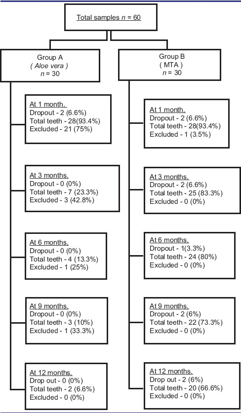
Table 2.
Summary of distribution of number of teeth with clinical and radiographic success or failure at various follow-up periods (1, 3, 6, 9, and 12 months)

Table 3.
Overall success rate (clinical and radiographic in percentage) observed between Group A (Aloe vera) and in Group B (mineral trioxide aggregate)
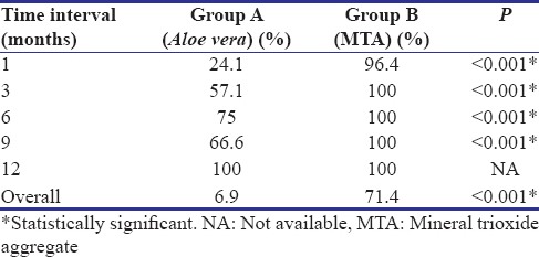
Figure 1.
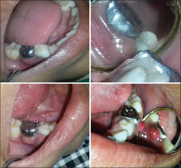
Composite digital image showing failure at 1-month follow-up interval of pulpotomized tooth treated with Aloe vera plant extract (swelling)
Figure 4.
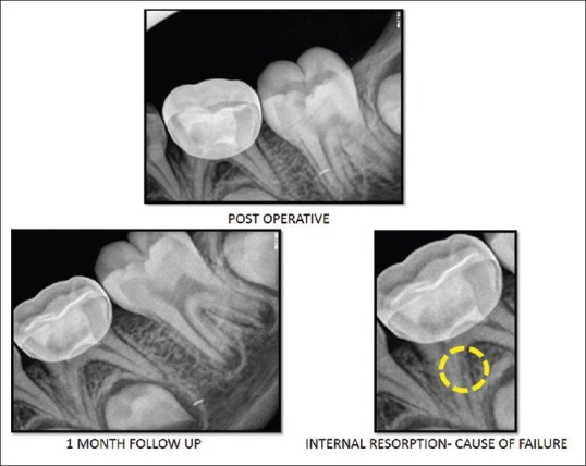
Composite digital radiographic image showing postoperative and failure within 1-month follow-up period of pulpotomized tooth treated with mineral trioxide aggregate (Group B)
Figure 2.
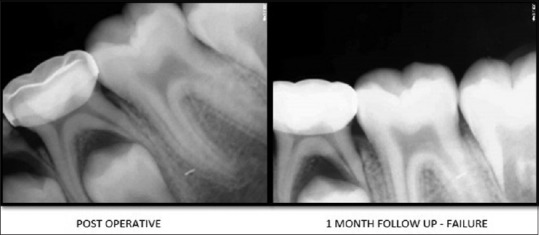
Composite digital radiographic image showing postoperative and failure within 1-month follow-up period of pulpotomized tooth treated with Aloe vera plant extract (Group A)
Discussion
The ideal pulp dressing material after pulpotomy should leave the radicular pulp vital, healthy and enclosed within an odontoblastic-lined dentine chamber. Along with that, it should be bactericidal, be harmless to the radicular pulp and surrounding tissues, should promote healing of the radicular pulp, and not interfere with the normal physiological process of root resorption. It should also show good clinical and radiographic success.[15] Many materials[2,3] have been investigated as possible replacements for FC because of concerns regarding its systemic distribution and potential for toxicity, carcinogenicity, and mutagenicity. Varying success rates and concerns regarding the safety of the application of these materials make it clear that additional research on the use of these pharmaco-therapeutic agents is necessary. Plants possessing medicinal properties have been widely employed in folk medicine as well as a therapeutic resource to primary medicine.[10] In the search of newer materials, there is a rapidly increasing interest and research toward these medicinal plants, considering that they will not be exerting any deleterious effect to the adjoining living tissues. Hence, the present study was undertaken to compare the clinical and radiographic outcomes of fresh A. barbadensis plant extract and MTA as pulpotomy medicaments in primary molar teeth.
The overall success rate of MTA pulpotomy at the end of 12-month follow-up was calculated as 71.4% after taking the dropout patients into considerations. The results were comparable to other studies such as reported by various authors.[5,16,17,18,19,20] In contrast, Sonmez et al.[21] have reported a lower success rate of 67%. The difference may be attributed to the restoration technique. They placed a wet cotton pellet over MTA for 1 day followed by the final restoration in the next day. The high success rate of MTA pulpotomy can be attributed to the property of dentin bridge formation, excellent biocompatibility, and alkalinity. However, good sealing ability without any sign of solubility forms the most distinctive feature. Only one failure was reported in case of MTA group at the end of 1st month follow-up. The failure was due to internal resorption in one of the root canals of the tooth [Figure 4]. The cause of failure can be attributed to the medicament itself and this is in accordance with the results obtained by Holan et al.[22]
From ancient times, A. vera plant extract has been extensively used by human beings as food, as well as medicine for the treatment of variety of systemic diseases. Around 2000 years ago, the Greek scientists regarded A. vera as the universal panacea.[9] Shelton[23] reviewed its chemical and therapeutic properties and concluded that A. vera gel is nontoxic, bactericidal, virucidal, and fungicidal against a broad range of microorganisms. It can be used as an anti-inflammatory agent, moisturizing agent as well as a wound-healing agent. In dentistry, it is used as a healing agent in treating aphthous ulcers, extraction socket, and chronic oral lesions as well as in the treatment of lichen planus. The anti-inflammatory role of steroids present in A. vera gel is well established, leading to the production of low levels of prostaglandins.[24,25,26] In view of various sanctified properties of A. vera, this natural product was selected for the study. The dental pulp is a specialized loose connective tissue, containing cells, fibers, ground substance, blood vessels, and nerve endings. The components found in the pulp tissue are very much similar to those found in dermal and epidermal layers of the skin. Hence, in the present study, pulpal stump was considered as an open wound, and fresh A. vera gel was applied directly over the pulp stump. There were no reported complications or side effects with A. vera being used in contact with animal[9] as well as human pulp tissue[1] and histologically.[1]
A. vera gel exhibits a limited shelf-life because of the rapid oxidation process when it is exposed to external environment as well as due to the microbial interaction which further degrades the gel and its constituents. The mucilaginous layer or the inner clear jelly substance of A. vera contains highest concentration of potentially beneficial components. They hold secret to the plant's medicinal properties and hence only this layer was placed onto the pulp stumps. Since the gel is semisolid in nature, an absorbable collagen sponge was placed over the gel. However, in contrast, we have observed 75% of failure (both clinical and radiographic) in A. vera group within 1 month of follow-up [Figures 1–3 and Table 2]. The overall success rate of A. vera pulpotomy at the end of 12-month follow-up was 6.9% after taking dropout patients into consideration. The results obtained in the present study were contradictory to Gupta et al.[1] who concluded that freshly extracted A. vera gel can be used as a successful pulpotomy agent. Gala-Garcia et al.[10] concluded that the application of A. vera placed in direct contact with the exposed rat pulp tissue has acceptable biocompatibility and can lead to tertiary dentin bridge formation. They attributed the reason behind this result to the various bioactive substances such as glycoprotein, polysaccharides, and beta-sitosterol in the stimulation of wound healing, cell proliferation, and angiogenesis.
Figure 3.
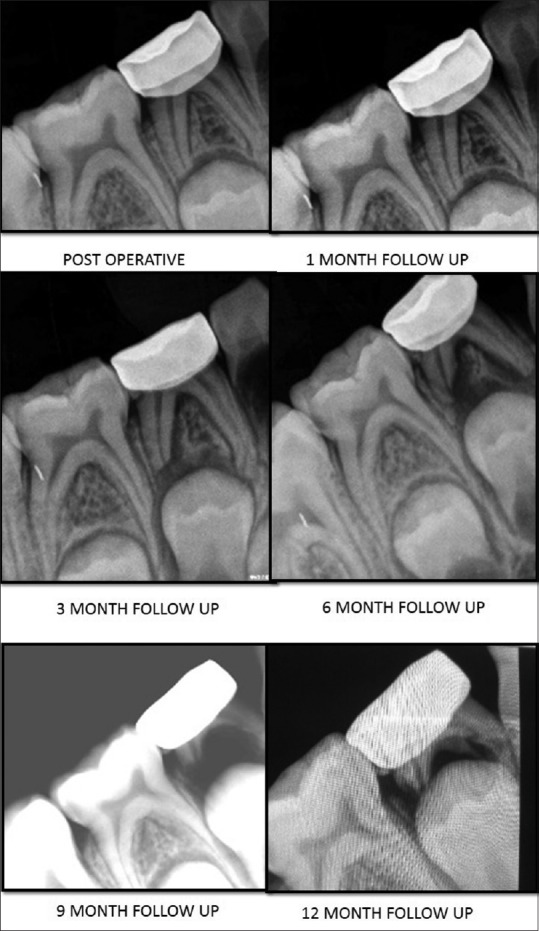
Composite digital radiographic image showing various follow-up periods of pulpotomized tooth treated with Aloe vera plant extract (Group A)
The teeth presented with only radiographic failures in this study were not treated immediately and were observed for further follow-up as they were asymptomatic and did not show any sign of clinical failure.[17] The difference between the overall success rate of A. vera and MTA pulpotomy was found to be statistically significant (P < 0.001) at all follow-up periods. Failures in pulpotomized teeth can be attributed to the medicament placed inside the pulp chamber because the changes produced inside the radicular pulp occur as a result of medicament–pulp interaction. Hence, further histological investigations should be conducted to ascertain the reaction between fresh A. vera gel and human dental pulp tissue. How a processed form of A. vera behaves in the vicinity of the human pulp tissue is also unknown and should be further explored in comparison with fresh A. vera plant extract.
Conclusions
MTA was found to be superior when compared to fresh A. barbadensis plant extract as a pulpotomy agent in primary molars. Even though A. vera gel is a natural and economically viable medicament, it has proved to be an unfavorable pulpotomy agent in primary teeth. Further studies are needed to ascertain the histological reaction between fresh A. vera gel and human dental pulp.
Financial support and sponsorship
Nil.
Conflicts of interest
There are no conflicts of interest.
References
- 1.Gupta N, Bhat M, Devi P, Girish Aloe-vera: A nature's gift to children. Int J Clin Pediatr Dent. 2010;3:87–92. doi: 10.5005/jp-journals-10005-1059. [DOI] [PMC free article] [PubMed] [Google Scholar]
- 2.Srinivasan V, Patchett CL, Waterhouse PJ. Is there life after Buckley's formocresol? Part I – A narrative review of alternative interventions and materials. Int J Paediatr Dent. 2006;16:117–27. doi: 10.1111/j.1365-263X.2006.00688.x. [DOI] [PubMed] [Google Scholar]
- 3.Yaman E, Görken F, Pinar Erdem A, Sepet E, Aytepe Z. Effects of folk medicinal plant extract Ankaferd Blood Stopper ® in vital primary molar pulpotomy. Eur Arch Paediatr Dent. 2012;13:197–202. doi: 10.1007/BF03262870. [DOI] [PubMed] [Google Scholar]
- 4.Torabinejad M, Pitt Ford TR, McKendry DJ, Abedi HR, Miller DA, Kariyawasam SP. Histologic assessment of mineral trioxide aggregate as a root-end filling in monkeys. J Endod. 1997;23:225–8. doi: 10.1016/S0099-2399(97)80051-9. [DOI] [PubMed] [Google Scholar]
- 5.Asgary S, Shirvani A, Fazlyab M. MTA and ferric sulfate in pulpotomy outcomes of primary molars: A systematic review and meta-analysis. J Clin Pediatr Dent. 2014;39:1–8. doi: 10.17796/jcpd.39.1.b454r616m2582373. [DOI] [PubMed] [Google Scholar]
- 6.Rao A, Rao A, Shenoy R. Mineral trioxide aggregate – A review. J Clin Pediatr Dent. 2009;34:1–7. doi: 10.17796/jcpd.34.1.n1t0757815067g83. [DOI] [PubMed] [Google Scholar]
- 7.Taheri JB, Azimi S, Rafieian N, Zanjani HA. Herbs in dentistry. Int Dent J. 2011;61:287–96. doi: 10.1111/j.1875-595X.2011.00064.x. [DOI] [PMC free article] [PubMed] [Google Scholar]
- 8.Tarro VE. The Honest Herbal: A Sensible Guide to the Use of Herbs and Related Remedies. 3rd ed. New York: Pharmaceutical Products Press; 1993. pp. 25–8. [Google Scholar]
- 9.Mohan R, Gundappa M. Aloe vera in dentistry – The herbal panacea. Indian J Dent Sci Res. 2013;29:15–20. [Google Scholar]
- 10.Gala-Garcia A, Teixeira KI, Mendes LL, Sobrinho AP, Santos VR, Cortes ME. Effect of Aloe vera on rat pulp tissue. Pharm Biol. 2008;46:302–8. [Google Scholar]
- 11.Pukallus ML, Plonka KA, Holcombe TF, Barnett AG, Walsh LJ, Seow WK. A randomized controlled trial of a 10 percent CPP-ACP cream to reduce mutans streptococci colonization. Pediatr Dent. 2013;35:550–5. [PubMed] [Google Scholar]
- 12.Ramachandran CT, Rao PS. Processing of Aloe vera leaf gel: A review. Am J Agric Biol Sci. 2008;3:502–10. [Google Scholar]
- 13.Fuks AB, Holan G, Davis JM, Eidelman E. Ferric sulfate versus dilute formocresol in pulpotomized primary molars: Long-term follow up. Pediatr Dent. 1997;19:327–30. [PubMed] [Google Scholar]
- 14.Durmus B, Tanboga I. In vivo evaluation of the treatment outcome of pulpotomy in primary molars using diode laser, formocresol, and ferric sulphate. Photomed Laser Surg. 2014;32:289–95. doi: 10.1089/pho.2013.3628. [DOI] [PMC free article] [PubMed] [Google Scholar]
- 15.Levin LG. Pulpal regeneration. Pract Periodontics Aesthet Dent. 1998;10:621–4. [PubMed] [Google Scholar]
- 16.Jayam C, Mitra M, Mishra J, Bhattacharya B, Jana B. Evaluation and comparison of white mineral trioxide aggregate and formocresol medicaments in primary tooth pulpotomy: Clinical and radiographic study. J Indian Soc Pedod Prev Dent. 2014;32:13–8. doi: 10.4103/0970-4388.127043. [DOI] [PubMed] [Google Scholar]
- 17.Neamatollahi H, Tajik A. Comparison of clinical and radiographic success rates of pulpotomy in primary molars using formocresol, ferric sulfate and mineral trioxide aggregate (MTA) J Dent. 2006;3:6–13. [Google Scholar]
- 18.Erdem AP, Guven Y, Balli B, Ilhan B, Sepet E, Ulukapi I, et al. Success rates of mineral trioxide aggregate, ferric sulfate, and formocresol pulpotomies: A 24-month study. Pediatr Dent. 2011;33:165–70. [PubMed] [Google Scholar]
- 19.Moretti AB, Sakai VT, Oliveira TM, Fornetti AP, Santos CF, Machado MA, et al. The effectiveness of mineral trioxide aggregate, calcium hydroxide and formocresol for pulpotomies in primary teeth. Int Endod J. 2008;41:547–55. doi: 10.1111/j.1365-2591.2008.01377.x. [DOI] [PubMed] [Google Scholar]
- 20.Niranjani K, Prasad MG, Vasa AA, Divya G, Thakur MS, Saujanya K. Clinical evaluation of success of primary teeth pulpotomy using mineral trioxide Aggregate®, Laser and Biodentine™ – An in vivo study. J Clin Diagn Res. 2015;9:ZC35–7. doi: 10.7860/JCDR/2015/13153.5823. [DOI] [PMC free article] [PubMed] [Google Scholar]
- 21.Sonmez D, Sari S, Cetinbas T. A Comparison of four pulpotomy techniques in primary molars: A long-term follow-up. J Endod. 2008;34:950–5. doi: 10.1016/j.joen.2008.05.009. [DOI] [PubMed] [Google Scholar]
- 22.Holan G, Eidelman E, Fuks AB. Long-term evaluation of pulpotomy in primary molars using mineral trioxide aggregate or formocresol. Pediatr Dent. 2005;27:129–36. [PubMed] [Google Scholar]
- 23.Shelton RM. Aloe vera. Its chemical and therapeutic properties. Int J Dermatol. 1991;30:679–83. doi: 10.1111/j.1365-4362.1991.tb02607.x. [DOI] [PubMed] [Google Scholar]
- 24.Zawahry ME, Hegazy MR, Helal M. Use of aloe in treating leg ulcers and dermatoses. Int J Dermatol. 1973;12:68–73. doi: 10.1111/j.1365-4362.1973.tb00215.x. [DOI] [PubMed] [Google Scholar]
- 25.Davis RH, Leitner MG, Russo JM. Topical anti-inflammatory activity of Aloe vera as measured by ear swelling. J Am Podiatr Med Assoc. 1987;77:610–2. doi: 10.7547/87507315-77-11-610. [DOI] [PubMed] [Google Scholar]
- 26.Vázquez B, Avila G, Segura D, Escalante B. Antiinflammatory activity of extracts from Aloe vera gel. J Ethnopharmacol. 1996;55:69–75. doi: 10.1016/s0378-8741(96)01476-6. [DOI] [PubMed] [Google Scholar]


